;:
Race Virus 101 - The Eugenics Investigation
(The Dysgenics Investigation)
Edited by Michael J. Ross
Published: May 12, 2019
Updated: June 28th, 2025
Website: MonsantoInvestigation.com
PollutionScience.com
-------------------------------------------------------------------------------------------------------------
Section 1: Chimera Viruses & DNA Specific Bio-Weapons
Section 2: Medical and Weaponized Gene Therapy
Section 3: Nanomites and Nanobots
Section 4: Archaea
Section 5: Bacteriophages & Nanotechnology
Section 6: Skin and Race
Section 7: Race, Virus & Disease
Section 8: Genetic Disorders and Diseases
Section 9: HIV
Section 10: Ancient Viruses ('Zombie Virus')
Section 11: Biohacking
Section 12: Zika
Section 13: Archaea
Section 14: Ocean Viruses
Section 15: Vaccines, bacteria & biofertilizers
Section 16: Azolla
Section 17: Chagas
Section 18: Trypanosoma Cruzi
Section 19: Poison, Bacteriophages & Viruses
Section 20: Cancer
Section 21: Pre-Historic & Ancient life
Section 22: Candida albicans
Section 23: Cryptococcus
Section 24: Genetically Modified Black Death & Plague
Section 25: Viruses and plants
Section 26: Space Viruses
Section 27: Staphylococcus
Section 28: Typhoid
Section 29: Rabies
Section 30: Ebola
Section 31: A Crimean-Congo hemorrhagic fever (CCHF)
Section 32: Marburg Virus
Section 33: SARS
Section 34: Coronavirus (COVID-19) & Middle East respiratory syndrome Coronavirus (MERS-CoV)
Section 35: MRSA
Section 36: Genetically Modified Viruses
Section 37: Foot and Mouth
Section 38: Bolivian Hemorrhagic Fever
Section 39: Machupo Virus
Section 40: Valley Fever
Section 41: Nipah Virus
Section 42: Junin Virus
Section 43: Miscellaneous Viruses
Section 44: Anthrax
Section 45: Clostridium
Section 46: Rickettsiae
Section 47: Tularemia
Section 48: Haplogroup
------------------
March 15th, 2022
https://archive.org/details/covid-news_202302
-----
Coronavirus Investigation News – Race Virus 201 – Part 1
March 15th, 2022
https://pollutionscience101.wordpress.com/2023/02/16/coronavirus-investigation-news-race-virus-201/
-----
Coronavirus Investigation News – Race Virus 201 – Part 2
March 15th, 2022
https://pollutionscience101.wordpress.com/2023/02/16/coronavirus-investigation-news-race-virus-201-part-2/
-----
Coronavirus Investigation News – Race Virus 201 – Part 3
March 15th, 2022
https://pollutionscience101.wordpress.com/2023/02/16/coronavirus-investigation-news-race-virus-201-part-3/
-----
Coronavirus Investigation News – Race Virus 201 – Part 4
March 15th, 2022
https://pollutionscience101.wordpress.com/2023/02/16/coronavirus-investigation-news-race-virus-201-part-4/
-----
Coronavirus Investigation News – Race Virus 201 – Part 5
March 15th, 2022
https://pollutionscience101.wordpress.com/2023/02/16/coronavirus-investigation-news-race-virus-201-part-5/
-----
Coronavirus Investigation News – Race Virus 201 – Part 6
March 15th, 2022
https://pollutionscience101.wordpress.com/2023/02/16/coronavirus-investigation-news-race-virus-201-part-6/
-----
Coronavirus Investigation News – Race Virus 201 – Part 7
March 15th, 2022
https://pollutionscience101.wordpress.com/2023/02/16/coronavirus-investigation-news-race-virus-201-part-7/
-----
Coronavirus Investigation News – Race Virus 201 – Part 8
March 15th, 2022
https://pollutionscience101.wordpress.com/2023/02/16/coronavirus-investigation-news-race-virus-201-part-8/
-----
Coronavirus Investigation News – Race Virus 201 – Part 9
March 15th, 2022
https://pollutionscience101.wordpress.com/2023/02/16/coronavirus-investigation-news-race-virus-201-part-9/
-----
Coronavirus Investigation News – Race Virus 201 – Part 10
March 15th, 2022
https://pollutionscience101.wordpress.com/2023/02/16/coronavirus-investigation-news-race-virus-201-part-10/
-----
Coronavirus Investigation News – Race Virus 201 – Part 11
March 15th, 2022
https://pollutionscience101.wordpress.com/2023/02/16/coronavirus-investigation-news-race-virus-201-part-11/
-----
Coronavirus Investigation News – Race Virus 201 – Part 12
March 15th, 2022
https://pollutionscience101.wordpress.com/2023/02/16/coronavirus-investigation-news-race-virus-201-part-12/
-----
4/15/2020 - Coronavirus Investigation News - Race Virus 201 - Pollution Science 101 (Covid-19 & SARS-CoV-2)
-----
Race Virus 101 Part 1
https://pollutionscience101.wordpress.com/2023/03/25/race-virus-101-part-1/
-----
Race Virus 101 – Part 2
https://pollutionscience101.wordpress.com/2023/03/26/race-virus-101-part-2/
-----
Race Virus 101 – Part 3
https://pollutionscience101.wordpress.com/2023/03/26/race-virus-101-part-3/
-----
Race Virus 301 – Coronavirus Investigation News – Part 1
https://pollutionscience101.wordpress.com/2023/03/26/race-virus-301-coronavirus-investigation-news-part-1/
-----
Race Virus 301 – Coronavirus Investigation News – Part 2
https://pollutionscience101.wordpress.com/2023/03/26/race-virus-301-coronavirus-investigation-news-part-2/
-----
Pollution Science 101 - Book Archive (Web Archive)
https://archive.org/details/@pollution_science?tab=web-archive
-----
Pollution Science 101 - Video Archive
https://archive.org/details/@pollution_science
-----
Monsanto Investigation.com - Video Archive
https://archive.org/details/@monsantoinvestigation
-----
Pollution Science 101 - Book Archive (Web Archive)
https://archive.org/details/@pollution_science?tab=web-archive
-----
------------------
-----------------
-----------------
Section 1: Chimera Viruses & DNA Specific Bio-Weapons
-----------------
-----------------
------------------
Genetically engineered viruses will WIPE OUT hundreds of millions, warns Bill Gates
2017
BIOTERRORISM has the potential to kill tens of millions of people and is a bigger threat to humanity than nuclear war, Bill Gates has warned.
https://www.express.co.uk/news/science/769636/Bill-Gates-microsoft-bioterrorism-munich-security-conference
-------------------------
Designing DNA Specific Bio-Weapons
2012
https://science.slashdot.org/story/12/10/29/0241252/designing-dna-specific-bio-weapons
"The Atlantic reports that experts in genetics and microbiology are convinced we may be only a few years away from the development of advanced, genetic bio-weapons able to target a single human being based on their DNA. The authors paint a scenario of the development of a virus that causes only mild flu in the general population but when the virus crosses paths with cells containing a very specific DNA sequence, the sequence would act as a molecular key to unlock secondary functions that would trigger a fast-acting neuro-destructive disease that produces memory loss and, eventually, death. The requisite equipment including gene sequencers, micro-array scanners, and mass spectrometers now cost over $1 million but on eBay, it can be had for as little as $10,000. According to Ronald Kessler, the author of the 2009 book In the President's Secret Service, Navy stewards gather bedsheets, drinking glasses, and other objects the president has touched—they are later sanitized or destroyed—in an effort to keep would-be malefactors from obtaining his genetic material. However no amount of Secret Service vigilance can ever fully secure the president's DNA, because an entire genetic blueprint can now be produced from the information within just a single cell. How to protect the President? The authors propose open-sourcing the president's genetic information to a select group of security-cleared researchers who could follow in the footsteps of the computer sciences, where 'red-team exercises,' are extremely common practices so a similar testing environment could be developed for biological war games. 'Advances in biotechnology are radically changing the scientific landscape. We are entering a world where imagination is the only brake on biology,' write the authors. 'In light of this coming synbio revolution, a wider-ranging relationship between scientists and security organizations—one defined by open exchange, continual collaboration, and crowd-sourced defenses—may prove the only way to protect the president.'"
------------------
Hacking the President’s DNA
2012
https://www.theatlantic.com/magazine/archive/2012/11/hacking-the-presidents-dna/309147/?single_page=true
The U.S. government is surreptitiously collecting the DNA of world leaders, and is reportedly protecting that of Barack Obama. Decoded, these genetic blueprints could provide compromising information. In the not-too-distant future, they may provide something more as well—the basis for the creation of personalized bioweapons that could take down a president and leave no trace.
------------------
Teacher Guide: How Do Viruses Recognize a Target Cell?
2002
https://teach.genetics.utah.edu/content/genetherapy/VirusesRecognizeTargetCell.pdf
Abstract: This activity demonstrates the specifi city of viral vectors for target cells in gene therapy delivery methods using two approaches: 1) STYROFOAM®models demonstrate viral ligand binding to receptor proteins on the surface of target cells; 2) Students use paper models of viruses and cells to fi nd the appropriate match between viral ligands and cell receptors.
Prior Knowledge Needed:Cell membranes contain protein receptors; virus structure; when a virus binds to a host cell, it injects its DNA or RNA into the host cell; gene therapy involves adding a normally functioning gene to cells to replace the function of a “faulty” gene.
------------------
Human gene targeting by viral vectors
2010
https://www.ncbi.nlm.nih.gov/pmc/articles/PMC3010411/
------------------
Targeting DNA
2012
https://www.the-scientist.com/features/targeting-dna-40937
-----------------
The Basics on Genes and Genetic Disorders
https://kidshealth.org/en/teens/genes-genetic-disorders.html
After 20 years of high-profile failure, gene therapy is finally well on its way to clinical approval.
Changing Genes
Sometimes scientists alter genes on purpose. For many years, researchers have altered the genes in plants to produce other plants with special characteristics, such as an increased resistance to disease and pests or the ability to grow in difficult environments. We call this genetic engineering.
Gene therapy is a promising new field of medical research. In gene therapy, researchers try to supply copies of healthy genes to cells with variant or missing genes so that the "good" genes will take over. Viruses are often used to carry the healthy genes into the targeted cells because many viruses can insert their own DNA into targeted cells.
But there are problems with gene therapy. Scientists still don't quite know what every gene in the human body does. Huge scientific efforts like The Human Genome Project and related projects have completed a map of the entire human genome (all of the genetic material on a living thing's chromosomes), but it will take many more years to find out what each gene does and how they interact with one another. For most diseases, scientists don't know if and how genes play a role. Plus, there are major difficulties inserting the normal genes into the proper cells without causing problems for the rest of the body.
There are also concerns that people might try changing genes for ethically troubling reasons, such as to make smarter or more athletic children. No one knows what the long-term effects of that kind of change would be.
Still, for many people who have genetic diseases, gene therapy holds the hope that they — or their children — will be able to live better, healthier lives.
------------------
How does gene therapy work?

https://ghr.nlm.nih.gov/primer/therapy/procedures
--------------------
Development of a Risk-Priority Score for Category A Bioterrorism Agents as an Aid for Public Health Policy
2006
https://watermark.silverchair.com/milmed.171.7.589.pdf?token=AQECAHi208BE49Ooan9kkhW_Ercy7Dm3ZL_9Cf3qfKAc485ysgAAAmEwggJdBgkqhkiG9w0BBwagggJOMIICSgIBADCCAkMGCSqGSIb3DQEHATAeBglghkgBZQMEAS4wEQQMrj80GfVh6vuYaNPYAgEQgIICFMr82xC2IeHYmko_Kz8iJDq7BKHkCzYdkr0Ge1X_XucdmMPEWSmU2r6I_6oe71IUZxkrqe_jac5Sb03Xp0mPUi7KzqP3zul0UbdilbpnMkDK5znKQT5IS4FBgdWvf0mEQijWQHmDDfYXevO1ZhjucwlMTjbZs4cSUjinud1RvCxYLGiTioPw7-Y9jkIXvvtQWfZGYoamsCzvGn4-WPYLQjX5o7OQ1H0i098G4p1_FUUxfGmitGF7N06O7gewyA7S2hLdNvm1udIAprxGQq_pdhty30EpcuHqMmDcT62qPwJr_R_Pmmw1UrQibTJ0WIi0hefM43D57dCICuapaBrwZFgsI3cSfZz2wAmJY13N4i8nSTwg4nGvVmbGGsC_qM5Luv587vyPQ3t6hwJViLwMWY0HlJely7mEDe3eGhntwKvVRFLLVjRI-6QCLiydn3RoaPXGP56ebS4AQkfI0DM-VtPfGh2QA3un0Nwo-K8eq1-jm7czz8yy-APmKOd-C_flxQG_QmOrCo7an0FClb89bjO2d92RncWiilIbCIRzxr1otusD72OL1zPWKIApwRGCVjeYyg-RNKdPIZHn2voZiq9IOgWLyo40kjGeNpDK8ZGVr9oBxaCLm_PkjhFBR2dexbh2F7XLG3Dtz8EiejFTa_2ONt-1x44qqhAnkxBoJad_oRQZTTEfhV5WEl2wzr1DNrO9wXk
--------------------------
10 Scariest Bioweapons
https://www.stufftoblowyourmind.com/blogs/10-scariest-bioweapons10.htm

#1 Chimera Viruses
Plague, smallpox, anthrax -- the world's deadliest biological agents aren't out to get you. Any harmful properties they possess are simply byproducts of their evolution. But what happens when scientists tinker with the genetic makeup of these organisms? What kind of horrors may come to life when we add the human desire to wage war to their natural design? Unfortunately, the creation of such life forms isn't just a page from a science fiction novel -- it's already happening.
In Greek and Roman mythology, the chimera combined elements of lion, goat and serpent into one monstrous form. Artists in the late medieval age often used the creature as a symbol to illustrate the complex nature of evil. In modern genetic science, a chimeric organism is a life form that contains genes from a foreign species. Given its namesake, you might expect all chimeric organisms to be awful examples of man twisting nature for nefarious ends. Fortunately, our increased understanding of genetic science has led to some beneficial creations. One such chimera, which combines the common cold with polio, may help cure brain cancer.
But as the war continues its forward momentum through human history, the abuse of such science is inevitable. Geneticists have already discovered the means to increase the lethality of such bioweapons as smallpox and anthrax by tweaking their genetic structure. By combining genes, however, scientists could theoretically create a virus that triggered two diseases at once. During the late 1980s, the Soviet Union's Chimera Project studied the feasibility of combining smallpox and Ebola into one super virus.
Other potential nightmare scenarios involve strains of viruses that require certain triggers. A stealth virus would remain dormant for an extended period until triggered by predetermined stimuli. Other possible chimeric bioweapons might require two components to become effective. Imagine a strain of botulinum toxin that, when combined with the botulinum toxin antidote, only becomes more lethal. Such a biological attack would not only result in a higher mortality rate, but might erode public trust in health initiatives, aid workers and government response to the outbreak.
From splitting the atom to cracking life's genomic riddles, the last century of scientific research has brought about tremendous potential for humans to build a better world -- or destroy the one they have.
#2 Nipah Virus
#3 Rinderpest
When Genghis Khan invaded Europe in the 13th century, he inadvertently unleashed a fearsome biological weapon in the wake of his conquest. The gray steppe cattle used by his supply trains introduced a deadly cattle plague, known throughout the world today by its German name, rinderpest.
Rinderpest is caused by a virus closely related to measles, and it affects cattle and other ruminant animals such as goats, bison and giraffes. The condition is highly contagious, causing fever, loss of appetite, dysentery and inflammation of the mucus membranes. The condition drags on for six to 10 days, when the animal typically succumbs to dehydration.
#4 Rice Blast
#5 Botulinum Toxin
#6 Tularemia
#7 Plague
#8 Ebola Hemorrhagic Fever
#9 Anthrax
#10 Smallpox
-------------------
Chimera viruses can help the fight against lymphomas
2017
https://www.sciencedaily.com/releases/2017/09/170914152251.htm
Researchers from Instituto de Medicina Molecular (iMM) Lisboa have created a chimera virus that allows the study of molecules to treat cancers caused by human herpes virus infection in mice models of disease.
There are several types of herpesvirus able to infect humans, such as herpes simplex, chickenpox, cytomegalovirus, Epstein-Barr and Kaposi Sarcoma herpesvirus. One of the main characteristics of herpesviruses is their ability to infect their hosts for life and in a small percentage of these people ultimately lead to cancer.
Cancers associated with Kaposi virus infection have an Achilles heel: their cells' viability is directly dependent on the survival of the virus, which means that if the virus were to be eliminated cancer cells would no longer proliferate, hence the cancer would be cured.
-------------------
Virus chimeras for gene therapy, vaccination, and oncolysis: adenoviruses and beyond.
2012
https://www.ncbi.nlm.nih.gov/pubmed/22633438
Abstract
Several challenges need to be addressed when developing viruses for clinical applications in gene therapy, vaccination, or viral oncolysis, including specific and efficient target cell transduction, virus delivery via the blood stream, and evasion of pre-existing immunity. With rising frequency, these goals are tackled by generating chimeric viruses containing nucleic acid fragments or proteins from two or more different viruses, thus combining different beneficial features of the parental viruses. These chimeras have boosted the development of virus-based treatment regimens for major inherited and acquired diseases, including cancer. Using adenoviruses as the paradigm and prominent examples from other virus families, we review the technological and functional advances in therapeutic virus chimera development and recent successful applications that can pave the way for future therapies.
-------------------
A Chimeric Dengue Virus Vaccine using Japanese Encephalitis Virus Vaccine Strain SA14-14-2 as Backbone Is Immunogenic and Protective against Either Parental Virus in Mice and Nonhuman Primates
2013

(In vitro characterization of ChinDENV. (A) Immunostaining of JEV-, ChinDENV-, DENV-2-, or mock-infected BHK cells with specific anti-JEV E, DENV-2 E, and JEV NS1 antibodies. Cells were infected with viruses at an MOI of 0.01. At 48 h postinfection, JEV or DENV-2 antigen was detected with monoclonal antibody 4AD5F5D5D6 (for JEV E protein), 2B8 (for DENV-2 E protein), and JN1 (for JEV NS1 protein). (B) Plaque morphology of JEV, ChinDENV, and DENV-2 on BHK-21 cells grown in 6-well plates were infected with a 10-fold serial dilution of viruses. The plates were incubated at 37°C for 1 h. Supernatant was removed and cells were overlaid with 1% low-melting-point agarose in DMEM containing 2% FBS. After further incubation at 37°C for 4 days, the cells were fixed with 4% formaldehyde and stained with 0.2% crystal violet to visualize the plaques. (C) Plaque morphology of ChinDENV passaged in PHK cells. The chimeric virus ChinDENV was passaged in PHK cells up to 15 times. The plaque phenotypes of passage 3, 9, and 15 viruses were examined on BHK-21 cells 4 days after infection by plaque assay. (D) Growth curves of the chimera ChinDENV and parental viruses JEV and DENV-2 in cell culture. Monolayers of Vero, C6/36, and PHK cells were infected with the indicated viruses at an MOI of 0.01. At each time point, the media were removed and virus titers in cell culture media were determined on BHK-21 cells by plaque assay).
https://jvi.asm.org/content/87/24/13694
-----------------------
An Alphavirus Replicon Particle Chimera Derived from Venezuelan Equine Encephalitis and Sindbis Viruses Is a Potent Gene-Based Vaccine Delivery Vector
2003
https://jvi.asm.org/content/77/19/10394
-------------------
Chimeric yellow fever 17D-Zika virus (ChimeriVax-Zika) as a live-attenuated Zika virus vaccine
2018
https://www.nature.com/articles/s41598-018-31375-9
-------------------
Generation of CD19-chimeric antigen receptor modified CD8+ T cells derived from virus-specific central memory T cells
2012
http://www.bloodjournal.org/content/119/1/72?sso-checked=true
------------------
Scientists are genetically engineering immune systems to attack and destroy HIV
2018
https://qz.com/1169727/genetically-engineered-car-t-cells-successfully-treated-hiv-in-monkeys-and-petri-dish-studies/
-------------------
Targeting of Influenza Viral Epitopes to Antigen-Presenting Cells by Genetically Engineered Chimeric Molecules in a Humanized NOD SCID Gamma Transfer Model
2018
https://www.liebertpub.com/doi/abs/10.1089/hum.2018.100
------------------
Chimera virus: How far science can go?
2016
http://outbreaknewstoday.com/chimera-virus-how-far-science-can-go-65815/
“In warfare there are no constant conditions. He, who can modify his tactics in relation to his opponent, will succeed and win. “
Sun Tzu
The biological weapons and the whole set of biological agents capable to be abused in some of the future asymmetric or even symmetric conflicts is a nightmare scenario. A long time ago, the idea of using some exotic virus or forgotten bacteria in a terrorist attack has sounded like a science fiction movie. Now it is our potential reality, the dimension of biological security which will never be the same. Bugs will be always around, ready to be militarized from humans and against humans.
--------------------------------
------------------------------
------------------------------
Section 2: Medical and Weaponized Gene Therapy
----------------------------
----------------------------
------------------------------
Synthesized Nanoparticles Act As Artificial Viruses for Gene Therapy
04/09/2015
Researchers of the Nanobiology Unit from the Universitat Autònoma de Barcelona (UAB) Institute of Biotechnology and Biomedicine, led by Antonio Villaverde, managed to create artificial viruses, protein complexes with the ability of self-assembling and forming nanoparticles which are capable of surrounding DNA fragments, penetrating the cells and reaching the nucleus in a very efficient manner, where they then release the therapeutic DNA fragments. The achievement represents an alternative with no biological risk to the use of viruses in gene therapy.
http://www.cemag.us/news/2015/04/synthesized-nanoparticles-act-artificial-viruses-gene-therapy
----------------------------
Gene therapy is ‘new weapon’ in fight against flu
https://www.channel4.com/news/gene-therapy-is-new-weapon-in-fight-against-flu
----------------------------
Gene Therapy May Be Our Newest — and Most Effective — Weapon Against Alzheimer’s Disease
2015https://singularityhub.com/2015/09/06/gene-therapy-may-be-our-newest-and-most-effective-weapon-against-alzheimers-disease/#sm.01grurjq199oe2n11of2oe3dqy4vq
----------------------------
Huntington's disease goes viral as UniQure inches ahead in gene therapy race
2019
FDA grants “Investigational New Drug” status to Huntingtin-lowering gene-therapy agent AMT-130, clearing path to human trials in Huntington’s Disease patients
--------------------------------
Gene Therapy Against HIV: Fighting The Virus In Disguise
2017https://www.bsgct.org/gene-therapy-against-hiv-fighting-the-virus-in-disguise/
----------------------------------
Gene Therapy (List)
https://www.sciencedaily.com/news/health_medicine/gene_therapy/
---------------------------------
Gene gun
https://en.wikipedia.org/wiki/Gene_gun
In genetic engineering, a gene gun or a biolistic particle delivery system, originally designed for plant transformation, is a device for delivering exogenous DNA (transgenes) to cells. The payload is an elemental particle of a heavy metal coated with DNA (typically plasmid DNA). This technique is often simply referred to as biolistics.
This device is able to transform almost any type of cell, including plants, and is not limited to transformation of the nucleus; it can also transform organelles, including plastids.

(A gene gun is used for delivery of exogenous DNA to cells. This method is known as 'biolistics'. Gene guns can be used effectively on most cells but are mainly used on plant cells).
Gene gun design
The gene gun was originally a Crosman air pistol modified to fire dense tungsten particles. It was invented by John C Sanford, Ed Wolf and Nelson Allen at Cornell University, and Ted Klein of DuPont, between 1983 and 1986. The original target was onions (chosen for their large cell size) and it was used to deliver particles coated with a marker gene. Genetic transformation was then proven when the onion tissue expressed the gene.
The earliest custom manufactured gene guns (fabricated by Nelson Allen) used a 22 caliber nail gun cartridge to propel an extruded polyethylene cylinder (bullet) down a 22 cal. Douglas barrel. A droplet of the tungsten powder and genetic material was placed on the bullet and shot down the barrel at a lexan "stopping" disk with a Petri dish below. The bullet welded to the disk and the genetic material blasted into the sample in the dish with a doughnut effect (devastation in the middle, a ring of good transformation and little around the edge). The gun was connected to a vacuum pump and was under vacuum while firing. The early design was put into limited production by a Rumsey-Loomis (a local machine shop then at Mecklenburg Rd in Ithaca, NY, USA). Later the design was refined by removing the "surge tank" and changing to nonexplosive propellants. DuPont added a plastic extrusion to the exterior to visually improve the machine for mass production to the scientific community. Biorad contracted with Dupont to manufacture and distribute the device. Improvements include the use of helium propellant and a multi-disk-collision delivery mechanism. Other heavy metals such as gold and silver are also used. Gold may be favored because it has better uniformity than tungsten and tungsten can be toxic to cells, but its use may be limited due to availability and cost.[citation needed]
--------------------------------
Why Gene-Editing Technology Just Made the Government's List of WMDs
2016 The U.S. government has pointed to a technique that scientists are using to modify genes as one of the top threats posed by weapons of mass destruction.
https://www.inc.com/graham-winfrey/the-controversial-technology-linking-designer-babies-and-wmd.html
---------------------------------
Genetically Engineered Bioweapons: A New Breed of Weapons for Modern Warfare
2013
Gram stained cerebrospinal fluid showing gram-positive anthrax baccilli (purple rods)
https://sites.dartmouth.edu/dujs/2013/03/10/genetically-engineered-bioweapons-a-new-breed-of-weapons-for-modern-warfare/
---------------------------------
Why the FBI and Pentagon are afraid of this new genetic technology
2015https://www.statnews.com/2015/11/12/gene-drive-bioterror-risk/
A powerful new genetic technology could eliminate scourges such as malaria and rid entire countries of destructive invasive species. But officials from the FBI to the Pentagon to the United Nations bioweapons office, STAT has learned, are concerned about the potential of “gene drives” to alter evolution in ways scientists can’t imagine, and even offer a devastating new tool to bioterrorists. Now they are scrambling to get ahead of it.
--------------------------------
Top U.S. Intelligence Official Calls Gene Editing a WMD Threat
2016
Easy to use. Hard to control. The intelligence community now sees CRISPR as a threat to national safety.
https://www.technologyreview.com/s/600774/top-us-intelligence-official-calls-gene-editing-a-wmd-threat/--------------------------------
Dangerous combination: Is CRISPR a potential weapon for terrorists?
2017
https://geneticliteracyproject.org/2017/06/08/dangerous-combination-crispr-potential-weapon-terrorists/---------------------------------
Could gene editing tools such as CRISPR be used as a biological weapon?
2017
https://theconversation.com/could-gene-editing-tools-such-as-crispr-be-used-as-a-biological-weapon-82187
----------------------------------
Rampage may stoke CRISPR fears, but scientists say it could be educational, too
2018
‘Hollywood loves to jump on new and potentially scary technologies’
https://www.theverge.com/2018/4/18/17248402/rampage-crispr-gene-editing-technology-hollywood-science-representation----------------------------------
Genome projects and gene therapy: gateways to next generation biological weapons.
2003https://www.ncbi.nlm.nih.gov/pubmed/14680038
-------------------------------
Gene Therapy and Genetic Engineering
https://medicine.missouri.edu/centers-institutes-labs/health-ethics/faq/gene-therapy
Introduction
The cells of a human being or other organism have parts called “genes” that control the chemical reactions in the cell that make it grow and function and ultimately determine the growth and function of the organism. An organism inherits some genes from each parent and thus the parents pass on certain traits to their offspring.
Gene therapy and genetic engineering are two closely related technologies that involve altering the genetic material of organisms. The distinction between the two is based on purpose. Gene therapy seeks to alter genes to correct genetic defects and thus prevent or cure genetic diseases. Genetic engineering aims to modify the genes to enhance the capabilities of the organism beyond what is normal.
Ethical controversy surrounds possible use of the both of these technologies in plants, nonhuman animals, and humans. Particularly with genetic engineering, for instance, one wonders whether it would be proper to tinker with human genes to make people able to outperform the greatest Olympic athletes or much smarter than Einstein.
Techniques of Genetic Alteration
Two problems must be confronted when changing genes. The first is what kind of change to make to the gene. The second is how to incorporate that change in all the other cells that are must be changed to achieve a desired effect.
There are several options for what kind of change to make to the gene. DNA in the gene could be replaced by other DNA from outside (called “homologous replacement). Or the gene could be forced to mutate (change structure – “selective reverse mutation.”) Or a gene could just be added. Or one could use a chemical to simply turn off a gene and prevent it from acting.
There are also several options for how to spread the genetic change to all the cells that need to be changed. If the altered cell is a reproductive cell, then a few such cells could be changed and the change would reach the other somatic cells as those somatic cells were created as the organism develops. But if the change were made to a somatic cell, changing all the other relevant somatic cells individually like the first would be impractical due to the sheer number of such cells. The cells of a major organ such as the heart or liver are too numerous to change one-by-one. Instead, to reach such somatic cells a common approach is to use a carrier, or vector, which is a molecule or organism. A virus, for example, could be used as a vector. The virus would be an innocuous one or changed so as not to cause disease. It would be injected with the genetic material and then as it reproduces and “infects” the target cells it would introduce the new genetic material. It would need to be a very specific virus that would infect heart cells, for instance, without infecting and changing all the other cells of the body. Fat particles and chemicals have also been used as vectors because they can penetrate the cell membrane and move into the cell nucleus with the new genetic material.
---------------------------
The gene therapy sector is experiencing an acceleration.
2019
https://www.risingtidebio.com/what-is-gene-therapy-uses/
The FDA, EMA and China are all beginning to approve gene therapy products. Every day there’s a new development. In this article, I attempt to make sense of this world.
I provide a brief background on the science and then discuss both the history and the future of gene therapy.
For the past 40 years, the scientific community has been learning to modify human DNA using viruses. This work is extremely challenging and progress has been slow but steady.
In the past, scientists have failed to design a gene therapy that can safely and effectively treat a disease.
All of this seems to be changing.
In 2017, we saw 2 gene therapy FDA approvals in the United States: Yescarta (Gilead) and Kymriah (Novartis).
Gene therapies have previously been approved in Europe and China (Glybera, Stremelis, Gendicine).
In 2018, we witnessed a landmark FDA approval for a gene therapy product called Luxturna. Spark Therapeutics is using an AAV virus to deliver a gene that is missing in patients suffering from an inherited eye disease. This treatment is restoring “functional vision” to the blind.
Now, in 2019, we are expecting a gene therapy approval from Novartis for a tragic condition called spinal muscular atrophy (SMA). Their clinical data looks good…and also, Novartis believes their treatment is cost-effective between $4-5 million/patient.
Yes, that’s right…$4-5M per treatment.
It can be a bit tricky to wrap your brain around this field. Most people I talk to (scientists and non-scientists) are not aware of the current state of gene therapy. Most people don’t know what has been done and what is coming down the pipeline.
----------------------------
The Next Generation Of Warfare: Genetically Engineered Viruses
2018
https://www.activistpost.com/2018/11/the-next-generation-of-warfare-genetically-engineered-viruses.html
-----------------------------
Genetically Engineered Viruses Are Next Generation Of Warfare
2018
https://www.technocracy.news/genetically-engineered-viruses-are-next-generation-of-warfare/
Nation-states would hesitate to use GMO viruses because of the ‘mutually assured destruction’ doctrine. However, any number of small groups of radical terrorists would not hesitate to release a plague on mankind.
-----------------------------
Advantages and disadvantages of genetically engineered live vaccines
2013
https://www.slideshare.net/singh_br1762/advantages-and-disadvantages-of-genetically-engineered-live-vaccines
----------------------------
---------------------------
---------------------------
Section 3: Nanomites and Nanobots
-------------------------
-------------------------
--------------------------
Nanomites from GI Joe Become a Reality
2014
https://www.youtube.com/watch?v=JQpIWU_DHWk
Researchers at Pennsylvania State University have inserted nanomotors inside living human cells, and manipulated them using ultrasound pulses. Advanced versions of these nanomotors could be used in the medical field for targeted therapy and other tasks. The video of the nanomotors inside the cell was taken under 1000x magnification. This is a truly remarkable achievement by PSU researchers.
----------------------------
Nanomotors controlled inside living human cells for the first time
2014
https://www.theverge.com/2014/2/16/5417266/nanomotors-controlled-inside-living-human-cells-for-the-first-time

Scientists at Penn State University have successfully controlled tiny nanomotors inside living human cells. Consisting of tiny, rocket-shaped bits of metal, the nanomotors were propelled by ultrasonic waves and steered with magnets. Researcher Tom Mallouk wasn't afraid to talk up potential future applications, saying that the technology could one day be used "to treat cancer and other diseases by mechanically manipulating cells from the inside." Once inside a living cell, the nanomotors could pulverize the cell's contents like an "egg beater" or just break the cell's membrane, Penn State's note about the research says, which could allow for targeted attacks on specific cells. More importantly, Mallouk says that the nanomotors were able to move independently of one another, instead of the "whole mass of them going in one direction."
----------------------------
Mechanical Force-Triggered Drug Delivery
2016
https://pubs.acs.org/doi/full/10.1021/acs.chemrev.6b00369?src=recsys
Advanced drug delivery systems (DDS) enhance treatment efficacy of different therapeutics in a dosage, spatial, and/or temporal controlled manner. To date, numerous chemical- or physical-based stimuli-responsive formulations or devices for controlled drug release have been developed. Among them, the emerging mechanical force-based stimulus offers a convenient and robust controlled drug release platform and has attracted increasing attention. The relevant DDS can be activated to promote drug release by different types of mechanical stimuli, including compressive force, tensile force, and shear force as well as indirect formats, remotely triggered by ultrasound and magnetic field. In this review, we provide an overview of recent advances in mechanically activated DDS. The opportunities and challenges regarding clinical translations are also discussed.
------------------------------
Progress in Nanomedicine and Medical Nanorobotics
http://www.nanomedicine.com/Papers/ProgressNM06.pdf
CONTENTS1.
Nanotechnology and Nanomedicine.................... 1
2. Medical Nanomaterials and Nanodevices................ 3
2.1. Nanopores................................. 3
2.2. Artificial Binding Sites and Molecular Imprinting...... 4
2.3. Quantum Dots and Nanocrystals.................. 4
2.4. Fullerenes and Nanotubes...................... 5
2.5. Nanoshells and Magnetic Nanoprobes.............. 6
2.6. Targeted Nanoparticles and Smart Drugs............ 7
2.7. Dendrimers and Dendrimer-Based Devices......... 10
2.8. Radio-Controlled Biomolecules.................. 11
3. Microscale Biological Robots........................ 13
3.1. Engineered Viruses.......................... 13
3.2. Engineered Bacteria.......................... 14
4. Medical Nanorobotics............................. 16
4.1. Early Thinking in Medical Nanorobotics........... 16
4.2. Nanorobot Parts and Components................ 16
4.3. Self-Assembly and Directed Parts Assembly......... 23
4.4. Positional Assembly and Molecular Manufacturing.... 29
4.5. Nanorobot Applications Designs and Scaling Studies . . 39
References..................................... 44
-----------------------------
An army of micro-robots can wipe out dental plaque
- Date:
- April 25, 2019
- Source:
- University of Pennsylvania
- Summary:
- A swarm of micro-robots, directed by magnets, can
break apart and remove dental biofilm, or plaque, from a tooth. The
innovation arose from a cross-disciplinary partnership among dentists,
biologists, and engineers.

https://www.sciencedaily.com/releases/2019/04/190425104323.htm
-----------------------------
Magnetic nanoparticles could stop blood clot-caused strokes
February 23, 2015
http://www.sciencedaily.com/releases/2015/02/150223122427.htm
-----------------------------
Magnetically guided virus stamping for the targeted infection of single cells or groups of cells
18 October 2019
https://www.nature.com/articles/s41596-019-0221-z
-----------------------------------
Magnets Turn Viruses Into Bacteria-Killers A team of international scientists has used phage-enhanced nanoparticles to kill bacteria that pollute water treatment systems.
Aug. 18, 2017
https://www.asianscientist.com/2017/08/tech/magnet-virus-bacteria-water-treatment/
-----------------------------------
Engineer Finds Way to Pull Diseases From Blood Using Magnets
November 12 2019
"In theory, you can go after almost anything. Poisons, pathogens, viruses, bacteria..."
https://futurism.com/neoscope/engineer-pull-diseases-blood-magnets
----------------------------------
12 magnets show how viruses are built
Apr 18, 2019
https://www.youtube.com/watch?v=3X6qEE2fHvE
----------------------------------
Magnets may one day cull deadly germs from blood
February 6, 2017
Tiny particles of antibody-covered iron would grab bacteria, then get mopped up by magnet
https://www.sciencenewsforstudents.org/article/magnets-may-one-day-cull-deadly-germs-blood
-----------------------------
Biology,
genetics, nanotechnology, neuroscience, materials science, biotech,
chemical engineering, 3 d, super computing, quantum physics, energy,
design, & sustainability
https://www.slideshare.net/BRuss10/biology-genetics-nanotechnology-neuroscience-materials-science-biotech-chemical-engineering-3-d-super-computing-quantum-physics-energy-design-sustainability
----------------------------
Genotoxic effect
https://www.slideshare.net/sreeremyasasi/genotoxic-effect
----------------------------
Nanorobots that hide in your blood like viruses could someday fight cancer
2014

https://qz.com/202221/nanorobots-that-hide-in-your-blood-like-viruses-could-someday-fight-cancer/
---------------------------
DNA Nanobots Can Fool the Immune System by Disguising Themselves as Viruses
2014
Scientists took a cue from the one of the most efficient genetic-code delivery systems in history or biology.
https://www.vice.com/en_us/article/xywy8a/dna-nanobots-can-fool-the-immune-system-by-disguising-themselves-as-viruses
---------------------------
‘Nanobot’ viruses tag and round up bacteria in food and water
2018
Tweaking DNA and adding magnetic nanoparticles creates a new tool to test for contaminants
https://www.sciencenews.org/article/nanobot-viruses-tag-and-round-bacteria-food-and-water
----------------------------
Future of lung treatment: Malaysian scientists join Harvard team creating safe, effective nano drugs
2016
Additional Malaysian nanoscience research includes converting greenhouse gases into energy source; 'Smart farming' nanosensors; New program aims for macro impact in health, energy, environment, agriculture, electronics
https://www.eurekalert.org/pub_releases/2016-01/tca-fol010516.php
--------------------------
Engineered mRNA-expressed antibodies prevent respiratory syncytial virus infection
2018
aPali is anchored to the membrane and inhibits RSV in cells. a Schematic of aPali anchored to the plasma membrane. b Schematic of aPali and sPali mRNA delivery and expression. c Cells were transfected with vehicle control or 1 μg of either aPali heavy chain only, aPali light chain, or both the heavy and light chain mRNAs. 24 h later, cells were fixed, permeabilized, and stained with a donkey anti-human secondary antibody (white).
https://www.nature.com/articles/s41467-018-06508-3
--------------------------
Adeno-associated Virus-mediated Transgene Expression in Genetically Defined Neurons of the Spinal Cord
2018
https://www.jove.com/video/57382/adeno-associated-virus-mediated-transgene-expression-genetically
Summary
Intraspinal injection of recombinase dependent recombinant adeno-associated virus (rAAV) can be used to manipulate any genetically labelled cell type in the spinal cord. Here we describe how to transduce neurons in the dorsal horn of the lumbar spinal cord. This technique enables functional interrogation of the manipulated neuron subtype.
--------------------------
Nanorobot takes on hepatitis C virus, wins
2012
https://newatlas.com/nanoparticles-hepatitis-c-university-florida/23379/
-------------------------
Nanorobot Hardware Architecture for Medical Defense
2008
https://www.ncbi.nlm.nih.gov/pmc/articles/PMC3675524/
-------------------------
Nanorobotic Applications in Medicine: Current Proposals and Designs
2014
https://www.ncbi.nlm.nih.gov/pmc/articles/PMC4562685/
------------------------
New DNA nanorobots successfully target and kill off cancerous tumors
2018
https://techcrunch.com/2018/02/12/new-dna-nanorobots-successfully-targeted-and-killed-off-cancerous-tumors/
---------------------------
New Vaccines Will Permanently Alter Human DNA
Why is the government so maniacal about injecting vaccines?
May 17, 2016
Consider this article in light of the accelerating push to mandate and enforce vaccination across the planet.
The reference is the New York Times, 3/15/15, “Protection Without a Vaccine.” It describes the frontier of research. Here are key quotes that illustrate the use of synthetic genes to “protect against disease,” while changing the genetic makeup of humans. This is not science fiction:
“By delivering synthetic genes into the muscles of the [experimental] monkeys, the scientists are essentially re-engineering the animals to resist disease.”
“’The sky’s the limit,’ said Michael Farzan, an immunologist at Scripps and lead author of the new study.”
“The first human trial based on this strategy — called immunoprophylaxis by gene transfer, or I.G.T. — is underway, and several new ones are planned.”
“I.G.T. is altogether different from traditional vaccination. It is instead a form of gene therapy. Scientists isolate the genes that produce powerful antibodies against certain diseases and then synthesize artificial versions. The genes are placed into viruses and injected into human tissue, usually muscle.”
Here is the punchline: “The viruses invade human cells with their DNA payloads, and the synthetic gene is incorporated into the recipient’s own DNA. If all goes well, the new genes instruct the cells to begin manufacturing powerful antibodies.”
Read that again: “the synthetic gene is incorporated into the recipient’s own DNA.” Alteration of the human genetic makeup. Permanent alteration.
https://www.infowars.com/new-vaccines-will-permanently-alter-human-dna/
-------------------------
Synthesized Nanoparticles Act As Artificial Viruses for Gene Therapy
04/09/2015
Researchers of the Nanobiology Unit from the Universitat Autònoma de Barcelona (UAB) Institute of Biotechnology and Biomedicine, led by Antonio Villaverde, managed to create artificial viruses, protein complexes with the ability of self-assembling and forming nanoparticles which are capable of surrounding DNA fragments, penetrating the cells and reaching the nucleus in a very efficient manner, where they then release the therapeutic DNA fragments. The achievement represents an alternative with no biological risk to the use of viruses in gene therapy.
http://www.cemag.us/news/2015/04/synthesized-nanoparticles-act-artificial-viruses-gene-therapy
------------------------------------------
PROTACs: A New Type of Drug That Can Target All Disease-Causing Proteins
June 11, 2015
http://scitechdaily.com/protacs-a-new-type-of-drug-that-can-target-all-disease-causing-proteins/
A newly published study from Yale University details the discovery of a new type of drug, called Proteolysis Targeting Chimeras (PROTACs), which can target all disease-causing proteins.
Current drugs block the actions of only about a quarter of known disease-causing proteins, but Yale University researchers have developed a technology capable of not just inhibiting, but destroying every protein it targets.
The new type of drug, called Proteolysis Targeting Chimeras (PROTACs), also can continue to destroy mutant proteins in mouse tumors, according to a new study published June 10 in the journal Nature Chemical Biology.
“This new drug modality culminates a decade of work in the field by my lab,” said Craig Crews, the Lewis B. Cullman Professor of Molecular, Cellular, and Developmental Biology and senior author of the paper, which was done in collaboration with scientists from GlaxoSmithKline and Arvinas, LLC.
Almost all current drugs are small molecules designed to fit into the folds of disease-causing proteins and inhibit their function. High doses are often needed to ensure that protein function is blocked sufficiently to produce therapeutic results, which in turn can produce harmful side effects.
In contrast, PROTACs engage the cells’ own protein degradation machinery to destroy targeted proteins by tagging them for removal and can do so multiple times, meaning it can work at lower doses, the authors say. This suggests this new type of drug has not only the potential to target proteins that are not currently “pharmaceutically vulnerable” but could do so safely, Crews said.
“This is a game-changer for drug development,” Crews said.
----------------------------------------
The future of medicine could be found in this tiny crystal ball
February 4, 2016
A Drexel University materials scientist has discovered a way to grow a crystal ball in a lab. Not the kind that soothsayers use to predict the future, but a microscopic version that could be used to encapsulate medication in a way that would allow it to deliver its curative payload more effectively inside the body.

http://phys.org/news/2016-02-future-medicine-tiny-crystal-ball.html?utm_source=menu&utm_medium=link&utm_campaign=item-menu
-------------------------
Researchers may have discovered fountain of youth by reversing aging in human cells
May 27, 2015
http://www.gizmag.com/reversal-of-aging-human-cell-lines/37721/
--------------------------
Researchers discover surprisingly wide variation across species in genetic systems that influence aging
May 28th, 2015
http://phys.org/news/2015-05-surprisingly-wide-variation-species-genetic.html
---------------------------
Use of genetically modified viruses and genetically engineered virus-vector vaccines: environmental effects.
2006
https://www.ncbi.nlm.nih.gov/pubmed/16982535
Abstract
Despite major therapeutic advances, infectious diseases remain highly problematic. Recent advancements in technology in producing DNA-based vaccines, together with the growing knowledge of the immune system, have provided new insights into the identification of the epitopes needed to target the development of highly targeted vaccines. Genetically modified (GM) viruses and genetically engineered virus-vector vaccines possess significant unpredictability and a number of inherent harmful potential hazards. For all these vaccines, safety assessment concerning unintended and unwanted side effects with regard to targeted vaccinees has always been the main focus. Important questions concerning effects on nontargeted individuals within the same species or other species remain unknown. Horizontal transfer of genes, though lacking supportive experimental or epidemiological investigations, is well established. New hybrid virus progenies resulting from genetic recombination between genetically engineered vaccine viruses and their naturally occurring relatives may possess totally unpredictable characteristics with regard to host preferences and disease-causing potentials. Furthermore, when genetically modified or engineered virus particles break down in the environment, their nuclei acids are released. Appropriate risk management is the key to minimizing any potential risks to humans and environment resulting from the use of these GM vaccines. There is inadequate knowledge to define either the probability of unintended events or the consequences of genetic modifications. The objective of this article is to highlight the limitations in environmental risk assessment and raise awareness of the potential risks involving the use of genetically modified viruses and genetically engineered virus-vector vaccines.
----------------------------
Self-replicating Nanobots could DESTROY all life on Earth, warn experts
2017
https://www.express.co.uk/news/science/825989/nanotechnology-nanobots-grey-goo-end-of-the-world
-----------------------------
---------------------------
---------------------------
Section 4: Archaea
--------------------------
--------------------------
-----------------------------
Nanobiomotors of archaeal DNA repair machineries: current research status and application potential
2014
https://www.ncbi.nlm.nih.gov/pmc/articles/PMC4080772/
Abstract
Nanobiomotors perform various important functions in the cell, and they also emerge as potential vehicle for drug delivery. These proteins employ conserved ATPase domains to convert chemical energy to mechanical work and motion. Several archaeal nucleic acid nanobiomotors, such as DNA helicases that unwind double-stranded DNA molecules during DNA damage repair, have been characterized in details. XPB, XPD and Hjm are SF2 family helicases, each of which employs two ATPase domains for ATP binding and hydrolysis to drive DNA unwinding. They also carry additional specific domains for substrate binding and regulation. Another helicase, HerA, forms a hexameric ring that may act as a DNA-pumping enzyme at the end processing of double-stranded DNA breaks. Common for all these nanobiomotors is that they contain ATPase domain that adopts RecA fold structure. This structure is characteristic for RecA/RadA family proteins and has been studied in great details. Here we review the structural analyses of these archaeal nucleic acid biomotors and the molecular mechanisms of how ATP binding and hydrolysis promote the conformation change that drives mechanical motion. The application potential of archaeal nanobiomotors in drug delivery has been discussed.
------------------------------------
Deep-Sea Viruses Destroy Archaea
2016
Viruses are responsible for the majority of archaea deaths on the deep ocean floors, scientists show.
https://www.the-scientist.com/daily-news/deep-sea-viruses-destroy-archaea-32707
-----------------------------------
35 Best Archaea images | Microbiology, Alchemy, Cell biology
https://www.pinterest.com/sidenmark/archaea/
----------------------------
Oil-eating Microbes Give Clue To Ancient Energy Source
2008
https://www.sciencedaily.com/releases/2008/09/080909204546.htm
----------------------------
How Microbes Clean Up Our Environmental Messes
The contamination cleanup strategy called bioremediation—using naturally occurring or genetically modified microbes to clean up our messes—is gaining steam, as scientists devise new ways to use bugs against mercury, oil spills, radioactive waste and more.
Genetic Tinkering
For one study published recently in the journal BMC Biotechnology, researchers at the Inter American University of Puerto Rico modified E. coli bacteria (a common lab bacteria) with genes that allowed the microorganisms to not only survive in mercury but to remove it from waste sites. The genes in question produce proteins called metallothionein and polyphosphate kinase that allow the bacterial cells to develop a resistance to mercury and to accumulate large amounts of the heavy metal within the organism, thereby isolating it."Mercury is really toxic, and there are no natural organisms that can bioremediate mercury," says Oscar Ruiz, one of the study's lead authors. However, there are a few organisms that make it more dangerous. They transform the ionic or elemental mercury, which is discharged from industrial sites such as coal-burning power plants, into the more toxic version, methylmercury. Methylmercury can accumulate in plants and animals, and is most toxic to those at the top of the food chain.
Ruiz's goal for his transgenic bacteria is for them to sequester mercury contamination before the natural bacteria have a chance to turn it into toxic methylmercury. The modified bacteria wouldn't be set loose in the wild, as there are strict government regulations about releasing genetically modified organisms into the environment. Instead, these bacteria would do their work in filters that can be brought to contaminated sites and used to filter the mercury out of water. It's possible they might even be able to recover the mercury for use in other industries.
https://www.popularmechanics.com/science/environment/a7176/how-microbes-will-clean-up-our-messes/
---------------------------
Microbes mine treasure from waste

(Arul Jayaraman’s team grew two species of bacteria together to form this mixed biofilm. One of the species is green and the other red. They are pictured here under a microscope).
https://www.sciencenewsforstudents.org/article/microbes-mine-treasure-waste
----------------------------
Archaea Are Eaten Too
2012
https://www.astrobio.net/extreme-life/archaea-are-eaten-too/
A team of scientists has documented for the first time that animals can and do consume Archaea – a type of single-celled microorganism thought to be among the most abundant life forms on Earth.
Archaea that consume the greenhouse gas methane were in turn eaten by worms living at deep-sea cold seeps off Costa Rica and the West Coast of the United States. Archaea perform many key ecosystem services including being involved with nitrogen cycling, and they are known to be the main mechanism by which marine methane is kept out of the atmosphere.
The finding of this new study adds a wrinkle to scientific understand of greenhouse gas cycles. Results of the study, which was funded by the National Science Foundation, have been published online in the International Society for Microbial Ecology Journal, a subsidiary of the journal Nature.
“This opens up a new avenue of research,” said Andrew Thurber, a post-doctoral researcher at Oregon State University and lead author on the study. “Archaea weren’t even discovered until 1977, and were thought to be rare and unimportant, but we are beginning to realize that they not only are abundant, but they have roles that have not fully been appreciated.”
Archaea are considered one of the three “domains of life” on Earth, along with bacteria and eukaryota (plants and animals). Despite their abundance, no member of the Archaea domain has been known to be part of a food web.
One of the basic questions scientists have asked is whether this life form could act as a food source for animals. To answer this, the researchers performed a laboratory study during which they fed two types of Archaea to the worms, as well as meals of bacteria, spinach or rice, and the worms thrived on all of the food sources, growing at the same rate.
“That showed us that Archaea can be a viable food source for at least some animals,” Thurber pointed out.
Thurber and his colleagues initially were looking at biological life forms at a cold seep in the deep ocean off Costa Rica, when they opened up a rock and found worms living within the crevices. They found that the worms had been feeding on Archaea, which had, in turn, been consuming methane. They were able to trace the isotopic signature of the methane from the Archaea to the worms.
From what they learned from the Costa Rican study, the scientists also discovered that worms of the same family as those found in the rocks consume methane-munching Archaea at cold seeps off northern California and at Hydrate Ridge off the central Oregon coast, west of Newport. The researchers think the family of worms, the Dorvilleids, uses its teeth to scrape the Archaea off rocks.
The consumption of Archaea by grazers, a process coined “archivory” by Thurber in the article, is particularly interesting because the only way it could be documented was by tracing the isotopic biomarkers from the methane. When the researchers attempted to trace consumption of Archaea through lipid types and other mechanisms, they failed because the chemicals and proteins broke down within the worms.
----------------------------
Oil-eating Microbes Give Clue To Ancient Energy Source
2008
https://www.sciencedaily.com/releases/2008/09/080909204546.htm
Microbes that break down oil and petroleum are more diverse than we thought, suggesting hydrocarbons were used as an energy source early in Earth's history, scientists heard at the Society for General Microbiology's Autumn meeting being held this week at Trinity College, Dublin. These microbes can change the composition of oil and natural gas and can even control the release of some greenhouse gases. Understanding the role of microbes in consuming hydrocarbons may therefore help us access their role in the natural control of climate change.
"Hydrocarbons like oil and natural gas are made up of carbon and hydrogen, they are among the most abundant substances on Earth," said Dr Friedrich Widdel from the Max Planck Institute for Marine Microbiology in Bremen, Germany. "Even though we use them as fuel sources, they are actually very unreactive at room temperature. This makes them difficult to use as a biological energy source, particularly if there is no oxygen around."
For over 100 years scientists have known that microbes such as bacteria can use hydrocarbons like oil and gas as nutrients. But this process usually requires supplies of oxygen to work at room temperature. "Scientists were always fascinated by the microbes that do this because hydrocarbons are so unreactive," said Dr Widdel. "But it is even more surprising to find an increasing number of microbes that can digest hydrocarbons without needing oxygen."
"The striking diversity of micro-organisms that can break down hydrocarbons may reflect the early appearance of these compounds as nutrients for microbes in Earth's history; Bacteria and archaea living with hydrocarbons therefore may have appeared early in the evolution of life," said Dr. Widdel.
These bacteria and archaea thrive in the hidden underworld of mud and sediments. You can find them in sunken patches of oil under the sea, in oil and gas seeping out underground, and maybe even in oil reservoirs. Their product, hydrogen sulphide, may nourish an unusual world of simple animal life around such seeps via special symbiotic bacteria.
Scientists have identified particular symbioses between archaea and bacteria that are capable of consuming the greenhouse gas methane before it can escape from the ocean's sediments. Others that have been discovered contribute to the bioremediation or cleaning up of petroleum contaminated water supplies in underground aquifers.
"This astounding oxygen-independent digestion of hydrocarbons is only possible via unique, formerly unknown enzymes," said Dr Widdel. "By getting a better understanding of the way these enzymes and microbes are functioning we will also have a better understanding of natural greenhouse gas control and the way hydrocarbons are naturally recycled into carbon dioxide."
----------------------------
There are microbes that eat and poo nothing but electricity
All living cells are ultimately powered by electrons. Most species get electrons from food, but some bacteria can survive on nothing but pure electricity

(Archaea were long mistaken for bacteria (Credit: Steve Gchmeissner/Science Photo Library)
http://www.bbc.com/earth/story/20160613-there-are-microbes-that-eat-and-poo-nothing-but-electricity
-----------------------
---------------------
---------------------
Section 5: Bacteriophages & Nanotechnology
---------------------
---------------------
-----------------------
Genetically modified bacteriophages for self-assembly

https://pf.is.mpg.de/research_fields/genetically-modified-bacteriophages
-------------------
The Deadliest Being on Planet Earth – The Bacteriophage
2018
https://www.youtube.com/watch?v=YI3tsmFsrOg
---------------------
MIT Creates Battery from Genetically Engineered Virus
2016
The bacteria selected is a bacteriophage.

https://interestingengineering.com/mit-creates-lithium-battery-from-genetically-engineered-virus
---------------------
Bacteriophage
Phages As Valuable Molecular Tools
Much of what has been learned about the mechanisms of viral infection in general has been discerned through the study of bacteriophage. They have proved to be valuable molecular tools for biotechnology, as they can be used as vehicles to move genetic material from one organism into another organism.
https://science.jrank.org/pages/716/Bacteriophage-Phages-valuable-molecular-tools.html
---------------------
Scientists Modify Viruses with CRISPR to Kill Antibiotic-Resistant Bacteria
2017
If the companies working on the tech have successful clinical trials, engineered viruses could provide humans with a powerful weapon in the fight against superbugs.
https://futurism.com/scientists-modify-viruses-with-crispr-to-kill-antibiotic-resistant-bacteria
---------------------
Genetic engineering of bacteriophages to be used for biofilm clearance and prevention in waste water treatment
https://www.findaphd.com/phds/project/genetic-engineering-of-bacteriophages-to-be-used-for-biofilm-clearance-and-prevention-in-waste-water-treatment/?p108711
---------------------
Navy researchers investigate phage therapy to treat periodontal infections
2017
https://www.jbsa.mil/News/News/Article/1172810/navy-researchers-investigate-phage-therapy-to-treat-periodontal-infections/
-------------------
‘Toolbox’ creates custom phages for killing pathogens
2018https://www.futurity.org/bacteriophages-engineered-phages-1681432-2/

A new technology platform lets scientists systematically modify and customize bacteriophages, viruses that can attack and kill specific bacteria.
----------------------------
Adsorption of Staphylococcus viruses S13' and S24-1 on Staphylococcusaureus strains with different glycosidic linkage patterns of wall teichoic acids
2017
https://www.microbiologyresearch.org/docserver/fulltext/jgv/98/8/2171_vir000865.pdf?expires=1557362354&id=id&accname=guest&checksum=EA8970D49FC6AE7D14997342E9B39186
----------------------------
Selection of Genetically Modified Bacteriophages Using the CRISPR-Cas System
2017
https://www.ncbi.nlm.nih.gov/pmc/articles/PMC5553622/
Abstract
We present a CRISPR-Cas based technique for deleting genes from the T7 bacteriophage genome. A DNA fragment encoding homologous arms to the target gene to be deleted is first cloned into a plasmid. The T7 phage is then propagated in Escherichia coli harboring this plasmid. During this propagation, some phage genomes undergo homologous recombination with the plasmid, thus deleting the targeted gene. To select for these genomes, the CRISPR-Cas system is used to cleave non-edited genomes, enabling isolation of the desired recombinant phages. This protocol allows seamless deletion of desired genes in a T7 phage, and can be expanded to other phages and other types of genetic manipulations as well.
--------------------------
Modified viruses deliver death to antibiotic-resistant bacteria
2017
Engineered microbes turn a bacterium's immune response against itself using CRISPR.

Phages (green) attack a bacterium (orange). Researchers are hoping to use engineered versions of these viruses to fight antibiotic resistance.
https://www.nature.com/news/modified-viruses-deliver-death-to-antibiotic-resistant-bacteria-1.22173
-------------------
Viruses discern, destroy E. coli in drinking water
September 19, 2018
https://cals.cornell.edu/news/viruses-discern-destroy-e-coli-drinking-water/
To rapidly detect the presence of E. coli in drinking water, Cornell food scientists now can employ a bacteriophage – a genetically engineered virus – in a test used in hard-to-reach areas around the world.

(A bacteriophage, or genetically engineered virus, developed by Cornell food scientists could help detect E. coli in tainted water samples).
Rather than sending water samples to laboratories and waiting days for results, this new test can be administered locally to obtain answers within hours, according to new research published by The Royal Society of Chemistry, August 2018.
“Drinking water contaminated with E. coli is a major public health concern,” said Sam Nugen ’99, Ph.D. ’08, Cornell associate professor of food science. “These phages can detect their host bacteria in sensitive situations, which means we can provide low-cost bacteria detection assays for field use – like food safety, animal health, bio-threat detection and medical diagnostics.”
The bacteriophage T7NLC carries a gene for an enzyme NLuc luciferase, similar to the protein that gives fireflies radiance. The luciferase is fused to a carbohydrate (sugar) binder, so that when the bacteriophage finds the E. coli in water, an infection starts, and the fusion enzyme is made. When released, the enzyme sticks to cellulose fibers and begins to luminesce.
After the bacteriophage binds to the E. coli, the phage shoots its DNA into the bacteria. “That is the beginning of the end for the E. coli,” said Nugen. The bacteriophage then lyses (breaks open) the bacterium, releasing the enzyme as well as additional phages to attack other E. coli.
Said Nugen: “This bacteriophage detects an indicator. If the test determines the presence of E. coli, then you should not be drinking the water, because it indicates possible fecal contamination.”
-------------------
Genetically modified bacteriophages in applied microbiology.
2016
Abstract
Bacteriophages represent a simple viral model of basic research with many possibilities for practical application. Due to their ability to infect and kill bacteria, their potential in the treatment of bacterial infection has been examined since their discovery. With advances in molecular biology and gene engineering, the phage application spectrum has been expanded to various medical and biotechnological fields. The construction of bacteriophages with an extended host range or longer viability in the mammalian bloodstream enhances their potential as an alternative to conventional antibiotic treatment. Insertion of active depolymerase genes to their genomes can enforce the biofilm disposal. They can also be engineered to transfer various compounds to the eukaryotic organisms and the bacterial culture, applicable for the vaccine, drug or gene delivery. Phage recombinant lytic enzymes can be applied as enzybiotics in medicine as well as in biotechnology for pathogen detection or programmed cell death in bacterial expression strains. Besides, modified bacteriophages with high specificity can be applied as bioprobes in detection tools to estimate the presence of pathogens in food industry, or utilized in the control of food-borne pathogens as part of the constructed phage-based biosorbents.
https://www.ncbi.nlm.nih.gov/pubmed/27321680
-----------------------------
Cryo-EM structure and in vitro DNA packaging of a thermophilic virus with supersized T=7 capsids
2019
https://www.pnas.org/content/116/9/3556
Significance
Understanding molecular events during virus assembly and genome packaging is important for understanding viral life cycles, and the functioning of other protein–nucleic acid machines. The model system developed for the thermophilic bacteriophage P23-45 offers advantages over other systems. Cryo-EM reconstructions reveal modifications to a canonical capsid protein fold, resulting in capsids that are abnormally large for this virus class. Structural information on the portal protein, through which the genome is packaged, demonstrates that the capsid influences the portal’s conformation. This has implications for understanding how processes inside and outside the capsid can be coordinated.
(Cryo-EM analysis. Micrographs showing (A) procapsids and (B) DNA-filled capsid (left), empty expanded capsid (center), and empty procapsid (right). (Scale bars, 50 nm.) (C and D) Icosahedral 3D reconstruction of the (C) procapsid and (D) empty expanded capsid, symmetry axes indicated. Pentons are in cyan, auxiliary proteins in red. (E and F) Internal views of the (E) procapsid and (F) expanded capsid, with one-quarter of the reconstruction shown as a central slice.)
Abstract
Double-stranded DNA viruses, including bacteriophages and herpesviruses, package their genomes into preformed capsids, using ATP-driven motors. Seeking to advance structural and mechanistic understanding, we established in vitro packaging for a thermostable bacteriophage, P23-45 of Thermus thermophilus. Both the unexpanded procapsid and the expanded mature capsid can package DNA in the presence of packaging ATPase over the 20 °C to 70 °C temperature range, with optimum activity at 50 °C to 65 °C. Cryo-EM reconstructions for the mature and immature capsids at 3.7-Å and 4.4-Å resolution, respectively, reveal conformational changes during capsid expansion. Capsomer interactions in the expanded capsid are reinforced by formation of intersubunit ß-sheets with N-terminal segments of auxiliary protein trimers. Unexpectedly, the capsid has T=7 quasi-symmetry, despite the P23-45 genome being twice as large as those of known T=7 phages, in which the DNA is compacted to near-crystalline density. Our data explain this anomaly, showing how the canonical HK97 fold has adapted to double the volume of the capsid, while maintaining its structural integrity. Reconstructions of the procapsid and the expanded capsid defined the structure of the single vertex containing the portal protein. Together with a 1.95-Å resolution crystal structure of the portal protein and DNA packaging assays, these reconstructions indicate that capsid expansion affects the conformation of the portal protein, while still allowing DNA to be packaged. These observations suggest a mechanism by which structural events inside the capsid can be communicated to the outside.
-----------------------------
Genetically Engineered Virus Spins Gold into Beads

RIVERSIDE, Calif. (www.ucr.edu) — The race is on to find manufacturing techniques capable of arranging molecular and nanoscale objects with precision.
Engineers at the University of California, Riverside, have altered a virus to arrange gold atoms into spheroids measuring a few nanometers in diameter. The finding could make production of some electronic components cheaper, easier, and faster.
“Nature has been assembling complex, highly organized nanostructures for millennia with precision and specificity far superior to the most advanced technological approaches,” said Elaine Haberer, a professor of electrical and computer engineering in UCR’s Marlan and Rosemary Bourns College of Engineering and senior author of the paper describing the breakthrough. “By understanding and harnessing these capabilities, this extraordinary nanoscale precision can be used to tailor and build highly advanced materials with previously unattainable performance.”
Viruses exist in a multitude of shapes and contain a wide range of receptors that bind to molecules. Genetically modifying the receptors to bind to ions of metals used in electronics causes these ions to “stick” to the virus, creating an object of the same size and shape. This procedure has been used to produce nanostructures used in battery electrodes, supercapacitors, sensors, biomedical tools, photocatalytic materials, and photovoltaics.
The virus’ natural shape has limited the range of possible metal shapes. Most viruses can change volume under different scenarios, but resist the dramatic alterations to their basic architecture that would permit other forms.
The M13 bacteriophage, however, is more flexible. Bacteriophages are a type of virus that infects bacteria, in this case, gram-negative bacteria, such as Escherichia coli, which is ubiquitous in the digestive tracts of humans and animals. M13 bacteriophages genetically modified to bind with gold are usually used to form long, golden nanowires.
Studies of the infection process of the M13 bacteriophage have shown the virus can be converted to a spheroid upon interaction with water and chloroform. Yet, until now, the M13 spheroid has been completely unexplored as a nanomaterial template.
Haberer’s group added a gold ion solution to M13 spheroids, creating gold nanobeads that are spiky and hollow.
“The novelty of our work lies in the optimization and demonstration of a viral template, which overcomes the geometric constraints associated with most other viruses,” Haberer said. “We used a simple conversion process to make the M13 virus synthesize inorganic spherical nanoshells tens of nanometers in diameter, as well as nanowires nearly 1 micron in length.”
The researchers are using the gold nanobeads to remove pollutants from wastewater through enhanced photocatalytic behavior.
The work enhances the utility of the M13 bacteriophage as a scaffold for nanomaterial synthesis. The researchers believe the M13 bacteriophage template transformation scheme described in the paper can be extended to related bacteriophages.
The paper, “M13 bacteriophage spheroids as scaffolds for directed synthesis of spiky gold nanostructures,” was published in the July 21 issue of Nanoscale. Other authors, all based at UCR, include Tam-Triet Ngo-Duc, a doctoral student in materials science and engineering; Joshua M. Plank, a doctoral student in electrical and computer engineering; Gongde Chen, a doctoral student in chemical and environmental engineering; Reed E. S. Harrison, a doctoral student in bioengineering; Dimitrios Morikis, a professor of bioengineering; and Haizhou Liu, a professor of chemical and environmental engineering.
The project is supported by award number N00014-14-1-0799 from the U.S. Office of Naval Research.
---------------------------
A Genetically Modified Tobacco Mosaic Virus that can Produce Gold Nanoparticles from a Metal Salt Precursor
2015
https://www.ncbi.nlm.nih.gov/pmc/articles/PMC4639705/
Abstract
We genetically modified tobacco mosaic virus (TMV) to surface display a characterized peptide with potent metal ion binding and reducing capacity (MBP TMV), and demonstrate that unlike wild type TMV, this construct can lead to the formation of discrete 10–40 nm gold nanoparticles when mixed with 3 mM potassium tetrachloroaurate. Using a variety of analytical physicochemical approaches it was found that these nanoparticles were crystalline in nature and stable. Given that the MBP TMV can produce metal nanomaterials in the absence of chemical reductants, it may have utility in the green production of metal nanomaterials.
FIGURE 1
Transmission electron microscope (TEM) images of (A) wild type (WT) tobacco mosaic virus (TMV) and (B) MBP TMV, with scale bars shown. Atomic force microscopy (AFM) images with scale bars of (C) WT TMV and (D) MBP TMV, and their respective 3D projections (E) and (F).
---------------------------
Genetically Tweaked Viruses Just Saved a Very Sick Teen
May 2019
https://www.wired.com/story/genetically-tweaked-viruses-just-saved-a-very-sick-teen/
------------------------------
A Tiny Problem with Huge Implications -Nanotech Agents as Enablers or Substitutes for Banned Chemical Weapons: Is a NewTreaty Needed?
https://ir.lawnet.fordham.edu/cgi/viewcontent.cgi?referer=&httpsredir=1&article=2198&context=ilj
------------------------------
---------------------------
---------------------------
Section 6: Skin and Race
----------------------------
----------------------------
------------------------------
Dermatologic Conditions in Skin of Color: Part I. Special Considerations for Common Skin Disorders
2013https://www.aafp.org/afp/2013/0615/p850.html
Part I of this two-part article discusses common skin conditions that require special consideration in skin of color, including basal cell carcinoma, squamous cell carcinoma, melanoma, acne, and postinflammatory hyperpigmentation. Part II is a review of skin conditions relatively unique to persons with skin of color, including dermatosis papulosa nigra, pseudofolliculitis barbae, acne keloidalis, keloids, and hair disorders.
Basal Cell Carcinoma
Basal cell carcinoma is the most common skin cancer in whites, Asians, and Hispanic persons. After squamous cell carcinoma, it is the second most common skin cancer in blacks. The primary risk factor for basal cell carcinoma is ultraviolet (UV) radiation. The incidence of basal cell carcinoma is lower in persons with darkly pigmented skin because the increased levels of melanin provide UV protection. However, the incidence is increasing in some Asian and Hispanic populations.Table 2.
Basal Cell Carcinoma Key Points
Most common skin cancer among whites, Asians, and Hispanic persons
|
Typical
characteristics include a solitary pearly papule with a rolled border
and telangiectasias; may be difficult to recognize in skin of color
|
Increased number of pigmented lesions in persons of color
|
In Asians, lesions are reported as brown to glossy black with a pearly appearance
|
Incidence is increasing in some Asian and Hispanic populations
|
Lesions most commonly present on sun-exposed areas of the head and neck
|
Physicians should encourage sun protection, including use of sunscreen and avoidance of tanning booths
|
Classically, basal cell carcinoma is described as a solitary pearly papule with a rolled border and telangiectasias. These features can be difficult to recognize in dark skin. Lesions may be papules, nodules, plaques, or ulcers; in persons with darker skin, lesions are often asymptomatic, translucent, and have central ulceration. Persons with skin of color have an increased number of pigmented basal cell carcinomas. About 50% of lesions may be pigmented in persons with skin of color compared with only 6% in whites. In Asians, basal cell carcinomas are reported as brown to glossy black with a pearly appearance. These different presentations may result in inaccurate diagnoses, such as seborrheic keratosis or melanoma. Regardless of skin type, basal cell carcinomas most commonly occur in sun-exposed areas of the head and neck. Generally, there is increased risk of basal cell carcinoma in scars and previously radiated sites, as well as in persons with albinism, with xeroderma pigmentosa, or who are immunosuppressed. In the Indian population, physical and thermal trauma are important risk factors for basal cell carcinoma.
Diagnosis requires a biopsy, and shave biopsy is usually adequate. Treatment modality depends on the size, location, and histopathologic type of the lesion. Superficial basal cell carcinoma may be treated with topical imiquimod (Aldara), topical fluorouracil, electrodesiccation and curettage, or surgical excision. Generally, Mohs micrographic surgery is the treatment of choice for recurrent lesions, lesions in high-risk areas (e.g., face, scalp, neck), or lesions with high-risk histopathologic subtypes. Basal cell carcinoma has a good prognosis. It rarely metastasizes, with rates of only 0.003% to 0.6%. Physicians play a role in managing skin cancer by encouraging sun-protective behaviors and avoidance of tanning booths.
Squamous Cell Carcinoma
Squamous cell carcinoma is the most common skin cancer in blacks and Indians, and the second most common in Chinese, Japanese, and Hispanic persons. UV exposure is a known risk factor in whites, with most lesions in sun-exposed areas. In comparison, the influence of UV light on the etiology of squamous cell carcinoma in skin of color is unclear, and lesions commonly occur in non–sun-exposed sites. There are increased rates of squamous cell carcinoma in Chinese persons who have fairer skin, compared with Indians of Singapore who have darker skin, suggesting that UV light does play a role in development. However, the most important risk factors in skin of color include chronic inflammation and scarring.Table 3.
Squamous Cell Carcinoma Key Points
Most common skin cancer in blacks and Indians
|
In skin of color, lesions commonly occur in non–sun-exposed sites, such as the lower extremities, anogenital area, and scalp
|
Chronic scarring and inflammation are risk factors in skin of color
|
Lesions are typically erythematous patches, plaques, or nodules and often have a varied appearance
|
-
Squamous cell carcinoma lesions are typically erythematous patches, plaques, or nodules, and often have a varied appearance. In one study of blacks, lesions were usually erythematous with degrees of scale and crust. These lesions could be confused with psoriasis, eczema, cutaneous infections, or trauma. There is an increasing incidence of squamous cell carcinoma among Asians older than 60 years. The peak incidence in blacks is 40 to 49 years of age. Squamous cell carcinoma often occurs at non–sun-exposed sites, such as the lower extremities, anogenital area, and scalp. In one study, 65% of lesions in blacks were in covered areas, including 15% in the anus. Development in chronic ulcers, lesions of discoid lupus erythematosus, and scars among blacks is well documented. Biopsy of all nonhealing lesions within or adjacent to chronic scarring or inflammation is important.
Shave biopsy is recommended. Treatment options include topical fluorouracil or imiquimod for superficial lesions, and routine surgical excision or Mohs micrographic surgery for invasive lesions. Squamous cell carcinoma has the potential to metastasize, especially for lesions associated with scars, discoid lupus erythematosus, non–sun-exposed areas, or mucous membranes.
Table 4.
Melanoma Key Points
Often diagnosed at later stages in persons with skin of color
|
Acral lentiginous melanoma is common among persons with skin of color
|
Skin examinations should include evaluation of the mucous membranes, hands, feet, and nails
|
Immediate biopsy of suspicious lesions is critical
|
ABCDEs of melanoma: asymmetry, border irregularity, color variation, diameter (> 6 mm), evolution
|
Table 5.
Acne Key Points
Increased potential for postinflammatory hyperpigmentation, scarring, and keloids among persons with darkly pigmented skin
|
Skin and hair care products may be comedogenic or contribute to irritation, leading to postinflammatory hyperpigmentation
|
Start retinoids at lower concentrations with infrequent dosing; cream formulations are better tolerated than gels
|
Use lower concentrations of benzoyl peroxide to limit postinflammatory hyperpigmentation
|
Azelaic
acid 20% cream (Azelex) and 15% gel (Finacea) are well-tolerated and
effective for acne and postinflammatory hyperpigmentation
|
Topical retinoids and chemical peels also may be useful in the treatment of acne and postinflammatory hyperpigmentation
|
Postinflammatory Hyperpigmentation
Postinflammatory hyperpigmentation can occur in anyone, but is most common in persons with dark skin. It is a result of inflammation from a condition such as eczema or acne, or a therapeutic intervention. Often, it causes more anxiety for the patient than the underlying inflammatory disorder.-------------------------
Skin conditions by the numbers
https://www.aad.org/media/stats/conditions/skin-conditions-by-the-numbers------------------------
Dark Skin Tones and Skin Cancer: What You Need to Know
https://www.skincancer.org/prevention/skin-cancer-and-skin-of-color
----------------------------
Understanding skin How does skin differ by ethnic group?
https://int.eucerin.com/about-skin/basic-skin-knowledge/skin-ethnics
----------------------------
Racial (ethnic) differences in skin properties: the objective data.
2003
https://www.ncbi.nlm.nih.gov/pubmed/14640777
Abstract
Racial (ethnic) differences in skin properties may explain racial disparities seen in dermatologic disorders and provide insight into appropriate differences in the management of these disorders. However, racial differences in skin have been minimally investigated by objective methods and the data are often contradictory. Objective methods studied include transepidermal water loss (TEWL), water content (WC), corneocyte variability, blood vessel reactivity, elastic recovery/extensibility, pH gradient, lipid content, surface microflora, microscopic evaluation of mast cell granules, and confocal microscopy. The majority of the evidence (six out of eight studies) indicates that TEWL is greater in Black skin compared with White skin. TEWL measurements of Asian skin are inconclusive as they have been found to be equal to Black skin and greater than Caucasian skin, equal to Caucasian skin, and less than all other ethnic groups in different studies. Racial differences in WC, as measured by resistance, capacitance, conductance and impedance, are also inconclusive as the data are contradictory. While the evidence regarding corneocyte desquamation is minimal, one clinically provocative observation is that Blacks have a 2.5 times greater spontaneous desquamation rate compared with Caucasians and Asians, possibly accounting for an increased frequency of xerosis seen clinically in Blacks. With regards to blood vessel reactivity, studies can not be compared to each other because each uses different vasoactive substances. However, each study, except for one study comparing Hispanics and Whites, and another comparing Japanese and German women, reveal some degree of racial variation in blood vessel reactivity. It has been demonstrated that the pH of Black skin is less than White skin; however, the studies that have demonstrated this have done so under different skin conditions and on different anatomic sites. Racial differences in lipid content are inconclusive. Additionally, there is insufficient and conflicting evidence to make conclusions regarding racial differences in skin biomechanics and skin microflora. Microscopic evaluation reveals that Black skin contains larger mast cell granules, and differences in stuctural properties and enzymes of mast cells compared with White skin, possibly accounting for differences in pruritus experienced by the individuals of these racial groups. There exists substantial evidence to support that Black skin has a higher TEWL, variable blood vessel reactivity, decreased skin surface pH, and larger mast cell granules compared with White skin. Although some deductions have been made about Asian and Hispanic skin, further evaluation needs to be done. Differences in WC, corneocyte desquamation, elastic recovery/extensibility, lipid content and skin microflora, although statistically significant, are inconclusive.
----------------------------
Ethnic skin disorders overview.
2003
https://www.ncbi.nlm.nih.gov/pubmed/12789168
Abstract
With the changing demographics of the US population, there is a need to understand the variety of dermatologic disorders that manifest in ethnic groups of non-Caucasian skin types. This article provides a review of presentations and current treatments of several common dermatologic diagnoses in black, Hispanic, and Asian racial groups and compares them with the presentations in Caucasian skin. The specific diagnoses discussed in the different racial groups include acne; pigmentary disorders such as postinflammatory hyperpigmentation and hypopigmentation, vitiligo, and melasma; and photoaging. Because the majority of the world's population already consists of people with pigmented skin and the population within the United States is approximately one-third non-Caucasian, physicians who practice in the field of dermatology today need a thorough understanding of non-Caucasian dermatoses.
----------------------------
Racial Differences Uncovered in Debilitating Itchy Skin Condition
2018
https://www.hopkinsmedicine.org/news/newsroom/news-releases/racial-differences-uncovered-in-debilitating-itchy-skin-condition
Johns Hopkins scientists advance knowledge of prurigo nodularis

An international team led by Johns Hopkins Medicine researchers has conducted what is believed to be the largest detailed published study of people with a poorly understood skin condition known as prurigo nodularis (PN). Such studies collect information on a whole subset of people at once and at a particular point in time.
Results of their analysis were highlighted in an oral presentation at the International Investigative Dermatology Conference in May 2018 and were published on May 4 by the Journal of the American Academy of Dermatology.
The prevalence of PN remains unknown as the condition has been understudied compared with other inflammatory skin diseases, and its origins, causes and treatment are still largely unexplored. However, says study leader Shawn Kwatra, M.D., assistant professor of dermatology at the Johns Hopkins University School of Medicine, PN’s impact is significant.
“These are patients whose lives have been devastated by an unbearable, persistent itch that dramatically reduces quality of life on par with the most severe diseases treated by our specialty,” he says.
Besides the immense discomfort from itching and scratching, Kwatra explains, PN interferes with sleep and affects social interactions with people who may be (needlessly) afraid of contracting this noninfectious disease. Despite its severe impact, he adds, PN has been largely ignored by scientists.
To learn more about it, he and his colleagues conducted a detailed study of data from 909 adults with PN over a five-year period. There were 475 females and 434 males. Of the participants, nearly half were African American, about 42% were white, and about 3% were Asian.
In addition to demographic information, the investigators extracted details of the patient group’s comorbidities, or other medical problems these patients had at the same time as PN. The majority of patients, nearly half, were between 51 and 65 years old. The findings also suggest that the condition disproportionately affects African-Americans between ages 50 and 65 as compared with the general population.
PN was more prevalent in African-Americans, but not whites, with HIV infection. African-Americans with PN in the patient group were 10.5 times more likely to have HIV than race-matched controls with atopic dermatitis and eight times more likely to have HIV than African-American patients with psoriasis who were treated during this time period.
PN patients were also 2.8, 4.7 and 9.9 times more likely to have diabetes, chronic kidney disease and hepatitis C than patients with atopic dermatitis, respectively. Further, patients with PN were nearly three times more likely to have clinical depression compared with patients with atopic dermatitis and 2.4 times more likely to have depression than patients with psoriasis.
The team suggests their information may be used not only in the clinical care of PN patients, but forms a basis for ongoing translational studies to develop novel treatments for the disorder, which has no FDA-approved therapy or consistently effective treatment.Kwatra cautions that such associations do not demonstrate that PN causes these disorders, or vice versa. More research will be necessary to tease out whether and why these various conditions are linked, he says, which could eventually lead to a better understanding of the mechanism of PN and better targeted treatments.
However, he adds, knowing the connections between PN and various health conditions could lead physicians to give patients more thorough evaluations to check for other conditions once they’re diagnosed with PN.
Current systemic treatments for PN include phototherapy, neuroactive agents such as gabapentin and pregabalin, and systemic immunosuppressants such as methotrexate and cyclosporine. As PN is associated with dermatologic conditions such as atopic dermatitis in African-Americans, as well as many systemic conditions, further study is needed to determine the exact prevalence.
-----------------------------
Skin Color Needs to Be Factored in When Discussing Skin Diseases
https://www.healthline.com/health-news/skin-color-when-discussing-skin-diseases#1
New programs are being started to educate dermatologists about how skin diseases can react to different types of skin color.
----------------------------
Skin Pigment Disorders
https://www.hopkinsmedicine.org/health/conditions-and-diseases/skin-pigment-disorders
----------------------------
Skin Differences, Needs, and Disorders across Global Populations
2008
https://www.sciencedirect.com/science/article/pii/S0022202X15526638
Clinical Manifestations of Differences
On a population level, several diseases have been preliminarily associated with specific skin types or complexions. One study showed that children with darker complexions are at risk for suboptimal vitamin D levels (Cornish et al., 2000). Acanthosis nigricans, thought to be a marker for insulin resistance, has been associated with black or mulatto populations as opposed to white populations by a Brazilian group (Araujo et al., 2002). Actinic prurigo, an inherited photodermatosis, is more prevalent in American Indians (Lane et al., 1993). Melasma is more common in skin types IV–VI, whereas solar lentigines are more often manifest in Caucasian and Asian skin. Post inflammatory hyperpigmentation is typically thought to be more of a problem in skin types IV–VI. Some authors have suggested that this differential response could be due to highly melanized melanosomes responding in an exaggerated way to cutaneous damage (Halder and Nootheti, 2003; Taylor, 2003).
The association between Fitzpatrick skin types I and II and the risk of developing skin cancer has been evaluated in many studies. Individuals who tan poorly and sunburn easily are at higher risk for developing skin cancer when exposed to artificial light (Stern and Momtaz 1984). Another study showed that skin type II had a hazard ratio of 3, signifying considerable increased risk for skin cancer in transplant patients with this skin type, although this study did not contain any subjects with type I skin (Fortina et al., 2000). Many studies have found a relationship between sun exposure and an increased risk of skin cancer (Mackie, 2006).
Similarly, skin aging does appear to be delayed in darker skin types due to increased protection. In darker skin, aging tends to manifest as deepening folds (primarily naso-labial fold) rather than the fine lines and wrinkling seen in lighter skin.
----------------------------
10 tips for clearing acne in skin of color
https://www.aad.org/public/diseases/acne-and-rosacea/acne-in-skin-of-color
The term “skin of color” refers to diverse skin types and complexions. It includes people of African, Asian, Latino, Mediterranean, Middle Eastern, and Native American descent.
People who have skin of color share some common skin care concerns. Near the top of this list is how to get rid of acne and the dark spots that often appear when acne clears.
The secret to seeing clearer skin lies in knowing what really works (and what doesn’t) when it comes to treating darker skin types. Below you’ll find 10 tips that science shows can lead to clearer skin.
--------------------------
Atopic dermatitis in different skin types. What is to know?
2014
https://onlinelibrary.wiley.com/doi/pdf/10.1111/jdv.12480
Atopic dermatitis (AD) affects patients from all over the world, including all races and skin types. Both environmental andgenetic factors play a role in influencing the main features of AD in different populations. For example, AD seems to bemore prevalent in Black and mixed race populations compared with Whites. Different genetic backgrounds for AD havebeen demonstrated in European, African and Asian populations. In this article, we will review the various aspects of ADaffecting different populations and racial skin types throughout the world.
-------------------------
Vitamin D and the Pathophysiology of Inflammatory Skin Diseases
2018
https://www.karger.com/Article/FullText/485132
--------------------------
The model who's bringing a rare skin condition into the open
2015
https://www.bbc.com/news/av/magazine-34129270/the-model-who-s-bringing-a-rare-skin-condition-into-the-open
------------------------
How a rare skin disease links South Africa to an 18th Century French seaman
https://theconversation.com/how-a-rare-skin-disease-links-south-africa-to-an-18th-century-french-seaman-77256
-------------------------
A Visible Difference: skin, race and identity 1720 -1820
https://www.history.ac.uk/1807commemorated/exhibitions/museums/visible.html
------------------------
Eczema in skin of color: What you need to know
https://nationaleczema.org/eczema-in-skin-of-color/
Atopic dermatitis can look different on a range of skin tones, and research shows that certain ethnic groups are more at risk. But the symptoms and treatment options for this common disease are universal to everyone.
While atopic dermatitis can affect anyone, research suggests that certain ethnic groups are at greater risk. The National Health and Nutrition Examination Survey, which evaluates the health and nutritional status of adults and children in the U.S., found that 19.3 percent of African-American children have atopic dermatitis, compared to 16.1 percent of white and 7.8 percent of Asian children. Another study found that African-American children are 1.7 times more likely to develop atopic dermatitis than white children.
Atopic dermatitis typically begins during infancy and early childhood (before age 5), but adults may also develop this condition. Adult-onset disease appears to be more common in Asian than Western countries.
Why are certain ethnic groups at greater risk for eczema?
Genetic and environmental factors influence one’s risk of developing atopic dermatitis. Typically, those with a family history of atopic dermatitis or other atopic diseases (asthma, hay fever) are more likely to have the condition.
This is because certain genetic mutations that affect the skin barrier cells and skin immune cells are passed from generation to generation. These mutations also tend to occur more often in some ethnic groups compared to others, which may help explain differences in the frequency and severity of eczema between whites, African Americans, Asians, Hispanics and others.
In addition, people who live in an urban setting or are exposed to certain environmental allergens (i.e. dust, mold) are at greater risk of developing atopic dermatitis.
------------------------
Why Are Black People Less Likely to Get Melanoma But More Likely to Die From It?
2018
https://www.self.com/story/black-people-melanoma-skin-cancer
--------------------------
Vitamin D and the Pathophysiology of Inflammatory Skin Diseases
2018
https://www.karger.com/Article/FullText/485132
------------------------
Skin Aging in the Asian Population
https://www.sciencedirect.com/topics/agricultural-and-biological-sciences/skin-type
------------------------
Why Asians Are More Susceptible To Severe Dengue
2018
Genetics makes Asians susceptible to dengue shock syndrome, according to a study by an international team of scientists. Read more from Asian Scientist Magazine at: https://www.asianscientist.com/2018/02/health/asian-susceptible-dengue-shock/
--------------------------
Genetics makes Asians, Europeans susceptible to dengue shock syndrome
2018
https://www.sciencedaily.com/releases/2018/02/180215141846.htm
As globalization and climate change spread tropical diseases around the globe, not all populations are equally susceptible to infection. Gene variants common in people of Asian and European ancestry, for instance, make them more prone than those of African origin to developing severe dengue shock syndrome, according to a new study.
---------------------------
Genetics may explain severe flu in Chinese people
2013
https://www.usatoday.com/story/news/world/2013/01/29/chinese-people-flu/1874045/
--------------------------
Got the Flu? It May Be Because of Your Genes
2017
Holes in Genetic Armor
One of the leading researchers in this field, Jean-Laurent Casanova of Rockefeller University in New York, focuses on how gene mutations can make certain everyday infections more life-threatening for some children.
https://www.getscience.com/disease-decoded/got-flu-it-may-be-because-your-genes
------------------------
The HBV Asian Epidemic You Don't Know About
http://www.natap.org/2007/HBV/070607_01.htm
A press conference in New York City last month addressed an issue that often goes ignored but is already affecting many Asian Americans in the 21st century - Chronic Hepatitis B.
A study released by pharmaceutical giants Idenix and Novartis revealed some alarming figures. A survey of 301 CHB (chronic hepatitis B) patients (55% of whom were Asian American) indicated that not enough CHB patients or the general population are properly informed about CHB and its causes, although the similarities to the HIV epidemic are obvious.
In the U.S. and estimated 1.25 million people are critically infected with HBV -- Asian Americans make up more than half of this number.
--------------------------
Molecular mechanisms in atopic eczema: insights gained from genetic studies
2016
https://onlinelibrary.wiley.com/doi/full/10.1002/path.4810
Conflict of interest statement The author has filed a patent application (GB 1602011.7) relating to a mechanism for the EMSY gene in skin, and has received honoraria for invited lectures at the American Academy of Asthma, Allergy and Immunology annual meetings.
Abstract
Atopic eczema (synonymous with atopic dermatitis) is a common heterogeneous phenotype with a wide spectrum of severity, from mild transient disease to a severe chronic disorder with atopic and non-atopic comorbidities. Eczema is a complex trait, resulting from the interaction of multiple genetic and environmental factors. The skin, as an organ that can be biopsied easily, provides opportunities for detailed molecular genetic analysis. Strategies applied to the investigation of atopic eczema include candidate gene and genome-wide studies, extreme phenotypes, and comparative analysis of inflammatory skin diseases. Genetic studies have identified a central role for skin barrier impairment in eczema predisposition and perpetuation; this has brought about a paradigm shift in understanding atopic disease, but specific molecular targets to improve skin barrier function remain elusive. The role of Th2-mediated immune dysfunction is also central to atopic inflammation, and has proved to be a powerful target for biological therapy in atopic eczema. Advances in understanding eczema pathogenesis have provided opportunities for patient stratification, primary prevention, and therapy development, but there remain considerable challenges in the application of this knowledge to optimize benefit for patients with atopic eczema in the era of personalized medicine. Copyright © 2016 Pathological Society of Great Britain and Ireland. Published by John Wiley & Sons, Ltd.
--------------------------
Self reported skin morbidity and ethnicity: a population-based study in a Western community
https://bmcdermatol.biomedcentral.com/articles/10.1186/1471-5945-7-4
-------------------------
Epidemiology of skin disease in people of color
2003
https://www.researchgate.net/publication/10776348_Epidemiology_of_skin_disease_in_people_of_color
Abstract
The epidemiology of skin diseases in people of color has not been extensively studied. Many skin diseases (eg, acne vulgaris; eczematous dermatitis; infections caused by bacteria, fungi, or viruses) are common to most people of color--blacks, Asians, Hispanics/Latinos, and Native Americans. Diseases of more cosmetic concern (eg, melasma, postinflammatory pigmentation, acne keloidalis nuchae, scalp and facial folliculitis, keloids, alopecias) occur more in skin of color than in white skin.
Blacks
Six practice population surveys have been conducted
for cutaneous diseases in black adults.
First, in a 1908 survey, Fox compared cases of skin
disease in 2200 blacks and 2200 whites. The 12 lead-
ing diseases in blacks were syphilis (595 blacks [B],
279 whites [W]), eczema (521 B, 490 W), scabies
(170 B, 243 W), impetigo contagiosa (154 B, 197 W),
acne vulgaris (101 B, 163 W), urticaria (62 B,
38 W), tinea capitis (56 B, 17 W), dermatitis (50 B,
89 W), herpes zoster (31 B, 28 W), seborrheic der-
matitis (28 B, 28 W), pruritus senilis (22 B, 11 W),
and pediculosis corporis (20 B, 15 W). Six of these
diseases are infectious; the other 6 are inflammatory.
Hazen conducted 2 surveys—the first, in 1914,
studied 2000 blacks and whites, and the second, in
1935, studied 11,729 blacks. Excluding syphilis,
which was epidemic at the time, the diseases in the
2 surveys are quite similar. The 1914 study reported
the following conditions: acne vulgaris (162 B,
180 W), scabies (211 B, 136 W), urticaria (125 B,
82 W), tinea tonsurans (81 B, 32 W), eczema (75 B,
78 W), eczema papulosum (71 B, 70 W), and
impetigo contagiosa (53 B, 78 W). Half the dis-
eases Hazen identified as occurring commonly in
US blacks in 1935—eczema, acne vulgaris, tinea
versicolor, urticaria, contact dermatitis, verrucae
vulgaris—were identified as common in a survey
conducted by Halder et al in 1983. Hazen also
listed diseases that appeared to be unique to blacks,
although the conditions were not in the top 10
diagnoses for blacks: chloasma (melasma), cicatri-
ces, dermatitis papillaris capillitii (acne keloidalis
nuchae [AKN]), dermatosis papulosa nigra, derma-
titis vegetans, erythema ab igne, fibroma, granuloma
inguinale, tinea tonsurans, keloids, pellagra, tinea
corporis, pityriasis faciei, pyoderma, scabies, mil-
iary tuberculosis, tuberculosis, and vitiligo. Of
these disorders, melasma, AKN, keloids, and
vitiligo continue to be the most common in blacks.
The fourth survey, written by Kenney in 1965,
tabulated the 12 most common dermatoses occur-
ring among 3860 black patients treated in his
Cleveland, Ohio, private practice in 1961. Kenney
compared his diagnoses with those reported in 1960
by Welton for 27,000 white patients. (Welton
gathered data from 50 dermatologists throughout
the United States.) The diagnoses compiled by
Kenney and Welton included atopic dermatitis
(11.7% B, ?3.0% W), fungal infections (10.8% B,
5.4% W), acne vulgaris (9.1% B, 18.0% W),
pityriasis rosea (5.9% B, ?3.0% W), eczematoid
dermatitis (5.5% B, 7.0% W), pyoderma (5.5% B,
3.7% W), seborrheic dermatitis (5.2% B, 3.3% W),
dermatitis venenata (4.7% B, 8.0% W), urticaria
(2.5% B, ?3.0% W), warts (2.0% B, 6.3% W), drug
eruption (1.6% B, 3.0% W), and vitiligo (1.6% B,
?3.0% W). Kenney also listed the diseases he
found occurring frequently in blacks; these diseases
included pigmentary changes (hypopigmentation,
hyperpigmentation), dermatosis papulosa nigra,
pseudofolliculitis of the beard, dermatitis papillaris
capillitii, and perifolliculitis abscedens et suffodiens
(dissecting cellulitis of the scalp).
All these diseases remain common in blacks.
In the fifth survey, published in 1983, Halder et al
tabulated the 12 most common diagnoses of 2000 black
patients in a private practice in Washington, DC.
These diagnoses were compared with 550 white
patients’ diagnoses derived from a private practice
in the same geographic area. The diagnoses included
acne vulgaris (27.7% B, 29.5% W), eczema (20.3% B,
10.7% W), pigmentary disorders other than vitiligo
(9.0% B, 1.7% W), seborrheic dermatitis (6.5% B,
1.8% W), alopecias (5.3% B), fungal infections
(4.3% B, 1.1% W), contact dermatitis (4.2% B,
2.2% W), verrucae vulgaris (3.1% B, 8.4% W), tinea
versicolor (2.4% B, 0.2% W), keloids (2.2% B),
pityriasis rosea (2.1% B), and urticaria (2.0% B,
1.2% W)(Table 1). As noted earlier, many of the
cutaneous diseases reported in the various surveys for
blacks have remained the same throughout the
20th century; these diseases include acne vulgaris,
eczema, seborrheic dermatitis, fungal infections,
urticaria, contact dermatitis, and warts. Furthermore,
the survey results of Halder et al showed a new
trend—more blacks pursuing dermatologic care for
diseases of more cosmetic concern (eg, pigmentary
disorders, alopecias, keloids).
In 1996, Child et al conducted the sixth survey.
They reported diagnoses from 274 consecutive black
(African, Afro Caribbean, mixed-race) adult
patients treated at the dermatology clinic at King’s
College Hospital in London, England. The 12 most
common cutaneous diagnoses were acne vulgaris
(13.7%); AKN/scalp folliculitis (13.7%); eczema
(9.6%); psoriasis (4.8%); keloids (4.1%); pityriasis
versicolor (3.8%); postinflammatory hyperpigmen-
tation (3.4%); alopecia areata (3.1%); dermatofibroma
(2.7%); and urticaria, pityriasis rosea, and
lichen simplex (1.7% each)(Table 1). Seven of these
diagnoses—acne vulgaris, AKN/scalp folliculitis,
eczema, psoriasis, keloids, pityriasis versicolor, and
postinflammatory hyperpigmentation—made up
more than 50% of the dermatoses encountered.
Despite differences in geographic location and num-
ber of patients, 9 of the 12 diagnoses in the survey by
Child et al overlap diagnoses in the survey by
Halder et al—acne vulgaris, eczema, pigmentary
disorders, alopecias, fungal infections, tinea versi-
color, keloids, pityriasis rosea, and urticaria.
In summary, published survey results have shown
a wide spectrum of skin diseases in black adults.
Results from at least 3 of the 6 surveys of diagnoses
in black adults have placed acne vulgaris, eczema,
fungal infections, urticaria, scabies, impetigo, sebor-
rheic dermatitis, contact dermatitis, and verrucae
vulgaris on the list of 12 leading cutaneous diseases.
By contrast, results from a survey of diagnoses in
US whites placed acne vulgaris, psoriasis, non-
melanoma skin cancer (basal cell carcinoma, squa-
mous cell carcinoma), verrucae vulgaris, and
dermatitis on the list of most commonly treated
dermatologic diseases (Table 1).
In addition, results of surveys conducted in the second half of
the 20th century showed a trend toward reporting
more diseases of more cosmetic concern (eg, acne
vulgaris, pigmentary disorders, alopecias, AKN/scalp
folliculitis, keloids).
Two surveys of cutaneous diseases in the black
pediatric population have been reported.
In Miami, Florida, Schachner et al
reported diseases in 1016 black children and 562 white children.
Of the 2043 cases reported, the 6 most common
diagnoses were atopic dermatitis (73.4% B, 26.6% W),
impetigo (84.6% B, 15.4% W), tinea capitis
(90.0% B, 10.0% W), acne vulgaris (49.3% B,
50.7% W), verrucae vulgaris (37.4% B, 62.6% W),
and seborrheic dermatitis (73.2% B, 26.8% W).
Child et al surveyed 187 consecutive black chil-
dren at King’s College Hospital in London. The
7 most common diagnoses were atopic eczema
(36.5%), tinea capitis (26.5%), pityriasis alba (3.7%),
viral warts (3.7%), keloids (2.6%), molluscum con-
tagiosum (2.1%), and alopecia areata (2.1%). In
the black pediatric population, atopic dermatitis
and 2 infectious diseases, tinea capitis and verrucae
vulgaris, were the 3 leading diagnoses reported in
the surveys by Schachner et al and Child et al.
Asians
Results of a survey of 74,589 Asian adults treated at
the National Skin Centre in Singapore showed the
spectrum of disease in this population.
12 Of these patients, 77.2% were Chinese, 9.9% were Indian,
7.6% were Malay, and 5.3% were other races. The
11 most common diagnoses were dermatitis (34.1%),
acne vulgaris (10.9%), viral infections (5.7%), fungal
infections (5.4%), urticaria (4.7%), contact derma-
titis (4.7%), psoriasis (3.3%), bacterial infections
(3.0%), alopecias (2.4%), nonvenomous insect bites
(2.3%), and postinflammatory hyperpigmentation
(1.9%)(Table 2).
Other reports in the literature and observations
made by practicing dermatologists indicate that
several other diseases occur in Asian populations.
Chloasma (melasma) is thought to be common, but
prevalence data are scant.
Lichen amyloidosis reportedly is more common in Chinese people than
in other ethnic groups.
Photodamage, another common cutaneous problem in the Far East, occurs
in various patterns within Asian groups.
Items on a list of traditional Korean treatments
are clues to some skin disorders common among
Koreans.
Korean spa waters are used for psoriasis
and eczema. Three healing methods are used for
vitiligo. Medicated pastes are used for furunculosis.
Topical applications and certain teas are used for
warts. Urine (containing the active ingredient urea)
is used for dry skin conditions including keratosis
pilaris, atopic dermatitis, ichthyosis, xerosis, and
chapped skin.
The epidemiology of skin diseases in Asian pop-
ulations is gleaned from survey data and published
reports of clinical experience. Asians are similar to
blacks in that diseases such as acne vulgaris; der-
matitis; urticaria; contact dermatitis; and fungal,
viral, and bacterial infections are common. Diseases
of more cosmetic concern (eg, alopecias, photodam-
age, chloasma, postinflammatory pigmentation) also
occur in Asians.
Skin diseases in Asian pediatric populations
treated at the National Skin Centre in Singapore
were also the subject of a retrospective clinical sur-
vey.
Survey results showed that, among 9273 chil-
dren, the 11 most common diagnoses were eczema
(49.3%), viral infections (6.5%), pigmentation dis-
orders (5.5%), bacterial infections (4.9%), insect bites
(4.8%), parasitic infections (3.8%), urticaria (3.8%),
acne vulgaris (3.1%), fungal infections (2.5%),
alopecias (1.8%), and psoriasis (1.1%). Eczema seems
to be the most common skin disease in Asian
pediatric populations.
Hispanics/Latinos
The epidemiology of skin diseases in Hispanics/
Latinos has not been formally monitored. Results of
a survey (M. Sanchez, MD, unpublished data) show
that the cutaneous diseases that commonly affect
Hispanics/Latinos also commonly affect the general
population—acne vulgaris, eczema, warts, and
fungal infections (Table 2). In addition, pigmented
basal cell carcinomas are more common in Hispanics
than in non-Hispanics.
Malignant melanoma
occurs in both southeastern and southwestern US
Hispanics.
According to 1988–1993 California
Cancer Registry data, age-adjusted rates of malig-
nant melanoma incidence were highest for non-
Hispanic whites, lowest for blacks and Asians, and
intermediate for Hispanics.
Several skin diseases that are uncommon in the
general population seemingly have a predilection for
Hispanics/Latinos. Hermansky-Pudlak syndrome, a
form of oculocutaneous albinism associated with
excessive bleeding and ceroid storage, primarily
affects Puerto Ricans. Erythema dyschromicum per-
stans or ashy dermatosis—an idiopathic disorder
marked by an ashy hypermelanosis—has been
reported in San Salvador, Venezuela, Colombia,
Mexico, northern Europe, and the United States.
Among Hispanics/Latinos, pigmentary disor-
ders (eg, melasma, postinflammatory hyperpig-
mentation) are of significant concern.
Sanchez indicated that pigmentary disorders are the third
most frequently occurring dermatologic problem
in this population. The incidence of melasma in
Hispanics/Latinos is as high as 80% (in pregnant
Mexican women). Likewise, postinflammatory
hyperpigmentation, the result of either various
inflammatory diseases or cutaneous injury, is
commonly treated by dermatologists serving
Hispanics/Latinos.
Native Americans
The epidemiology of skin diseases in Native Americans
has not been formally studied or monitored. As with
other people of color, the spectrum of diseases in
Native Americans is discerned from reports in the
literature and from observations made by practicing
dermatologists. In 1958, Goldman, practicing on
a Navaho reservation, reported treating impetigo,
verrucae vulgaris, nevi, seborrheic keratoses, head
lice, acne vulgaris, vitiligo, atopic dermatitis, mol-
luscum contagiosum, and erythema nodosum,
among other diseases (Table 2). Frequently reported
in Native Americans are photodermatoses, which
include hereditary polymorphic light eruptions,
familial polymorphous light eruptions, and actinic
prurigo.
Collagen vascular disease, particularly
systemic sclerosis (scleroderma), also has been
reported.
Acanthosis nigricans was found in 38%
of the Alabama-Coushatta tribe of Texas and in
19% of Omaha and Winnebago tribal children.
Conclusion
The spectrum of cutaneous diseases occurring in peo-
ple of color is broad. Many skin diseases (eg, acne vul-
garis; pigmentary disorders; eczematous dermatitis;
infections caused by bacteria, fungi, or viruses) are
common to most people of color—blacks, Asians,
Hispanics/Latinos, and Native Americans. Over
recent years, diseases of more cosmetic concern—
pigmentary disorders (eg, melasma, postinflammatory
pigmentation), AKN/scalp and facial folliculitis,
keloids, alopecias, and photoaging—have emerged.
Identification of cutaneous diseases affecting these
rapidly growing populations will help us to focus our
research and clinical resources appropriately.
-------------------------
Hereditary and Acquired Ichthyosis Vulgaris
2018https://emedicine.medscape.com/article/1112753-overview
Background
Hereditary ichthyosis vulgaris and acquired ichthyosis vulgaris, members of a group of cutaneous disorders of keratinization, appear similar both clinically and histologically. The term ichthyosis is derived from the ancient Greek root ichthys, meaning fish. Although the resemblance is rather fanciful, it nevertheless conveys the characteristic features of these diseases. References to ichthyosis have been found in ancient medical texts aged more than 2000 years. Robert Wilan first made the most accurate description of ichthyosis in the English literature in 1808. Later modifications classified the diseases into hereditary and acquired forms.------------------------
A - Z of Skinformation
https://www.britishskinfoundation.org.uk/pages/category/a-z-skinformation
------------------------
Mongolian spot
https://en.wikipedia.org/wiki/Mongolian_spot
Slate grey nevus (congenital dermal melanocytosis, Mongolian spot) is a benign, flat, congenital birthmark, with wavy borders and an irregular shape. In 1883, it was described and named after Mongolians by Erwin Bälz, a German anthropologist based in Japan, who erroneously believed it to be most prevalent among his Mongolian patients.[2][3][4][5] It normally disappears three to five years after birth and almost always by puberty. The most common color is blue, although they can be blue-gray, blue-black or deep brown.
-------------------------
In life of mysteries, Jackson's changed color baffled public
The singer denied changing his skin color for vanity reasons and repeatedly asserted that he had a disease called vitiligo, in which the immune system attacks cells that produce melanin, the pigment that determines skin color. The condition results in milky white spots.
2009
http://www.cnn.com/2009/HEALTH/07/06/skin.color.vitiligo/
------------------------
Vitiligo
https://kidshealth.org/en/teens/vitiligo.html
Fair, dark, or any shade in between — most of us have skin that is generally the same color all over our bodies. But this isn't the case for people who have a condition called vitiligo.
What Is Vitiligo?
Vitiligo is a loss of skin pigment that causes white spots or patches to appear on the skin. No one knows exactly why this happens, but it affects people of all races, many of them kids and teens.
Because vitiligo affects a person's appearance, it can be upsetting. But it isn't medically dangerous. It's not a form of skin cancer. It's not an infection like MRSA. And it's definitely not contagious, so you can't catch it from someone else. In fact, most of the people who have vitiligo are every bit as healthy as everyone else.
What Happens?
To explain vitiligo, it helps to know a bit about how skin gets its color in the first place. Skin color is determined by cells called melanocytes. They produce a pigment called melanin, which gives skin its color and helps protect it from the sun.
Skin color is determined not by how many melanocytes someone has (we're all born with a similar amount), but rather by how active the cells are. Dark-skinned people have cells that naturally produce a lot of melanin, while light-skinned people produce much less.
Sometimes, the skin suddenly stops producing melanin. At first, this might cause a small spot, called a macule, that's lighter in color than the skin around it. In time these white patches may spread and grow to cover a larger portion of the body. Sometimes these white patches spread quickly at first and then remain stable for years. Other times the spread is slower, occurring over a longer period of time.
Although vitiligo affects people of all races equally, the spots tend to be more noticeable on darker skin.
Different Types of Vitiligo
There are three types of vitiligo, depending on how many patches someone has and where they are on the body:
Focal vitiligo. A person has a few vitiligo spots in a single area.
Generalized vitiligo. A person has many vitiligo patches all over the body and they tend to affect the right and left sides of the body in a symmetrical pattern, like a mirror image. This is the most common type of vitiligo.
Segmental vitiligo. A person has vitiligo patches on one part or side of the body and usually nowhere else. This is the least common type of vitiligo.
Vitiligo can happen anywhere on the body, but it's more likely to develop in some areas:
skin that's exposed to the sun, such as the face or hands
skin that has folds, such as the elbows, knees, or groin
skin around the eyes, nostrils, belly button, and genital areas
Because pigment cells give color to hair as well as skin, some teens with vitiligo may notice graying of the hair or a loss of color on the lips.
Possible Causes
Experts don't know exactly what causes vitiligo, but they do have theories. Some think it's an and that the immune system is mistakenly attacking healthy melanocytes. Others think it's genetic.
Scientists do know that the risk of developing vitiligo increases in people with a family history of thyroid disease, diabetes, and certain conditions like alopecia (an autoimmune disease that causes hair loss).
----------------------
Albinism
http://albinism.ohchr.org/about-albinism.html
----------------------------
---------------------------
--------------------------
Section 7: Race, Virus & Disease
---------------------------
----------------------------
-----------------------------
Synthetic Biology Could Bring a Pox on Us All
2019
https://www.wired.com/story/synthetic-biology-vaccines-viruses-horsepox/
At around 11:30 am on July 1, 2014, a scientist from the Food and Drug Administration went inside Room 3C16, a cold-storage area at the National Institutes of Health Labs in Bethesda, Maryland.
The FDA had been using the space since the early 1990s to store samples for biological research but had been cleaning it out in preparation for a move to a nearby campus in Silver Spring.
The scientist who entered saw 12 mysterious cardboard boxes on a crowded shelf in the far left corner of the storage space and pried one open to see what it contained. Inside, dozens of long vials were packed in rolls of white cotton and sealed with melted glass; many of the labels were worn to the point of illegibility. The scientist noticed one vessel that held some loose, freeze-dried material. Its label bore a single decipherable word: “variola,” another word for smallpox—a disease that the 19th-century British historian Thomas Babington Macaulay deemed “the most terrible of all the ministers of death.”
The highly contagious virus spreads through close contact, bodily fluids, or contaminated objects. It starts like chicken pox: The victim runs a high fever and is prone to vomiting. A rash develops in the mouth and spreads quickly over the entire body, like tiny marbles pushing up from under the skin. Some 30 percent of people who contract the virus die within two weeks. Those who survive are often scarred, blinded, or disfigured.
Smallpox ravaged the world for centuries. It wasn’t until 1796 that English physician Edward Jenner famously discovered how to turn the immune system against the disease. Even so, it took centuries for the vaccine he created to be fully deployed. Smallpox killed an estimated 500 million people in the 19th and 20th centuries before it was finally eradicated worldwide in 1980. Yet here in this cluttered Maryland lab were six forgotten vials of the dreaded poxvirus, including at least two live samples still capable of growing and infecting untold masses.
During a two-year investigation into the origin of the vials, the FDA determined that they dated to February 10, 1954. But the agency couldn’t figure out how or why they ended up in a cold-storage room at the NIH. The incident triggered a government-wide search for other dangerous materials that may have been overlooked and led to revisions in the FDA’s policies on the storage of infectious agents. The 60-year-old smallpox strains were destroyed under the watch of World Health Organization officials.
In June 2015, thanks in part to research by Evans and his colleagues into synthetic biology, public health advisers issued a report warning of smallpox’s potential return. “With the increasing availability of DNA fragments that can be synthesized from simple chemicals, it would be possible to re-create variola virus,” the report found, “and that could be done by a skilled laboratory technician or by undergraduate students working with viruses in a relatively simple laboratory.”
The following year, the US national intelligence director at the time, James Clapper, cited bioengineered pandemics as one of his agencies’ biggest concerns; the Worldwide Threat Assessment report added genome editing to its appraisal of current weapons of mass destruction and proliferation—alongside North Korea’s nukes, Syria’s chemical weapons, and Russia’s cruise missiles. As Bill Gates warned in 2017 at the Munich Security Conference, “the next epidemic could originate on the screen of a terrorist intent on using genetic engineering to create a synthetic version of the smallpox virus.”
If that wasn't enough, a disturbing mystery emerged out of Russia. The Siberian Times reported in early 2017 that professor Ilya Drozdov, the 63-year-old microbiologist who ran the state research facility where Russia’s sole smallpox sample is held, vanished from his hometown of Saratov in southwestern Russia. No further information has been made public. A WHO spokeswoman said it was not in the organization’s “mandate to confirm or deny the existence of an investigation.”
For years, Evans had been urging his colleagues to upgrade their smallpox defenses. But it wasn’t until he met Seth Lederman that he found a like-minded scientist with the will and resources to do something about it. The CEO and cofounder of a New York company called Tonix Pharmaceuticals, Lederman was interested in funding research to develop biodefense technologies and drugs.
Lederman shared Evans’ apprehension about the potential for a smallpox epidemic. “There’s an urgent need for a new vaccine,” he says. Smallpox vaccinations ended in 1978, meaning that the roughly 5 billion people worldwide under the age of 40 have not been inoculated.
Lederman, a former associate professor of medicine at Columbia University, was prepared to commit his company to coming up with a solution. He was convinced that the secret to a better vaccine could be found in horsepox, a lesser-known cousin of smallpox. Horsepox isn’t known to be harmful to humans, but its genetic makeup is closely related to smallpox. In theory, the closer one can get to a virus’s origins, the more effective the vaccine that can be derived.
Evans was intrigued. But the Centers for Disease Control maintains a single sample of horsepox, extracted from an infected horse in Mongolia in 1976, and Evans said it was unlikely he would be able to use the sample for commercial purposes. There was another option for getting their hands on some horsepox, Evans told Lederman: They could re-create the virus from scratch using synthetic DNA, similar to the way researchers had synthesized polio a decade earlier. The horsepox genome sequence had been published by researchers in 2006, offering up a road map for the virus’s revival.
Evans didn’t know if he could succeed. Despite his Cassandra warnings, no one had ever engineered a virus in the smallpox family. Lederman decided the attempt was worth the gamble. He offered Evans’ lab $200,000 to try to bring horsepox back to life.
When I ask Evans if he had any doubts about re-creating a cousin of smallpox, he hesitates. “You do think about that,” he says, “I don’t like controversy.” He had seen what had happened when polio was synthesized and had spoken with those researchers. Evans accepted that many would not agree with his choice. But he also believed, emphatically, that people already knew how to create such a virus–it was just that no one had achieved it yet. This was his chance, then, to prove that a synthetic version of a poxvirus was not only conceivable but a looming reality. “As long as people kept debating whether it was possible,” Evans notes, “nothing was ever going to be done about it.” It was time to put those questions to rest.
In 2016, with approval from the University of Alberta’s biosafety office, Evans purchased 10 DNA fragments from GeneArt, a DNA synthesis company based in Regensburg, Germany. The synthetic DNA, which arrived by FedEx as vaporized powder, was harmless. “If you wanted to, you could eat it,” Evans says, “My guess is that it would have a fizzy tang, like Pop Rocks.”
The arduous job of assembling the horsepox genome fell to Evans’ research associate, a young microbiologist named Ryan Noyce. Noyce wears his dark hair short and favors socks that read “Get shit done.” Like Evans, he has devoted his career to studying the nuances of viruses.
Building a virus from scratch is like assembling Lego blocks. A decade ago, Evans had improved on a process that uses a “helper virus”—another form of a poxvirus—to kick-start the replication of DNA. In this case, once the helper virus started growing inside a cell, Noyce would use pipettes to introduce a solution containing the horsepox DNA. “You’re laying down a piece here, a piece here,” Evans says, “mortaring them together.” The fragments affix to each other using an enzyme called DNA ligase, which acts as a kind of glue. If the DNA fragments are introduced into a cell in the right way, under just the right conditions, they’ll join together through a natural biological process and hopefully grow into a virus.
Noyce had to get every step of the process exactly right, from the sequence of the fragments to the timing of their insertion into the cell. If any part of the chain fails, the entire process falls apart. “It takes a tremendous amount of planning and timing and design work,” Evans explains.
Every weekday morning at 7:30, Noyce crossed the University of Alberta campus to reach Evans’ dimly lit lab. He’d don his long white lab coat, then spend 10 hours moving between his computer and a microscope, stitching DNA fragments together based on horsepox’s previously published genome sequence.
----------------------------
Modified chickenpox in children immunized with the Oka/Merck varicella vaccine.
1993
https://www.ncbi.nlm.nih.gov/pubmed/8416499
---------------------
Evolution of Cocirculating Varicella-Zoster Virus Genotypes during a Chickenpox Outbreak in Guinea-Bissau
https://jvi.asm.org/content/88/24/13936
---------------------
Viral protein may help chickenpox virus spread within the body
2017
Protein may manipulate immune system to influence white blood cell movement
https://www.sciencedaily.com/releases/2017/05/170525141607.htm
------------------
Genetically Modified Chickens Approved By The FDA To Treat Rare Disease
2015
https://www.iflscience.com/health-and-medicine/genetically-modified-chickens-approved-fda/
-----------------
Viral protein may help chickenpox virus spread within the body
2017
https://medicalxpress.com/news/2017-05-viral-protein-chickenpox-virus-body.html
The virus that causes chickenpox—varicella zoster virus (VZV)—possesses a protein that could enhance its ability to hijack white blood cells and spread throughout the body, according to new research published in PLOS Pathogens.
------------------
Scabies Mites Alter the Skin Microbiome and Promote Growth of Opportunistic Pathogens in a Porcine Model
2014
https://www.ncbi.nlm.nih.gov/pmc/articles/PMC4038468/
------------------
In Vitro Efficacy of Moxidectin versus Ivermectin against Sarcoptes scabiei
https://aac.asm.org/content/61/8/e00381-17ABSTRACT
Mitochondrial Genome Sequence of the Scabies Mite Provides Insight into the Genetic Diversity of Individual Scabies Infections
2016
https://journals.plos.org/plosntds/article?id=10.1371/journal.pntd.0004384
-----------------
Skin Aging in the Asian Population
2009
https://www.sciencedirect.com/topics/agricultural-and-biological-sciences/skin-type
9.3 How is Asian Skin Different?
Skin type is typically classified by the Fitzpatrick score on the predicted reaction of skin to sunlight and ultraviolet radiation. The majority of the Asian population has relatively darker skin, usually type IV and above (Table 9.1), compared to Westerners. Furthermore, there are distinct differences between different ethnic groups in Asia. Asians can be subdivided into North East Asians (Chinese, Japanese, Koreans), Southeast Asians (Indonesians, Malaysians, Singaporeans, Thais, Cambodians, Vietnamese) and South or Central Asians (Indians, Pakistanis, Sri Lankans, Bangladeshis). The skin of people from Northeast Asia tend to be lighter and exhibit more seasonal variation compared with Southeast Asians who inhabit countries receiving more sunlight all year round since they are geographically closer to the equator. For example, people with skin type II, commonly associated with Caucasians, have been reported in Korea and even in Thailand.
In fact, this diversity only originates from two different skin types. Indians and inhabitants of Central Asia are Caucasian in origin, while Japanese, Koreans, Chinese, and South East Asians are primarily of Mongolian descent, with typical Mongol features. Thus, at the very basic level, differences in skeletal structure exist which could affect the progress of intrinsic aging. Along with this, Asian skin is reported to have a thicker dermis containing more collagen and epidermally, there is an increased pigmentation in skin providing greater protection against UV exposure. Conversely, Asian faces are subjected to greater gravitational force due to the weaker skeletal support and heavier soft tissue which could make this group more prone to skin sagging during aging. Although Caucasian skin is firmer, the better photoprotective property of Asian skin, owing to the high pigment content, preserves Asian skin, with it showing fewer signs of wrinkling and less sagging.
Ethnic differences have also been found in the stratum corneum and the barrier properties it provides to skin, between blacks, Asians and Caucasians, although the data are contradictory. Some scientists find Asian skin to have a thinner stratum corneum, indicating that as a consequence it can be compromised more easily and is more susceptible to barrier damage. Others, however, report finding no ethnic differences in Japanese compared with German women or a better barrier function in Asians related to the higher ceramide content.
----------------------------
Is China Ground Zero for a Future Pandemic?
2017
Hundreds there have already died of a new bird flu, putting world health authorities on high alert
https://www.smithsonianmag.com/science-nature/china-ground-zero-future-pandemic-180965213/
----------------------------
Asia is traditional cradle of influenza
2004
http://www.nbcnews.com/id/4135122/ns/health-infectious_diseases/t/asia-traditional-cradle-influenza/#.XNAPgKR7ldg
----------------------------
Genetics may explain severe flu in Chinese people
2013
https://medicalxpress.com/news/2013-01-genetics-severe-flu-chinese-people.html
People with this genetic predisposition to severe flu should be treated more aggressively than others, a researcher says.
LONDON — A genetic variant commonly found in Chinese people may help explain why some got seriously ill with swine flu, a discovery scientists say could help pinpoint why flu viruses hit some populations particularly hard and change how they are treated.
Less than 1% of Caucasians are thought to have the gene alteration, which has previously been linked to severe influenza. Yet about 25% of Chinese people have the gene variant, which is also common in Japanese and Korean people.
British and Chinese researchers analyzed 83 patients admitted to a Beijing hospital during the 2009-2010 swine flu pandemic. Of those with serious complications like pneumonia, respiratory or kidney failure, 69% had the genetic alteration. Among patients with mild illness, only 25% did.
"It doesn't mean you should panic if you have this gene variant," said Andrew McMichael, director of the Weatherall Institute of Molecular Medicine at Oxford University, one of the study's authors. "Most people who have it won't run into any trouble at all."
----------------------------
What Is Swine Fever? 38,000 Pigs Killed as Virus Spreads to China
2018
https://www.newsweek.com/38000-pigs-killed-swine-fever-spreads-china-1101811
----------------------------
Genetically modified pigs are protected from classical swine fever virus
2018
https://journals.plos.org/plospathogens/article?id=10.1371/journal.ppat.1007193
Abstract
Classical swine fever (CSF) caused by classical swine fever virus (CSFV) is one of the most detrimental diseases, and leads to significant economic losses in the swine industry. Despite efforts by many government authorities to stamp out the disease from national pig populations, the disease remains widespread. Here, antiviral small hairpin RNAs (shRNAs) were selected and then inserted at the porcine Rosa26 (pRosa26) locus via a CRISPR/Cas9-mediated knock-in strategy. Finally, anti-CSFV transgenic (TG) pigs were produced by somatic nuclear transfer (SCNT). Notably, in vitro and in vivo viral challenge assays further demonstrated that these TG pigs could effectively limit the replication of CSFV and reduce CSFV-associated clinical signs and mortality, and disease resistance could be stably transmitted to the F1-generation. Altogether, our work demonstrated that RNA interference (RNAi) technology combining CRISPR/Cas9 technology offered the possibility to produce TG animal with improved resistance to viral infection. The use of these TG pigs can reduce CSF-related economic losses and this antiviral strategy may be useful for future antiviral research.-------------------------
African Swine Fever Virus Georgia Isolate Harboring Deletions of MGF360 and MGF505 Genes Is Attenuated in Swine and Confers Protection against Challenge with Virulent Parental Virus
2015https://jvi.asm.org/content/89/11/6048
-------------------------
Directed genetic modification of African horse sickness virus by reverse genetics
2015
http://www.scielo.org.za/scielo.php?script=sci_arttext&pid=S0038-23532015000400020
-------------------------
-----------------------------
Researchers Race to Figure Out the Flu’s Wild Origins—Before It’s Too Late
2018
To understand how and why new influenza viruses may grip us again, infectious disease specialists need to go far beyond human hospitals and into the wild.
https://www.thedailybeast.com/researchers-race-to-figure-out-the-flus-wild-originsbefore-its-too-late
----------------------------
Phylogenetic Evidence against Evolutionary Stasis and Natural Abiotic Reservoirs of Influenza A Virus
https://jvi.asm.org/content/82/7/3769
----------------------------
Protective Immunity and Safety of a Genetically Modified Influenza Virus Vaccine
2014
Abstract
Recombinant influenza viruses are promising viral platforms to be used as antigen delivery vectors. To this aim, one of the most promising approaches consists of generating recombinant viruses harboring partially truncated neuraminidase (NA) segments. To date, all studies have pointed to safety and usefulness of this viral platform. However, some aspects of the inflammatory and immune responses triggered by those recombinant viruses and their safety to immunocompromised hosts remained to be elucidated. In the present study, we generated a recombinant influenza virus harboring a truncated NA segment (vNA-Δ) and evaluated the innate and inflammatory responses and the safety of this recombinant virus in wild type or knock-out (KO) mice with impaired innate (Myd88 -/-) or acquired (RAG -/-) immune responses. Infection using truncated neuraminidase influenza virus was harmless regarding lung and systemic inflammatory response in wild type mice and was highly attenuated in KO mice. We also demonstrated that vNA-Δ infection does not induce unbalanced cytokine production that strongly contributes to lung damage in infected mice. In addition, the recombinant influenza virus was able to trigger both local and systemic virus-specific humoral and CD8+ T cellular immune responses which protected immunized mice against the challenge with a lethal dose of homologous A/PR8/34 influenza virus. Taken together, our findings suggest and reinforce the safety of using NA deleted influenza viruses as antigen delivery vectors against human or veterinary pathogens.
http://journals.plos.org/plosone/article?id=10.1371/journal.pone.0098685
------------------------------
U.S. Hog Farmers See A Real Threat As Deadly Pig Virus Races Through China, Europe
2019
https://www.harvestpublicmedia.org/post/us-hog-farmers-see-real-threat-deadly-pig-virus-races-through-china-europe
----------------------------
Swine influenza viruses: an Asian perspective.
2013
https://www.ncbi.nlm.nih.gov/pubmed/22266639
Abstract
Swine influenza viruses (SIVs) are respiratory viral pathogens of pigs that are capable of causing serious global public health concerns in human. Because of their dual susceptibility to mammalian and avian influenza A viruses, pigs are the leading intermediate hosts for genetic reassortment and interspecies transmission and serve as reservoirs of antigenically divergent human viruses from which zoonotic stains with pandemic potential may arise. Pandemic influenza viruses emerging after the 1918 Spanish flu have originated in asia. Although distinct lineages of North American and European SIVs of the H1N1, H3N2, and HiN2 subtypes have been widely studied, less is known about the porcine viruses that are circulating among pig populations throughout Asia. The current review understanding of Contemporary viruses, human infection with SIVs, and the potential threat of novel pandemic strains are described, Furthermore, to best use the limited resources that are available for comprehensive genetic assessment of influenza, consensus efforts among Asian nations to increase epidemiosurveillance of swine herds is also strongly promoted.
----------------------------
Deadly swine virus in south-west China threatens Asian neighbours
2018https://www.straitstimes.com/asia/east-asia/deadly-swine-virus-in-southwest-china-threatens-asian-neighbours
---------------------------
Swine influenza
https://en.wikipedia.org/wiki/Swine_influenzaContents
1.1 Humans
2 Virology
2.1 Transmission
2.2 Structure
3 Diagnosis
4 Prevention
4.1 Swine
4.2 Humans
4.3 Surveillance
5 Treatment
5.1 Swine
5.2 Humans
6 History
6.1 1918 pandemic
6.2 1976 U.S. outbreak
6.3 1988 U.S. outbreak
6.4 2007 Philippine outbreak
6.5 2009 Northern Ireland outbreak
6.6 2015 and 2017 India outbreaks
6.7 2015 Nepal outbreak
6.8 2016 Pakistan outbreak
6.9 2017 Maldives outbreak
6.10 H1N1 virus pandemic history
7 Notes
8 Further reading
9 External links
--------------------------
The Asian flu arrives in Canada
Broadcast Date:
Sept. 29, 1957
https://www.cbc.ca/archives/entry/the-asian-flu-arrives-in-canada
The Story
It's been nearly 40 years since the last major outbreak of influenza swept the world, but the deadly memories still persist. Now the Asian flu, the latest viral threat to human health has reached North America and public health officials are scrambling to head off another devastating pandemic. This report from CBC Television assesses the damage across Canada and looks at what's being done at UN headquarters in New York City. The Asian flu (also known as the oriental flu) is believed to have originated in northern China in February 1957. It hit Canada in the fall of that year, forcing the closure of schools, public gathering places and eventually killing an estimated 2,000 people. By the time it had run its course in the spring of 1958, this strain had claimed an estimated two million lives worldwide, making it the second most fatal flu pandemic in history.
-----------------------------
{We need to stop East Indians and Chinese people from coming over to America, Canada and Europe}.
----------------------------
Why are East Asians more susceptible to several infection-associated cancers (carcinomas of the nasopharynx, stomach, liver, adenocarcinoma of the lung, nasal NK/T-cell lymphomas)?
2012
https://www.ncbi.nlm.nih.gov/pubmed/23079399
Abstract
There are at least five cancers with uniquely high incidence amongst East and Southeast Asian ethnic groups - namely nasopharyngeal carcinoma (NPC); gastric carcinoma; hepatocellular carcinoma (HCC); adeno-carcinoma of the lung in female non-smokers and nasal NK/T-cell lymphomas. They all appear to be related to an infective cause (Epstein Barr Virus, Helicobacter pylori, hepatitis B virus). We hypothesize that a genetic bottleneck 30,000years ago at the Last Glacial Maximum could have resulted in unique genetic polymorphisms in Toll-like receptor 8, making East Asians more vulnerable to these infective associated cancers. This bottleneck could have been caused by the presence of malaria in the southern Himalayan conduit between central and East Asia; and only those with an attenuated innate immune response to the malarial parasite (perhaps reflected by the TLR8 polymorphism) were spared the ravages of cerebral malaria; allowing these people to cross into east Asia, but then rendering them susceptible to later endemic infections and their associated cancers.
----------------------------
Health Disparities in HIV/AIDS, Viral Hepatitis, STDs, and TB
Asians
Health disparities are differences in the incidence, prevalence, and mortality of a disease and the related adverse health conditions that exist among specific population groups. These groups may be characterized by gender, age, race or ethnicity, education, income, social class, disability, geographic location, or sexual orientation. These health disparities are one reason why HIV/AIDS, viral hepatitis, STDs, and TB take a greater toll in one population group over another. Find information about how these diseases affect Asian populations.
Syphilis
In 2014, the rate of primary and secondary (P&S) syphilis among Asians was 2.8 cases per 100,000 population. The 2014 rate of P&S syphilis for Asians was 0.8 times the rate for whites. This difference is larger for Asian women (0.4 times the rate among white women) than for Asian men (0.9 times the rate among white men).
Acute Hepatitis A
Although rates of acute hepatitis A among Asian/Pacific Islanders have continued to decline, this group has had the highest rate since 2008. In 2014 the rate of hepatitis A for Asian/Pacific Islanders was 0.73 per 100,000 population in 2014.
https://www.cdc.gov/nchhstp/healthdisparities/asians.html
Chronic Hepatitis B
In 2014, among the 1784 cases for whom race/ethnicity was known, Asian/Pacific Islanders accounted for the highest number of chronic HBV cases (n=1,784, 60%) reported from all 7 funded sites .
Tuberculosis (TB)
Overall, 9,421 TB cases were reported to CDC from the 50 states and the District of Columbia in 2014. In 2014, Asians accounted for 32% of the total number of reported TB cases in the United States. Asians born outside the United States represented 46% of the TB cases among foreign-born persons in 2014.
----------------------------
Tuberculosis in South Asians Living in the United States, 1993-2004
2008
https://jamanetwork.com/journals/jamainternalmedicine/fullarticle/414211
Abstract
Background Patients with tuberculosis (TB) in the United States are often described in 2 broad categories, US-born and foreign-born, which may mask differences among different immigrant groups. We determined characteristics of patients born in South Asia and diagnosed as having TB in the United States.
Methods All 224 101 TB cases reported to the US National Tuberculosis Surveillance System from the 50 states and the District of Columbia from 1993 to 2004 were included. We used descriptive analysis and logistic regression to explore differences among patients born in South Asia, other foreign-born, and US-born TB patients.
Results Half of the South Asian TB patients (50.5%) in our study were in the 25- to 44-year-old age group, compared with 40.1% of other foreign-born TB patients and 31.8% of US-born TB patients. Compared with other foreign-born TB patients, South Asians were more likely to have extrapulmonary disease (odds ratio [OR], 1.7), more likely to be uninfected with human immunodeficiency virus (HIV) (OR, 5.8) but also more likely not to be offered HIV testing (OR, 9.4) or not to accept an HIV test if offered (OR, 11.8), and more likely not to be homeless (OR, 2.9) or not to use drugs or excess alcohol (OR, 2.7).
-----------------------------
Asian Americans and Pacific Islanders and Chronic Hepatitis B
https://www.cdc.gov/hepatitis/populations/api.htm<br />
Key Facts
Asian Americans and Pacific Islanders (AAPIs) make up less than 5% of the total population in the United States, but account for more than 50% of nearly one million Americans living with chronic hepatitis B.
The burden of chronic hepatitis B in the US is greater among people born in regions of the world with high or moderate prevalence of chronic hepatitis B, including much of Asia and the Pacific Islands.
Nearly 70% of Asian Americans are foreign-born and estimates have found that approximately 58% of foreign-born people with chronic hepatitis B are from Asia
Left untreated, approximately 15% to 25% of those with chronic hepatitis B infection develop serious liver disease, including cirrhosis, liver damage, and even liver cancer
Asian Americans and Pacific Islanders are 8-13 times more likely to develop liver cancer than other groups, primarily due to hepatitis B infection
The liver cancer death rate is 60% higher for Asian Americans and Pacific Islanders than Caucasians
----------------------------
Why Asians Are More Susceptible To Severe Dengue Genetics makes Asians susceptible to dengue shock syndrome, according to a study by an international team of scientists.
2018
https://www.asianscientist.com/2018/02/health/asian-susceptible-dengue-shock/
Genetics makes Asians susceptible to dengue shock syndrome, according to a study by an international team of scientists.
The researchers studied the genetics of 411 patients admitted with dengue virus infection to three hospitals in Thailand between 2000 and 2003. 290 healthy individuals admitted to the same hospitals during the same time period were used as controls. The researchers identified two genes related to blood vessel inflammation that elevate the risk of DSS in Asians, and four genes related to drug metabolism that raise the risk of DF in Asian populations. Moreover, the prevalence of the genetic variations varied based on ancestry. “The particular genetic risk conferred by these genes indicates that Southeast and Northeast Asians are highly susceptible to both phenotypes [DSS and DF], while Africans are best protected against DSS. Europeans are best protected against DF but are most susceptible to DSS,” the researchers said.
----------------------------
Genetics makes Asians, Europeans susceptible to dengue shock syndrome
2018
https://www.sciencedaily.com/releases/2018/02/180215141846.htm
----------------------------
Broadly Neutralizing DENV Antibodies Found The discovery of antibodies that can neutralize all four dengue strains has sparked new hopes for a vaccine.
2014
https://www.asianscientist.com/2014/12/health/broadly-neutralizing-denv-antibodies/
----------------------------
Risk Factors for Laryngeal and Hypopharyngeal Cancers
https://www.cancer.org/cancer/laryngeal-and-hypopharyngeal-cancer/causes-risks-prevention/risk-factors.html
Genetic syndromes
People with syndromes caused by inherited gene defects (mutations) have a very high risk of throat cancer, including cancer of the hypopharynx.
Fanconi anemia: People with this syndrome often have blood problems at an early age, which may lead to leukemia or aplastic anemia. They also have a very high risk of cancer of the mouth and throat.
Dyskeratosis congenita: This genetic syndrome can cause aplastic anemia, skin rashes, and abnormal fingernails and toenails. People with this syndrome have a very high risk of developing cancer of the mouth and throat when they are young.
Workplace exposures
Long and intense exposures to wood dust, paint fumes, and certain chemicals used in the metalworking, petroleum, plastics, and textile industries can increase the risk of laryngeal and hypopharyngeal cancers.
Asbestos is a mineral fiber that was often used as an insulating material in many products in the past. Exposure to asbestos is an important risk factor for lung cancer and mesothelioma (cancer that starts in the lining of the chest or abdomen). Some studies have suggested a link between asbestos exposure and laryngeal cancer, but not all studies agree.
Gender
Cancers of the larynx and hypopharynx are about 4 times more common in men than women. This is likely because the main risk factors - smoking and heavy alcohol use - are more common in men. But in recent years, as these habits have become more common among women, their risks for these cancers have increased as well.
Age
Cancers of the larynx and hypopharynx usually develop over many years, so they are not common in young people. Over half of patients with these cancers are 65 or older when the cancers are first found.
Race
Cancers of the larynx and hypopharynx are more common among African Americans and whites than among Asians and Latinos.
----------------------------
Nasopharyngeal carcinoma
https://www.mayoclinic.org/diseases-conditions/nasopharyngeal-carcinoma/symptoms-causes/syc-20375529
Overview
Nasopharyngeal (nay-zoh-fuh-RIN-jee-ul) carcinoma is cancer that occurs in the nasopharynx, which is located behind your nose and above the back of your throat.
Nasopharyngeal carcinoma is rare in the United States. It occurs much more frequently in other parts of the world — specifically Southeast Asia.
Nasopharyngeal carcinoma is difficult to detect early. That's probably because the nasopharynx isn't easy to examine and symptoms of nasopharyngeal carcinoma mimic those of other, more-common conditions.
Treatment for nasopharyngeal carcinoma usually involves radiation therapy, chemotherapy or a combination of the two. You can work with your doctor to determine the exact approach depending on your particular situation.
Risk factors
Researchers have identified some factors that appear to increase your risk of developing nasopharyngeal carcinoma, including:
Sex.
Nasopharyngeal carcinoma is more common in men than it is in women.
Race.
This type of cancer more commonly affects people in parts of China, Southeast Asia and northern Africa. In the United States, Asian immigrants have a higher risk of this type of cancer than do American-born Asians. Inuits in Alaska also have an increased risk of nasopharyngeal cancer.
Age. Nasopharyngeal cancer can occur at any age, but it's most commonly diagnosed in adults between the ages of 30 and 50.
Salt-cured foods.
Chemicals released in steam when cooking salt-cured foods, such as fish and preserved vegetables, may enter the nasal cavity, increasing the risk of nasopharyngeal carcinoma. Being exposed to these chemicals at an early age may increase the risk even more.
Epstein-Barr virus. This common virus usually produces mild signs and symptoms, such as those of a cold. Sometimes it can cause infectious mononucleosis. The Epstein-Barr virus is also linked to several rare cancers, including nasopharyngeal carcinoma.
Family history. Having a family member with nasopharyngeal carcinoma increases your risk of the disease.
Alcohol and tobacco.
Heavy alcohol intake and tobacco use can raise your risk of developing nasopharyngeal carcinoma.
----------------------------
Moyamoya disease
https://www.mayoclinic.org/diseases-conditions/moyamoya-disease/symptoms-causes/syc-20355586
Moyamoya disease is a rare, progressive blood vessel (vascular) disorder in which the carotid artery in the skull becomes blocked or narrowed, reducing blood flow to your brain. Tiny blood vessels then open up at the base of the brain in an attempt to supply the brain with blood. The word "moyamoya" means "puff of smoke" in Japanese, a term describing the appearance of this cluster of tiny blood vessels.
These tiny clusters of blood vessels cannot supply the necessary blood and oxygen to the brain, resulting in temporary or permanent brain injury.
The condition may cause a ministroke (transient ischemic attack, or TIA), stroke, bulge or ballooning in a blood vessel (aneurysm) or bleeding in the brain. It can also affect how well your brain functions and cause cognitive and developmental delays or disability.
Moyamoya disease most commonly affects children, but adults may have the condition. Moyamoya disease is found all over the world, but it's more common in East Asian countries, especially Korea, Japan and China and in people of East Asian descent.
Risk factors
Though the cause of moyamoya disease is unknown, certain factors may increase your risk of having the condition, including:
Being of Asian descent. Moyamoya disease is found all over the world, but it's more common in East Asian countries, especially Korea, Japan and China. This may possibly be due to certain genetic factors in those populations. Higher rates of moyamoya disease have been documented among Asians living in western countries.
Having a family history of moyamoya disease. If you have a family member with moyamoya disease, your risk of having the condition is 30 to 40 times higher than the general population — a factor that strongly suggests a genetic component to the disease and may justify screening of family members.
Having a certain medical condition. Moyamoya disease sometimes occurs in association with another disorder, including neurofibromatosis type 1, sickle cell anemia and Down syndrome, among others.
Being female. Females have a slightly higher incidence of moyamoya disease.
Being young. Though adults can have moyamoya disease, children younger than 15 years old are most commonly affected.
Complications
Most complications from moyamoya disease are associated with the effects of stroke, such as:
Vision problems. As a result of stroke, some people with moyamoya disease experience visual disturbances.
Weakness (hemiparesis).
Language disturbance (aphasia).
Movement disorders. Though rare, some people with moyamoya disease experience involuntary movement of certain muscles.
Learning or developmental issues. Following a stroke, a child may have problems with mental processing, which can affect schoolwork as well as cause emotional difficulties and low self-esteem. Adults may experience memory decline as well as challenges with other areas of cognitive function.
Seizures.
----------------------------
Why some people don't get the Flu
2011
http://www.nbcnews.com/id/44275043/ns/health-cold_and_flu/t/why-some-people-dont-get-flu/#.XM9hI6R7ldg
Why do some people end up in bed feverish, hacking and sneezing for days from the flu — when others seem to never get sick?
To answer that question, University of Michigan researchers did the first study of its kind: They infected 17 healthy people with the flu virus and discovered that everyone who is exposed to the flu actually is affected by it, but their bodies just have a different way of reacting to it. Half of the study participants got sick; the other half didn’t notice a thing.
“Many people might conclude that if you are exposed to a virus and you don’t get sick, it’s because the virus didn’t stick or it was so weak, it just passed right through your system and your system didn’t notice. That’s not a correct notion,” says Alfred Hero, professor at the University of Michigan College of Engineering and author of the study, which was published Thursday in the journal PLoS Genetics.
He continues, “There is an active immune response which accounts for the resistance of certain people getting sick, and that response is just as active as the response we all know and hate, which is being sick with the sniffles, fever, coughing and sneezing. It’s just that the responses are different.”
Hero, along with scientists from Duke University Medical Center and the Duke Institute for Genome Sciences & Policy, studied participants’ gene expression to watch how the immune system reacted to the flu virus. The analysis reviewed 22,000 genes and 267 blood samples, and used a pattern recognition algorithm and several other methods to discover the genomic signatures associated with the immune response in people who get flu symptoms and those who do not.
They found significant and complex immune responses in the people who got sick and the people who didn’t. Scientists noticed changes in their blood 36 hours before some people actually felt sick. Although they understand that some people’s immune systems resist the virus, they still don’t know how or why that happens.
“There is a behind the scene active immune response even when you don’t get sick,” Hero says. “What we found were differences in their biological metabolism and gene expression. These differences had to do with antioxidants.”
----------------------------
Why do we have blood types?
2014
http://www.bbc.com/future/story/20140715-why-do-we-have-blood-types
Crash diet
In 1996 a naturopath named Peter D’Adamo published a book called Eat Right 4 Your Type. D’Adamo argued that we must eat according to our blood type, in order to harmonise with our evolutionary heritage.
Blood types, he claimed, “appear to have arrived at critical junctures of human development.” According to D’Adamo, type O blood arose in our hunter-gatherer ancestors in Africa, type A at the dawn of agriculture, and type B developed between 10,000 and 15,000 years ago in the Himalayan highlands. Type AB, he argued, is a modern blending of A and B.
From these suppositions D’Adamo then claimed that our blood type determines what food we should eat. With my agriculture-based type A blood, for example, I should be a vegetarian. People with the ancient hunter type O should have a meat-rich diet and avoid grains and dairy. According to the book, foods that aren’t suited to our blood type contain antigens that can cause all sorts of illness. D’Adamo recommended his diet as a way to reduce infections, lose weight, fight cancer and diabetes, and slow the ageing process.
D’Adamo’s book has sold seven million copies and has been translated into 60 languages. It’s been followed by a string of other blood type diet books; D’Adamo also sells a line of blood-type-tailored diet supplements on his website. As a result, doctors often get asked by their patients if blood type diets actually work.
The best way to answer that question is to run an experiment. In Eat Right 4 Your Type D’Adamo wrote that he was in the eighth year of a decade-long trial of blood type diets on women with cancer. Eighteen years later, however, the data from this trial have not yet been published.
Recently, researchers at the Red Cross in Belgium decided to see if there was any other evidence in the diet’s favour. They hunted through the scientific literature for experiments that measured the benefits of diets based on blood types. Although they examined over 1,000 studies, their efforts were futile. “There is no direct evidence supporting the health effects of the ABO blood type diet,” says Emmy De Buck of the Belgian Red Cross-Flanders.
After De Buck and her colleagues published their review in the American Journal of Clinical Nutrition, D’Adamo responded on his blog. In spite of the lack of published evidence supporting his Blood Type Diet, he claimed that the science behind it is right. “There is good science behind the blood type diets, just like there was good science behind Einstein’s mathmatical [sic] calculations that led to the Theory of Relativity,” he wrote.
--------------------------
New Documentary Alleges CDC Withheld Proof of Link Between Vaccines and Autism in Black Boys
Jan 26, 2016
A controversial scientist says the data was omitted from an official CDC report a decade ago. The agency says the link doesn't exist.
https://www.colorlines.com/articles/new-documentary-alleges-cdc-withheld-proof-link-between-vaccines-and-autism-black-boys
--------------------------
The HPV Vaccine and the Case for Race-Based Medicine
Nov 1, 2013
The human papillomavirus (HPV) vaccine, approved in 2006, protects against strains of the virus responsible for 70% of cervical cancers. But what about the remaining 30%?
It turns out that those strains circulate more frequently among African-American and non-white Hispanic women, meaning that even if they are properly immunized, these populations aren’t protected against the sexually transmitted virus and the cancer it can cause...
http://healthland.time.com/2013/11/01/the-hpv-vaccine-and-the-case-for-race-based-medicine/
--------
Study: Whites with muscular dystrophy live up to 12 years longer than blacks
Sep 13, 2010
Whites with muscular dystrophy live up to 12 years longer than their African American counterparts, according to a study published Monday in Neurology.
Although medical advancements over a period of 20 years increased the life span of patients with the debilitating muscle disease, those improvements haven’t been equal among different groups.White women with muscular dystrophy had a median death age of 63, versus 51 for African American women. For men, their median age at death was 33, versus 23 for African American males.
http://thechart.blogs.cnn.com/2010/09/13/study-whites-with-muscular-dystrophy-live-up-to-12-years-longer-than-blacks/
--------------------------
-------------------------
-------------------------
Section 8: Genetic Disorders and Diseases
-------------------------
-------------------------
--------------------------
List of genetic disorders
https://en.wikipedia.org/wiki/List_of_genetic_disorders
------------------------------
Genetic Disorders in Arab Populations
http://www.cags.org.ae/cbc02ga.pdf
Genetic disorders in Arab populations as for OMIM.
http://www.cags.org.ae/cbc02ga.pdf
------------------------------
Medical genetics of Jews
https://en.wikipedia.org/wiki/Medical_genetics_of_Jews
The medical genetics of Jews is the study, screening, and treatment of genetic disorders more common in particular Jewish populations than in the population as a whole. The genetics of Ashkenazi Jews have been particularly well-studied, resulting in the discovery of many genetic disorders associated with this ethnic group. In contrast, the medical genetics of Sephardic Jews and Mizrahi Jews are more complicated, since they are more genetically diverse and consequently no genetic disorders are more common in these groups as a whole; instead, they tend to have the genetic diseases common in their various countries of origin. Several organizations, such as Dor Yeshorim, offer screening for Ashkenazi genetic diseases, and these screening programs have had a significant impact, in particular by reducing the number of cases of Tay–Sachs disease.
Ashkenazi diseases
The most detailed genetic analysis study of Ashkenazi was published in September 2014 by Shai Carmi and his team at Columbia University. The results of the detailed study show that today's 10 million Ashkenazi Jews descend from a population of only 350 individuals who lived about 600–800 years ago. That population derived from both Europe and the Middle East. There is evidence that the population bottleneck may have allowed deleterious alleles to become more prevalent in the population due to genetic drift. As a result, this group has been particularly intensively studied, so many mutations have been identified as common in Ashkenazis. Of these diseases, many also occur in other Jewish groups and in non-Jewish populations, although the specific mutation which causes the disease may vary between populations. For example, two different mutations in the glucocerebrosidase gene causes Gaucher's disease in Ashkenazis, which is their most common genetic disease, but only one of these mutations is found in non-Jewish groups. A few diseases are unique to this group; for example, familial dysautonomia is almost unknown in other populations.
Genetic disorders common in Ashkenazi Jews
| Disease | Mode of inheritance | Gene | Carrier frequency |
|---|---|---|---|
| Favism | X-linked | G6PD | |
| Bloom syndrome |
Autosomal recessive | BLM | 1/100 |
| Breast cancer and ovarian cancer |
Autosomal dominant | BRCA1 or BRCA2 | 1/100 and 1/75, respectively |
| Canavan disease | Autosomal recessive | ASPA | 1/60 |
| Congenital deafness | Autosomal recessive | GJB2 or GJB6 | 1/25 |
| Cystic fibrosis | Autosomal recessive | CFTR | 1/25 |
| Haemophilia C | Autosomal recessive | F11 | 1/12 |
| Familial dysautonomia | Autosomal recessive | IKBKAP | 1/30 |
| Familial hypercholesterolemia | Autosomal dominant | LDLR | 1/69 |
| Familial hyperinsulinism | Autosomal recessive | ABCC8 | 1/125–1/160 |
| Fanconi anemia C | Autosomal recessive | FACC | 1/100 |
| Gaucher disease | Autosomal recessive | GBA | 1/7–1/18 |
| Glycogen Storage Disease type 1a | Autosomal recessive | G6PC | 1/71 |
| Mucolipidosis IV | Autosomal recessive | MCOLN1 | 1/110 |
| Niemann–Pick (type A) | Autosomal recessive | SMPD1 | 1/90 |
| Nonclassical 21 OHase deficiency | Autosomal recessive | CPY21 | 1/6 |
| Parkinson's disease | Autosomal dominant | LRRK2 | 1/42 |
| Tay–Sachs | Autosomal recessive | HEXA | 1/25–1/30 |
| Torsion dystonia | Autosomal dominant | DYT1 | 1/4000 |
| Usher syndrome | Autosomal recessive | PCDH15 | 1/72 |
Tay–Sachs disease
Tay–Sachs disease, which can present as a fatal illness of children that causes mental deterioration prior to death, was historically more prevalent among Ashkenazi Jews, although high levels of the disease are also found in some Pennsylvania Dutch, southern Louisiana Cajun, and eastern Quebec French Canadian populations. Since the 1970s, however, proactive genetic testing has been quite effective in eliminating Tay–Sachs from the Ashkenazi Jewish population.
Lipid transport diseases
Gaucher's disease, in which lipids accumulate in inappropriate locations, occurs most frequently among Ashkenazi Jews; the mutation is carried by roughly one in every 15 Ashkenazi Jews, compared to one in 100 of the general American population. Gaucher's disease can cause brain damage and seizures, but these effects are not usually present in the form manifested among Ashkenazi Jews; while sufferers still bruise easily, and it can still potentially rupture the spleen, it generally has only a minor impact on life expectancy.
Ashkenazi Jews are also highly affected by other lysosomal storage diseases, particularly in the form of lipid storage disorders. Compared to other ethnic groups, they more frequently act as carriers of mucolipidosis and Niemann–Pick disease, the latter of which can prove fatal.
The occurrence of several lysosomal storage disorders in the same population suggests the alleles responsible might have conferred some selective advantage in the past. This would be similar to the hemoglobin allele which is responsible for sickle-cell disease, but solely in people with two copies; those with just one copy of the allele have a sickle cell trait and gain partial immunity to malaria as a result. This effect is called heterozygote advantage.
Some of these disorders may have become common in this population due to selection for high levels of intelligence (see Ashkenazi intelligence). However, other research suggests no difference is found between the frequency of this group of diseases and other genetic diseases in Ashkenazis, which is evidence against any specific selectivity towards lysosomal disorders.
Familial dysautonomia
Diseases inherited in an autosomal recessive pattern often occur in endogamous populations. Among Ashkenazi Jews, a higher incidence of specific genetic disorders and hereditary diseases have been verified, including:
Colorectal cancer due to hereditary nonpolyposis colorectal cancer
Congenital adrenal hyperplasia (nonclassical form)
Congenital insensitivity to pain with anhidrosis
Crohn's disease (the NOD2/CARD15 locus appears to be implicated)
Joubert syndrome type 2 is disproportionately frequent among people of Jewish descent; this has been attributed to the resistance to intermarriage of this population.
Kaposi's sarcoma
Maple syrup urine disease
Mucolipidosis IV
Nonsyndromic hearing loss and deafness, DFNB1 (connexin 26)
Parkinson's disease (G2019S/LRRK2 mutation; The LRRK2 mutation on the main haplotype, shared by Ashkenazi Jews, North Africans, and Europeans, initially arose in the Near East at least 4000 years ago. Because of a founder effect, the ancestors of present-day Ashkenazi Jews may have kept the low-frequency G2019S mutation through the different diasporas, whereas Near Eastern daughter populations lost the mutation. The mutation might then have been "reintroduced by recurrent gene flow from Ashkenazi populations to other Jewish, European, and North African populations. The present-day frequency of the mutation in control populations (0.05% in Europeans, 0.5% in North-African Arabs and 1% in Ashkenazi Jews) may support this scenario".)
Pemphigus vulgaris
Schizophrenia (DNST3 gene variation)
Von Gierke disease
Zellweger syndrome
Non-Ashkenazi disorders
In contrast to the Ashkenazi population, Sephardic and Mizrahi Jews are much more divergent groups, with ancestors from Spain, Portugal, Morocco, Tunisia, Algeria, Italy, Libya, the Balkans, Iran, Iraq, India, and Yemen, with specific genetic disorders found in each regional group, or even in specific subpopulations in these regions.
| Disease | Mode of inheritance | Gene or enzyme | Carrier frequency | Populations |
|---|---|---|---|---|
| Oculocutaneous albinism | Autosomal recessive | TYR | 1/30 | Morocco |
| Ataxia telangiectasia | Autosomal recessive | ATM | 1/80 | Morocco, Tunisia |
| Creutzfeldt–Jakob disease | Autosomal dominant | PRNP | 1/24,000 | Libya |
| Cerebrotendinous xanthomatosis | Autosomal recessive | CYP27A1 | 1/70 | Morocco |
| Cystinuria | Autosomal recessive | SLC7A9 | 1/25 | Libya |
| Familial Mediterranean fever | Autosomal recessive | MEFV | 1/5–7 | All MENA (Middle Eastern and North African countries). |
| Glycogen storage disease III | Autosomal recessive | AGL | 1/35 | Morocco, North Africa |
| Limb girdle muscular dystrophy | Autosomal recessive | DYSF | 1/10 | Libya |
| Tay–Sachs | Autosomal recessive | HEXA | 1/110 | Morocco |
| 11-β-hydroxylase deficiency | Autosomal recessive | CYP11B1 | 1/30–1/128 | Morocco |
| Disease | Mode of inheritance | Gene or enzyme | Carrier frequency | Populations |
|---|---|---|---|---|
| Beta-thalassemia | Autosomal recessive | HBB | 1/6 | Iran, Iraq, Kurdistan |
| Factor VII deficiency | Autosomal recessive | F7 | 1/40 | Iran |
| Familial Mediterranean fever | Autosomal recessive, but heterozygous carriers also can show clinical manifestations. | MEFV | 1/5–1/7 | Iraq, Iran, Armenia, North African Jews, Ashkenazi |
| Glucose-6-phosphate dehydrogenase deficiency | X-linked | G6PD | 1/4 | Iraq, esp. Kurdistan, Syria and all MENA countries. Female heterozygotes can also show clinical symptoms due to lyonization (X-inactivation) especially during pregnancy. |
| Inclusion body myopathy | Autosomal recessive | GNE | 1/12 | Iran |
| Metachromatic leukodystrophy | Autosomal recessive | ARSA | 1/50 | Yemen |
| Oculopharyngeal muscular dystrophy | Autosomal, recessive or dominant | PABPN1 | 1/7 | Bukhara |
| Phenylketonuria | Autosomal recessive | PAH | 1/35 | Yemen |
https://en.wikipedia.org/wiki/Medical_genetics_of_Jews
-----------------------------------------------------
Ashkenazi Jewish Genetic Panel (AJGP) - What Are Ashkenazi Jewish Genetic Diseases?
Ashkenazi Jewish genetic diseases are a group of rare disorders that occur more often in people of Eastern European (Ashkenazi) Jewish heritage than in the general population. Even though most of these diseases are severe and can cause early death, some can be treated to reduce symptoms and prolong life. Some of these diseases can be found during pregnancy through chorionic villus sampling (CVS) or amniocentesis. This testing is done usually if one or both parents are carriers of a genetic disease.
Diseases in this group include:
Bloom syndrome. Babies with this disease are born small and remain shorter than normal as they grow. Their skin may look red, and they have more lung and ear infections than children normally have.
Canavan disease. This disease gradually destroys brain tissue.
Cystic fibrosis. This disease causes very thick mucus in the lungs and problems with digesting food.
Familial dysautonomia (FD). People with this problem cannot feel pain, they sweat a lot, and they have trouble with speech and coordination.
Fanconi anemia. People with this problem do not have enough blood cells and have problems with the heart, kidneys, arms, or legs. They also are more likely to get cancer.
Gaucher disease. This disease causes a type of fat called glucocerebroside to build up in certain cells of the liver, spleen, and bone marrow.
Mucolipidosis IV. This problem causes the nervous system to deteriorate, or break down, over time.
Niemann-Pick disease (type A). This disease causes a type of fat called sphingomyelin to build up in cells of the liver, spleen, lymph nodes, and bone marrow.
Tay-Sachs disease. This disease causes a type of fat called ganglioside to build up in the cells of the brain and nervous system.
Torsion dystonia. People with this problem have ongoing spasms that twist the muscles in their arms, legs, and sometimes their body. Testing for this condition may not always be done.
About 1 out of 4 people of Ashkenazi Jewish heritage is a carrier of one of these genetic conditions, most commonly of Gaucher disease, cystic fibrosis, Tay-Sachs disease, familial dysautonomia, or Canavan disease.1
http://www.webmd.com/children/tc/ashkenazi-jewish-genetic-panel-ajgp-what-are-ashkenazi-jewish-genetic-diseases
-----------------------------
Jewish Genetic Disease Consortium (JGDC)
Sephardic and Mizrahi Diseases
There is no single preconception carrier-screening panel for people of Sephardic or Mizrahi background. Carrier screening is dependent upon country of origin. People of Sephardic or Mizrahi background should seek genetic counseling.
http://www.jewishgeneticdiseases.org/jewish-genetic-diseases/
---------------------------------
All About Genetic Diseases That Strike Sephardic Jews
The Forward Staff has compiled a guide to the most common heritable “Sephardic Jewish diseases,” with information on symptoms, causes and carrier rates for each, as well as the geographic regions from which affected Jewish populations originate.
These diseases are mostly caused by recessive genetic mutations, meaning that mutations must be present in both copies (alleles) of the gene for the associated condition to be expressed. When both parents carry a given mutation, each child of theirs has a 25% of developing the associated disease. This is why couples with at least one partner of Sephardic or Mizrahi origin are encouraged to undergo screening if they plan to have children.
Unlike Ashkenazi Jews, who share ethnic commonalities regardless of country of origin,” Sephardi” is a broad label. Subgroups like Moroccan Jews or Iranian Jews have distinct characteristics, making universal screening panels for inherited genetic diseases for all Sephardic and Mizrahi Jews impractical. Therefore, it’s best to discuss one’s family heritage with a doctor or genetic counselor in order to receive screening recommendations.
The Sephardic Health Organization for Referral and Education recommends that non-Ashkenazi Jewish couples get tested for the 19 most common Ashkenazi Jewish diseases as well — because some of the diseases, such as cystic fibrosis and spinal muscular atrophy, can also be found among non-Ashkenazi populations. Screenings usually require blood samples.
Data on the estimated carrier frequency and the affected Jewish population are courtesy of the Jewish Genetic Disease Consortium in New York.
Dr. Adele Schneider, the medical director of the Einstein Victor Center for the Prevention of Jewish Genetic Diseases in Philadelphia, Pennsylvania, has contributed to this section.
(1) Alpha Thalassemia
(2) Ataxia Telangiectasia
(3) Beta Thalassemia
(4) Corticosterone Methyloxidase Type 2 Deficiency
(5) Costeff Optical Atrophy
(6) Cystic Fibrosis
(7) Familial Mediterranean Fever
(8) Glycogen Storage Disease Type 3
(9) G6PD Deficiency
(10) Hereditary Inclusion Body Myopathy
(11) Limb-Girdle Muscular Dystrophy Type 2B
(12) Metachromatic Leukodystrophy
(13) Normophosphatemic Familial Tumoral Calcinosis
(14) Polyglandular Deficiency Syndrome
(15) Pseudocholinesterase Deficiency
(16) Spinal Muscular Atrophy Type 1A
(17) Wolman's Disease
http://forward.com/culture/203321/all-about-genetic-diseases-that-strike-sephardic-j/
-----------------------------------------------
Genetic Diseases
Jewish
There are several genetic disease mutations that occur at increased frequencies in the Ashkenazi Jewish (Central & Eastern European), Sephardi Jewish (Southern European and Northern African), and Mizrahi Jewish (Middle Eastern/Arab) populations. The Mount Sinai Comprehensive Jewish Carrier Screening Panel covers 96 conditions that fall into this category. Some disorders are specific to one of the 3 sub-populations; however, there are certain diseases that are relevant to all Jewish sub-groups. Because these disorders are inherited in an autosomal recessive or X-linked manner, if you are of Jewish descent you may be at risk for being a carrier for a genetic disorder without even knowing it. Some of the most common diseases are listed below.Abetalipoproteinemia
Alport Syndrome
Arthrogryposis Multiplex Congenita
Bardet-Biedl Syndrome
Bloom Syndrome
Canavan Disease
Carnitine Palmitoyltransferase II Deficiency
Congenital Amegakaryocytic Thrombocytopenia
Congenital Disorder of Glycosylation Ia
Dyskeratosis Congenita
Ehlers-Danlos VIIC
Familial Dysautonomia
Familial Hyperinsulinism
Fanconi Anemia
Galactosemia (also more frequent among people of Irish descent)
Gaucher Disease (Type I)
Glycogen Storage Disease Type 1a
Joubert Syndrome 2
Lipoamide Dehydrogenase Deficiency (E3)
Maple Syrup Urine Disease
Mucolipidosis Type IV
Nemaline Myopathy
Niemann-Pick Type A
3-Phospoglycerate Dehydrogenase Deficiency
Polycystic Kidney Disease
Retinitis Pigmentosa 59
Smith-Limli-Opitz Syndrome
Spinal Muscular Atrophy
Tay-Sachs Disease (also more frequent among French Canadians, Cajuns, and people of Irish/British descent)
Tyrosinemia I (also more frequent in Norwegians, Finnish, French Canadians)
Usher Syndrome (IF & III)
Walker-Warburg Syndrome
Wilson Disease
Zellweger Syndrome
http://www.geneticdiseasefoundation.org/genetic-diseases/
-----
The population genetics of the Jewish people
Oct 2012
https://www.ncbi.nlm.nih.gov/pmc/articles/PMC3543766/
----
Creutzfeldt-Jakob Disease among Libyan Jews
Sep 1991https://www.jstor.org/stable/3520745?seq=1#page_scan_tab_contents
-------------------
Jewish Genetics: 75% of Jews Are Lactose Intolerant and 11 Other Facts
July 8, 2015
Almost half of Ashkenazim carry at least one of 38 genetic diseases, and our closest genetic relatives are Druze, Bedouin, Palestinians - and Italians.
Is there such a thing as a “Jewish gene”? No, there isn't.
http://www.haaretz.com/israel-news/science/.premium-1.664967
-----------------------
Genetic studies on Arabs
The Centre for Arab Genomic Studies (CAGS) oversees genetic analyses on the populations of the Arab world. Based in Dubai, United Arab Emirates, it indicates that Arab countries have among the highest rates of genetic disorders in the world. Some 906 pathologies are endemic to the Arab states, including thalassaemia, Tourette's syndrome, Wilson's disease, Charcot-Marie-Tooth disease, mitochondrial encephalomyopathies and Niemann-Pick disease.
Genetic diseases Databases in Arabic countries and studies
Several organizations maintain genetic databases for each Arabic country. The Centre for Arab Genomic Studies (CAGS) is the main organization based in the United Arab Emirates. It initiated a pilot project to construct the Catalogue for Transmission Genetics in Arabs (CTGA) database for genetic disorders in Arab populations. At present, the CTGA database is centrally maintained in Dubai, and hosts entries for nearly 1540 Mendelian disorders and related genes. This number is increasing as researchers are joining the largest Arab scientific effort to define genetic disorders described in the region. The Center promotes research studies on these emergent disorders. Some of the genetic disorders endemic to the Arab world are: hemoglobinopathy, sickle cell anemia, glucose-6-phosphate dehydrogenase deficiency, and fragile X syndrome (FXS), which is an inherited genetic condition with critical consequences. The Centre provide information about specific countries, and maintain a list of Genomic diseases.
Specific rare autosomal recessive diseases are high in Arabic countries like Bardet Biedl syndrome, Meckel syndrome, congenital chloride diarrhea, severe childhood autosomal recessive muscular dystrophy (SMARMD) Lysosomal storage diseases and PKU are high in the Gulf states. Dr Teebi's book provides detailed information and by country. Even the Middle East respiratory syndrome coronavirus (MERS-CoV) that was first identified in Saudi Arabia last year, it has infected 77 people, mostly in the Middle East and Europe. Forty of them - more than half - have died. But MERS is not yet a pandemic, could become pervasive in genetic disease patient.
Dr Thurman' guidebook about Rare genetic diseases another book Arabic genetic disorders layman guide Suadi Journal article about genetic diseases in Arabic countries The highest proportion of genetic disorders manifestations are: congenital malformations followed by endocrine metabolic disorders and then by Neuron disorders (such as Neuromotor disease)and then by blood immune disorders and then neoplasms. The Mode of Inheritance is mainly autosomal recessive followed by autosomal dominant. Some of the diseases are beta-thalassemia mutations, sickle-cell disease, congenital heart-disease, glucose-6-phosphate dehydrogenase deficiency, alpha-thalassemia, molecular characterization, recessive osteoperosis, gluthanione-reducatsafe DEf. A study about sickle cell anemia in Arabs article about Birth defects 6Glucose Phisphate isomere deficiency responsible for unexpected hemolytic episodes. one of late Dr Teebi's syndromes. flash cards guide. NY times article In Palestinian Arabs study study about potential on pharmacology another study on Arab Palestinians Database of Genetic disorders in Arabs study In Palestinians new general study about databases Database for B thalassemia in Arabs Israeli National genetic bank contains genetic mutations of Arabs Teebi database 2002 2010 genes responsible for genetic diseases among Palestinian Arabs The next Pan-Arab conference Nov 2013
https://en.wikipedia.org/wiki/Genetic_studies_on_Arabs
---------------
Genetic disease in the Arab world
Oct, 2006
https://www.ncbi.nlm.nih.gov/pmc/articles/PMC1618432/
-------------------
Sickle-cell Anemia and Consanguinity among the Saudi Arabian Population
June 15, 2016
Abstract
Sickle Cell Disease (SCD) is one of the most common severe autosomal recessively inherited blood disorders. In Saudi Arabia, the prevalence of this disease is significantly varied in different regions of the country, and the highest prevalence in the Eastern province of the country. A consanguineous marriage has been linked to the high incidence and prevalence of Sickle Cell Anemia (SCA), which, accounts more than 50%, with the rate of marriage between first cousins ranging from 40% to 50%. However, the last few years showed no increase in the prevalence of sickle cell disease among Saudi’s. This might be related to the remarkable scientific progress in the understanding of the complex pathophysiology of the disease, improving knowledge regarding SCA among community, better medical care, and the efforts of Saudi’s government to provide genetic counselling services and implementing of mandatory premarital screening program. This review therefore is about the epidemiology, history of SCA among Saudi’s, clinical complications, and consanguinity marriage and SCA, with a focus on its local premarital screening program.
http://www.archivesofmedicine.com/medicine/sicklecell-anemia-and-consanguinity-among-the-saudi-arabian-population.php?aid=9701
----------------
Sickle Cell Anemia: It's Not a 'Black Disease'
How Sickle Cell is Acquired - Inheritance
As we've become more knowledgeable about sickle cell anemia we've discovered that it is not infectious but rather genetic. In other words you can't get sickle cell from exposure to a toxin, infection, virus, or parasite. People with sickle cell are born with the disease. It is inherited when parents pass it on to their children.
Location of Sickle Cell Carriers
Sickle Cell in the United States
We've also discovered that sickle cell is, in the United States, very prevalent among dark skinned people and almost completely absent in "white" populations. This is why sickle cell anemia has been, for a very long time, associated with people of dark skin color. This association has been based on the partially correct assumption that sickle cell originates in Africa and those who are of African descent (and therefore very often dark skinned) are the only people who can carry the gene for the disease and pass it on genetically.
Sickle Cell in the World
While it is true that sickle cell is very prevalent in much of Africa it is entirely untrue that it is confined just to that region. In fact sickle cell is prevalent in parts of all of the following areas:
Africa
Mediterranean countries (such as Greece, Turkey, and Italy)
The Arabian peninsula
India
Spanish-speaking regions (South America, Central America, and parts of the Caribbean)
In each region both dark and light skinned people have been found to be sickle cell carriers. The explanation for this particular distribution lies in explanation for the survival of sickle cell over time.
http://www.netwellness.org/healthtopics/sicklecell/sicklecellblackdisease.cfm
----------
The epidemiology of thyroid diseases in the Arab world: A systematic review
2015
The review showed that the prevalence of different types of thyroid disease varied between the
reported studies in Arab world ranging from 6.18 to 47.34% prevalence of goiter reported by
several studies conducted in Arab world, such as Egypt, Algeria and Bahrain with 25.25, 86 and 1.7%, respectively. Gender, dietary factors, iodine deficiency, family history, diabetes and x-ray radiation were reported as risk factors associated with different type of thyroid diseases.
The most prevalence of thyroid disease was concluded to be thyroid lesions which varied in different regions of Arab and the burden of thyroid cancer is very high and very common in different Arab region, and further longitudinal studies are still needed to investigate the prognosis and determinants of these thyroid diseases in the Arab world.
http://www.academicjournals.org/journal/JPHE/article-full-text-pdf/0C73CDC56758
{Eating Iodine can help fight thyroid cancer, seaweed has a lot of Iodine}.
----------
Centre for Arab Genomic Studies, Dubai, United Arab Emirates
GENETIC DISORDERS IN ARABS
Genetic and inherited disorders have accompanied humanity since its earliest existence. Many prehistoric and historic sites have revealed archeological remains with pathologies suggestive of inherited disorders. Paleopathology studies - the identification of pathological conditions in ancient skeletal remains - from many world sites revealed the presence of
various hereditary or congenital conditions including Paget’s disease, neurofibromatosis, cleft lip and cleft palate, juvenile kyphosis (Scheuermann’s disease), scoliosis, spina bifida, achondroplasia, Hurler syndrome (mucopolysaccharidosis type I), Hunter’s syndrome,
(mucopolysaccharidosis type II), Morquio’s syndrome (mucopolysaccharidosis type IV), osteogenesis imperfecta (types III and IV), cleidocranial dysostosis, osteopetrosis,
diaphyseal sclerosis (Camuratoi-Engelmann disease), osteopoikilosis, and many others (reviewed in Ortner, 2003). One of the oldest of such records includes a 1.5
million year old fossil of a 2-year-old Homo erectus child with amelogenesis imperfecta (
Zilberman et al., 2004). In Indonesia, the skeleton of a 25-30 year-old Homo
floresiensis, discovered in 2003 on the island of Flores, featured a small skull that could be due to microcephaly (Jacob et al., 2006). In Egypt, scientific investigation of mummies from the huge necropolis of Thebes-West in Upper Egypt revealed osseous manifestations suggestive of metabolic and chronic anemia in high frequencies in populations of the “Middle Kingdom” (2050-1750 BCE;
Nerlich et al., 2002). In addition, bizarre physical features were shared by many members of Egypt’s 18th Dynasty, including the Pharaoh Akhenaten, suggestive of possible familial disorders possibly including the aromatase excess syndrome, the sagittal craniosynostosis syndrome, or a variant of the Antley-Bixler syndrome (Braverman et al., 2009). Interestingly, ancient DNA analysis revealed
a b-thalassemia mutation in the skeletal remains of an Ottoman child with severe porotic hyperostosis (Filon et al., 1995).
The Early Farmers
Around 12,000 years ago, Neolithic human populations adapted agricultural technologies that allowed them to establish permanent sizeable settlements and to adapt a far-reaching shift in subsistence and lifestyle. Undoubtedly, improvement of the climatic conditions in the area along with the practice of agriculture helped in the establishment of major historical settlements with sizeable densities that could have contributed enormously to the genetic makeup of modern Arab populations. Yet, farming was almost always associated with settlements near mosquito-infested soft and marshy soil causing large malarial outbreaks (Grmek, 1994; de Zulueta, 1994; Joy et al., 2003). These outbreaks imposed selective pressure on the human genome and amplified the frequencies of
several genetic disorders including sickle cell disease, b-thalassemia, and glucose-6-phosphate dehydrogenase (G6PD) deficiency (Angel, 1966 ;Carter and Mendis, 2002 Kwiatkowski, 2005).
http://www.cags.org.ae/cb404c1.pdf
------------
Lifestyle disorders top health issues in Arab world
January 2014
PARIS: Heart disease and stroke have replaced infectious disease as the top causes of early death in the Arab world, tracking the West in a trend toward lifestyle disorders, The Lancet reported Monday.
An international consortium of scientists compared the state of health in the 22 countries of the Arab League in 1990 and in 2010, using data from a vast study — the 2010 Global Burden of Diseases report.
In 1990, respiratory infection headed the list of concerns, accounting for 11 percent of deaths, while stillbirths and poor nutrition also featured high on the mortality list.
These problems still persist in the low-income countries of the Comoros, Djibouti, Mauritania, Somalia and Yemen, the investigators found.
http://www.arabnews.com/news/515126
----------------
Arab countries living longer but battling chronic disease
In the region as a whole, important changes occurred over those two decades. Burden attributable to non-communicable disease, including ischemic heart disease, mental disorders such as depression and anxiety, and musculoskeletal disorders increased, while the premature death and disability from most newborn, nutritional, and maternal disorders decreased. Basically, there were tremendous improvements in what is killing people but not in what is ailing them.
Of the 10 leading causes of health loss, combining both premature mortality and years lived with disability, between 1990 and 2010, lower respiratory infections remained the first, while ischemic heart disease rose to second. Major depressive disorder rose from eighth to fifth place, and low back pain, which was not among the top causes of health loss in 1990, was ranked seventh in 2010. The rise of non-communicable disease in the Arab world mirrors similar changes in the US, Western Europe, and Canada.
http://www.healthdata.org/news-release/arab-countries-living-longer-battling-chronic-disease
-----------------------
Frequently asked questions on Middle East respiratory syndrome coronavirus (MERS-CoV)
May, 2017
1. What is Middle East respiratory syndrome (MERS)?
Middle East respiratory syndrome (MERS) is a viral respiratory illness caused by a coronavirus (Middle East respiratory syndrome coronavirus, or MERS-CoV) that was first identified in Saudi Arabia in 2012. Coronaviruses are a large family of viruses that can cause diseases in humans, ranging from the common cold to Severe Acute Respiratory Syndrome (SARS).
http://www.who.int/csr/disease/coronavirus_infections/faq/en/
-----------
Population structure and the burden of disease in the United Arab Emirates
2012
http://www.sciencedirect.com/science/article/pii/S2210600612000214
-------------
Saudi Arabia Awakes to the Perils of Inbreeding
MAY 1, 2003
Health officials and genetic researchers here say there is no way to stop inbreeding in this deeply conservative Muslim society, where marrying within the family is a tradition that goes back hundreds of years.
Today, when most unions are still arranged by parents, marrying into wealth and influence often means marrying a relative. Social lives are so restricted that it is virtually impossible for men and women to meet one another outside the umbrella of an extended family. Courtships without parental supervision are rare.
Among more educated Saudis, marrying relatives has become less common and younger generations have begun to pull away from the practice. But for the vast majority, the tradition is still deeply embedded in Saudi culture.
http://www.nytimes.com/2003/05/01/world/saudi-arabia-awakes-to-the-perils-of-inbreeding.html
------------
Hemoglobinopathies in the United Arab Emirates
Genetic Disorders in the UAE Autosomal recessive disorders are common in the UAE.
Hemoglobinopathies are one of the most common disorders among the UAE nationals. Other
diseases include congenital abnormalities, cancers, metabolic disorders, chromosomal aberrations and mental retardation. Monogenic diseases such as cystic fibrosis, fragile-X and G6PD also exist at appreciable levels. During the last two years alone, the author's laboratory has carried out mutational identification and characterization of more than 50 cases of cystic
Fibrosis, predominantly among the UAE nationals.
ß-Thalassemia
ß-Thalassemia (ß-thal) is one of the most common single gene disorders affecting almost all the countries in the Mediterranean Basin, the Middle East, SouthbEast Asia, Far East, Australasia, the Americas and Africa. It is characterized by the deficiency or absence of ß-globin chain production. More than 200 different mutations have so far been reported that result in
ß- thalassemia.
a-Thalassemia and HbH Disease
a-Thalassemia (a-thal) is generally caused by the deletion of one (-a/
a a) or both (-a/-a or --/a) func-tional a-globin genes leading to a-thal-2
(-a/a) and a-thal-1 (--/a) conditions, respectively. Individuals who inherit two or three functional a-genes (-a/a; -a-a; --/) have a-thal trait with a mild hypochromic, microcytic anemia. Those who inherit a single a-gene (--/-a) have HbH (ß4) disease, a moderately severe hemolytic anemia with a variable clinical course. HbH Disease is the most severe form of the a-thal syndromes compatible with life. Hb Bart's Hydrops Fetalis syndromes arise from total absence of four a-globin genes (--/--) and such condition is incompatible with life. The majority of a-thal and HbH cases in the Gulf Region are caused by point mutations characterized by relatively severe phenotype.
HbH disease is a moderately severe hemolytic anemia with microcytosis, hypochromia, low HbA and HbF levels, and varying quantities of HbH (ß4; 2-30%).
Most of the HbH syndromes were thought to be caused by the deletion or inactivation of three of the four a-globin genes. However, in the last decade, numerous reports have been published demonstrating an increasing number of non-deletional (aT)a-thal as
the molecular basis for many of the HbH syndromes, particularly from the Middle East.
For decades, hematological evaluation and gene mapping techniques have been used to
identify these anomalies at the molecular level. More recently, novel techniques such as PCR have been devised which enable the molecular characterization of such patients rapidly, easily and accurately.
Several studies were conducted in the author's laboratory in an attempt to elucidate the frequency of a-thal in the UAE. Cord blood samples were collected from 418 consecutive UAE national newborns. The PCR-based analysis of the a
-globin gene status demonstrated that the incidence of a-thal, particularly the -a3.7 deletion, was extremely high.
The DNA-based newborn screening survey demonstrated that 49 % of the neonates had
α-thal, one of the highest in the world. The distribution of mutations was as follows:
αα/αα: 51%; -α3.7/αα:34%; -α3.7/-α3.7: 11%; -
α4.2/αα: 1.0% and one newborn was compound heterozygous for the -α3.7/-α4.2geno-
type. The remaining 3% of the chromosomes were
identified with the non-deletional type of αthal (αT). Four different αT alleles were identified; PolyA-1[αPA-1(AATAAA→AATAAG)], PolyA-2 [αPA-2(AATAAA→AATGAA)], Hb Constant Spring [HbCS(αCSα/αCSα) TAA→CAA] and pentanucleotide deletion [α-5nt del(GAGGTGAGG→GAGG)]
https://www.dha.gov.ae/en/SpecialtyCentres/GeneticsCenter/Documents/Hemoglobinopathies%20in%20the%20UAE%20By%20Dr.%20E.%20Baysal.pdf
------------------
In Middle East and North Africa, Health Challenges are Becoming Similar to Those in Western Countries
September 4, 2013
WASHINGTON, September 4, 2013 - In the Middle East and North Africa region, non-communicable diseases such as heart disease (up by 44%), stroke (up 35%), and diabetes (up 87%) are causing more premature death and disability than they did in the past. Potentially preventable risk factors such as poor diets, high blood pressure, high body mass index (an indicator of obesity and overweight), and smoking are contributing to the growing burden of non-communicable diseases in the region.
http://www.worldbank.org/en/news/press-release/2013/09/04/middle-east-north-Africa-health-challenges-similar-western-countries
---------------------
Cardiovascular diseases on the increase in Arab states
March 2012
Children in the Arab Gulf region are more at risk of developing cardiovascular diseases (CVD) than those in other Arab states, according to a new report from the World Heart Federation.
The rapid urbanisation of Arab Gulf states means children are increasingly living in densely populated cities and suffering exposure to air and water pollution. Many are being denied access to green spaces and their health is further compromised by passive smoking and fast food.
Kuwait is the most urbanised Arab state, with 98% of its population living in cities, followed closely by Qatar with 96%. Neighbouring Saudi Arabia and the United Arab Emirates (UAE) come next, with 84% and 78% of people living in cities respectively.
http://www.natureasia.com/en/nmiddleeast/article/10.1038/nmiddleeast.2012.36
-----------
About the Epidemiology of IBD
June, 2012
Epidemiology is the study of the frequency and distribution of diseases in the population.
It is estimated that 1.4 million Americans suffer from Crohn's disease or ulcerative colitis (collectively known as inflammatory bowel diseases, or IBD). The search for risk factors in IBD has been frustrating, and the difficulty in diagnosing these diseases has been a further hindrance. However, epidemiologists have gathered enough information to know a good deal about the distribution of IBD in the United States and Western Europe. Current evidence suggests that both genetic and environmental factors contribute to these diseases.
Genetics
In 2001, Nod2, the first gene linked to Crohn's disease, was discovered. This breakthrough was funded in part by a CCFA research grant. Most researchers agree that there is a strong genetic component in IBD. If a person has a relative with the disease, his/her risk is estimated to be at least 10 times that of the general population -- 30 times greater if the relative is a sibling. New technologies, including a genome-wide search, are helping researchers to close in on the genes that predispose people to IBD.
Race and Ethnicity
American Jews of European descent are four to five times more likely to develop IBD than the general population.
IBD has long been considered a predominantly white disease. The prevalence rate among whites is 149 per 100,000. Among African Americans, however, there has been a steady increase in reported cases of both Crohn's disease and ulcerative colitis. An HMO with two million members reported hospitalization rates per 100,000 by race, over a six-year period, as:
10.2 - Whites
10.2 - African Americans
According to this study, prevalence rates among Hispanics and Asians were lower than those for whites and African Americans.
http://www.crohnscolitisfoundation.org/resources/epidemiology.html
-----------
Prevalence and Risk Factors of Postpartum Depression in Middle Eastern/Arab Women
Postpartum Depression (PPD) affects women around the world and it is estimated that its prevalence runs at about 10-15% (Fuggle, Glover, Khan & Haydon, 2002). Some studies show that PPD may affect up to 30% of all women after delivery (Evins, Theofrastous & Galvin, 2000; WHO, 2003), and has a significant impact on the mother and long-term consequences on the cognitive and emotional development of children (Tammentie, Tarka, Astedt-Kurki & Paavilainen, 2002). It is generally also agreed that while this illness can turn into major depression and carries substantial risk of morbidity and death, it is an underdiagnosed and underrated illness. Mathers & Loncar (2006) project that by the year 2030, depression will be one of the top three leading causes of death in the world; yet PPD is one of the least addressed types of depression today. Additionally, for women who have experienced PPD, up to 50% will face a reoccurrence during subsequent pregnancies (Nonacs and Cohen, 2000).\
http://quod.lib.umich.edu/j/jmmh/10381607.0009.104/--prevalence-and-risk-factors-of-postpartum-depression?rgn=main;view=fulltext
-----------
Indian genetic disease database
Indians, representing about one-sixth of the world population, consist of several thousands of endogamous groups with strong potential for excess of recessive diseases. However, no database is available on Indian population with comprehensive information on the diseases common in the country. To address this issue, we present Indian Genetic Disease Database (IGDD) release 1.0 (http://www.igdd.iicb.res.in)—an integrated and curated repository of growing number of mutation data on common genetic diseases afflicting the Indian populations. Currently the database covers 52 diseases with information on 5760 individuals carrying the mutant alleles of causal genes. Information on locus heterogeneity, type of mutation, clinical and biochemical data, geographical location and common mutations are furnished based on published literature. The database is currently designed to work best with Internet Explorer 8 (optimal resolution 1440?×?900) and it can be searched based on disease of interest, causal gene, type of mutation and geographical location of the patients or carriers. Provisions have been made for deposition of new data and logistics for regular updation of the database. The IGDD web portal, planned to be made freely available, contains user-friendly interfaces and is expected to be highly useful to the geneticists, clinicians, biologists and patient support groups of various genetic diseases.
https://www.ncbi.nlm.nih.gov/pmc/articles/PMC3013653/
----------
Prevalence of common disease-associated variants in Asian Indians
2008
Trevor J Pemberton,
Niyati U Mehta,
David Witonsky,
Anna Di Rienzo,
Hooman Allayee,
David V Conti and
Pragna I PatelEmail author
BMC Genetics20089:13
DOI: 10.1186/1471-2156-9-13
© Pemberton et al; licensee BioMed Central Ltd. 2008
Background
Asian Indians display a high prevalence of diseases linked to changes in diet and environment that have arisen as their lifestyle has become more westernized. Using 1200 genome-wide polymorphisms in 432 individuals from 15 Indian language groups, we have recently shown that: (i) Indians constitute a distinct population-genetic cluster, and (ii) despite the geographic and linguistic diversity of the groups they exhibit a relatively low level of genetic heterogeneity.
Results
We investigated the prevalence of common polymorphisms that have been associated with diseases, such as atherosclerosis (ALOX5), hypertension (CYP3A5, AGT, GNB3), diabetes (CAPN10, TCF7L2, PTPN22), prostate cancer (DG8S737, rs1447295), Hirschsprung disease (RET), and age-related macular degeneration (CFH, LOC387715). In addition, we examined polymorphisms associated with skin pigmentation (SLC24A5) and with the ability to taste phenylthiocarbamide (TAS2R38). All polymorphisms were studied in a cohort of 576 India-born Asian Indians sampled in the United States. This sample consisted of individuals whose mother tongue is one of 14 of the 22 "official" languages recognized in India as well as individuals whose mother tongue is Parsi, a cultural group that has resided in India for over 1000 years. Analysis of the data revealed that allele frequency differences between the different Indian language groups were small, and interestingly the variant alleles of ALOX5 g.8322G>A and g.50778G>A, and PTPN22 g.36677C>T were present only in a subset of the Indian language groups. Furthermore, a latitudinal cline was identified both for the allele frequencies of the SNPs associated with hypertension (CYP3A5, AGT, GNB3), as well as for those associated with the ability to taste phenylthiocarbamide (TAS2R38).
Conclusion
Although caution is warranted due to the fact that this US-sampled Indian cohort may not represent a random sample from India, our results will hopefully assist in the design of future studies that investigate the genetic causes of these diseases in India. Our results also support the inclusion of the Indian population in disease-related genetic studies, as it exhibits unique genotype as well as phenotype characteristics that may yield new insights into the underlying causes of common diseases that are not available in other populations.
https://bmcgenet.biomedcentral.com/articles/10.1186/1471-2156-9-13
---------
Why does Japan care so much about Blood Types?
2018
Blood types are like Zodiac signs to many Japanese people.
https://www.youtube.com/watch?v=GPhEgtChaag
------------
Diet, Genetics, and Disease: A Focus on the Middle East and North Africa Region
2012
https://www.hindawi.com/journals/jnme/2012/109037/
----------
An Overview of Human Genetic Disorders with Special Reference to African Americans
October 27, 2015
Diabetes
Hypertension
Pancreatic Cancer
Prostate Cancer
Alzheimer’s Disease
Sickle Cell Disease
Kidney Disease
Inflammatory Bowel Disease
AIDS
Sarcoidosis
[Glucose-6-Phosphate Dehydrogenase Deficiency]
[Beta-thalassemia]
https://www.omicsonline.org/open-access/an-overview-of-human-genetic-disorders-with-special-reference-to-africanamericans-2155-9821-1000e139.php?aid=63273
-----------
Genetic disorders in Southern Africa.
Abstract
Certain uncommon genetic disorders occur relatively frequently in the various population groups of Southern Africa. Prominent among these are porphyria, colonic polyposis and sclerosteosis in the Afrikaner community, Huntington's chorea in the British, Gaucher's and Tay-Sachs diseases in the Jewish population, glucose-6-phosphate dehydrogenase deficiency (G-6-PD deficiency) and thalassaemia in the Greek community, various skeletal dysplasias in the Black group, lipoid proteinosis and cleidocranial dysostosis in the Cape Coloured population, diabetes mellitus in the Indian community and retinitis pigmentosa in the Tristan da Cunha islanders. In addition, 'private' syndromes have been encountered in virtually every group. Awareness of the ethnic distribution of unusual genetic conditions is of considerable practical importance during the differential diagnosis of obscure disease.
https://www.ncbi.nlm.nih.gov/pubmed/959924
---------------------------------------------
GENETIC ANALYSIS OF AFRICAN
POPULATIONS: HUMAN EVOLUTION
AND COMPLEX DISEASE
http://bioinformatics.bc.edu/~marth/BI820-2004S/files/Tishkoff-AfricanPop-NRG-2002.pdf
---------------------------------------------------
Moroccan Genetic Disease Database
Database content
| Disease | 325 |
|---|---|
| Gene | 389 |
| Mutation | 532 |
| Polymorphism | 305 |
| Article | 399 |
http://mgdd.pasteur.ma/
--------------------------------
Genetics and genomic medicine in Morocco: the present hope can
make the future bright
http://onlinelibrary.wiley.com/doi/10.1002/mgg3.255/pdf
-------------------------
Consanguinity and genetic disorders in Egypt
Jan 2012http://journals.lww.com/mejmedgen/Fulltext/2012/01000/Consanguinity_and_genetic_disorders_in_Egypt.3.aspx
------------------------
Profile of genetic disorders prevalent in northeast region of Cairo, Egypt
February 2012http://www.sciencedirect.com/science/article/pii/S1110863011000620
---------------------------
The genetic affinities of Ethiopians
http://blogs.discovermagazine.com/gnxp/2011/01/the-genetic-affinities-of-ethiopians/#.WXDzxullDIU
-------------------------
Hereditary neurodegenerative disorders in Nigerian Africans.
1984https://www.ncbi.nlm.nih.gov/pubmed/6230542
--------------------
Oldest genetic disorder few Kenyans know about
April 16th 2017
https://www.standardmedia.co.ke/health/article/2001236551/oldest-genetic-disorder-few-kenyans-know-about
--------------------
Congo’s Uncharted Territory
Aug 19, 2013http://blogs.discovermagazine.com/bodyhorrors/2013/08/19/congos-neglected-tropical-diseases/#.WXDxSOllDIU
--------------------
Genetic Disorders in Sudan
April 2010https://link.springer.com/chapter/10.1007/978-3-642-05080-0_20/fulltext.html
---------------------
Blacks More Prone to Colon Cancers That Arise Between Colonoscopies: Study
May 22, 2017
A new study finds that older black Americans are far more likely than whites to develop a colon cancer in the decade-long gap between these screenings.
Some of this may be due to where black patients receive their colonoscopy, the researchers said.
"Blacks and other minorities more frequently received colonoscopies from physicians with lower [colon] polyp detection rates, suggesting there was lower quality of care," said study lead author Stacey Fedewa, a researcher with the American Cancer Society.
http://health.usnews.com/health-care/articles/2017-05-22/blacks-more-prone-to-colon-cancers-that-arise-between-colonoscopies-study
------------
African Americans at Increased Risk for Eye Diseases
Cataracts
Cataracts are a clouding of the lens of the eye. African Americans are 1.5 times more likely to develop cataracts than the general population and five times more likely to develop related blindness.
Glaucoma
Glaucoma refers to a family of diseases that affect the optic nerve and cause vision loss. African Americans are five times more likely than whites to develop glaucoma and four times more likely to develop related blindness.
Diabetes
African American adults are twice as likely as non-Hispanic whites to be diagnosed with diabetes and twice as likely to develop and die from diabetes-related complications. Diabetes can cause diabetic retinopathy, a condition that can lead to retinal damage and permanent vision loss.
Hypertension
Even though hypertension may not seem to be related to the eyes, high blood pressure can cause vision problems and vision loss. African American adults are more likely to be diagnosed with hypertension but less likely to have the condition under control (Source: Vision Problems).
Black History Month is truly a time to celebrate, so what better way to celebrate than to schedule a comprehensive eye exam? An eye exam does much more than evaluate the clarity of your vision; it can serve as a window into your overall health. Celebrate good health this February by getting a thorough vision screening!
http://yoursightmatters.com/african-americr-eye-diseases/
--------------------
Glaucoma in the African American and Hispanic Communities
Yvonne Ou, MD
University of California, San Francisco, UCSF Medical Center
Thursday, January 1, 2015
African Americans and Hispanics are at increased risk of developing glaucoma. Find out why and learn about important steps that can prevent vision loss from this eye disease.
http://www.brightfocus.org/glaucoma/article/glaucoma-african-american-and-hispanic-communities
--------
Blacks Seem More Vulnerable to Deadly Blood Infection
By Jenifer Goodwin
HealthDay Reporter
TUESDAY, June 22 (HealthDay News) -- Black patients are more likely to develop the life-threatening blood infection sepsis and have a greater chance of dying from it than whites, new research suggests.
http://www.medicinenet.com/script/main/art.asp?articlekey=117445
-----------
High Blood Pressure in African-Americans
High blood pressure, also known as hypertension, affects African-Americans in unique ways:
African-Americans develop high blood pressure at younger ages than other groups in the U.S.
African-Americans are more likely to develop complications associated with high blood pressure. These problems include stroke, kidney disease, blindness, dementia, and heart disease.
http://www.webmd.com/hypertension-high-blood-pressure/guide/hypertension-in-african-americans#1
------------
Blacks still dying more from cancer than whites
February 18, 2009
Three years ago, the American Cancer Society (ACS) broke some exciting news: for the first time in decades, U.S. cancer deaths fell. The trend continued the following year. But new research today shows that the milestone has been a mixed bag for one segment of the population, African-Americans. They’re also dying less of cancer—in some cases, their gains are coming at a faster pace than for whites—but the disease still kills them more often.
https://blogs.scientificamerican.com/news-blog/blacks-still-dying-more-of-cancer-t-2009-02-18/
------------
Prostate cancer risk in African Americans
African Americans are more likely to develop — and die from — prostate cancer than others. But why?
This year, the American Cancer Society estimates that nearly 1.5 million Americans will be diagnosed with some form of cancer — and that figure doesn’t even include more than 1 million cases of certain skin cancers. The organization estimates that cancer will also claim 562,340 lives in 2009. Scientific evidence shows that about one-third of those deaths could have been prevented by making lifestyle changes. Smoking, being overweight or obese, not exercising, and eating a poor diet — all modifiable risk factors — have been linked to cancer (as well as heart disease, diabetes, and many other conditions).
http://www.harvardprostateknowledge.org/prostate-cancer-risk-in-african-americans
------------
Racial Differences in Reported Lyme Disease Incidence
2000
In the United States, the incidence of Lyme disease is considered to be disproportionately high among Whites because of risk of exposure. For assessment of racial differences in Lyme disease incidence and the role of risk
exposure, incidence rate ratios (IRRs) for Lyme disease and its manifestations between Whites and African Americans in Maryland and in its focus of endemicity, the Upper Eastern Shore, were calculated. Calculations were based on reported cases of Lyme disease in Maryland during the years 1992–1996. The IRR for Lyme disease between Whites and African Americans was 6.3 (95% confidence interval (CI): 5.0, 8.0), decreasing to 1.8 (95% CI: 1.2, 2.7) for the Upper Eastern Shore. Statewide, there was a significant difference between the White to African American IRR for erythema migrans and for Lyme disease-associated arthritis, at 17.7 (95% CI: 11.2, 27.8) and 2.3 (95% CI: 1.7, 3.2), respectively. On the Upper Eastern Shore, the IRR for arthritis
reversed, indicating higher incidence among African Americans than among Whites: IRR = 5.7 (95% CI: 2.4,13.9) for erythema migrans and IRR = 0.7 (95% CI: 0.4, 1.1) for arthritis. White patients were more likely to have erythema migrans (risk ratio = 2.8, 95% CI: 1.9, 4.1) and less likely to have arthritis than were African Americans(risk ratio = 0.4, 95% CI: 0.3, 0.5). Among all patients, there was a significant negative association between arthritis and erythema migrans. Although much of the racial disparity in incidence rates diminishes in a rural, endemic area, consistent with exposure risk being responsible for much of the variation, a difference remains.
This may be due to failure to recognize early disease (erythema migrans) among African Americans, resulting in increased rates of late manifestations. Geographic spread of the disease warrants efforts to increase awareness of Lyme disease and its manifestations among people of color and the health care providers who serve them.
https://oup.silverchair-cdn.com/oup/backfile/Content_public/Journal/aje/152/8/10.1093/aje/152.8.756/2/756.pdf?Expires=1500258112&Signature=F1DoMlshZnNEHWu~2fUwXIR1oKQq4~R2DNIiCIc5GXfui4gPyBRd0~98cDAx74Sda~2DqPTtQAOshONXI1fZHWYUIWMj8LbDZkFeFupFdQfZ-ceP-Akto8-CrB4Uq7B-4wd1qDq0zKQrhks0vKDFJGd3Cvxe7ySA-b2L1zzRdiRDEEQJwYOpl5Wvuf9MIsQ5BSzzlQPA~woJXRfkZJ3p0Cno1o4erPBQiAOs4ngIKLYjafnTnVIhPnve2YvMt54lXG7kWfKLDioAhxZMxup9fCo5dPj5Fa8gXi-iCnvusIW62cb9ytVGhBwicoNIKETVwz9wEhkKZUYo7bOKZ~0YGQ__&Key-Pair-Id=APKAIUCZBIA4LVPAVW3Q
-----------------------------
Alzheimer's Affects Races Differently: Researchers
Thursday, 16 Jul 2015
Alzheimer's disease seems to develop differently in the brains of black patients than in whites.
And, black people seem more likely to suffer different types of brain changes that also contribute to dementia, a new study reports.
Alzheimer's disease dementia is generally associated with a build-up of substances known as plaques and tangles inside the brain.
But, there are other brain changes that can also contribute to dementia, the study authors noted.
For example, the brains of people with dementia sometimes contain infarcts -- tiny areas of dead tissue caused by micro-strokes, the researchers explained.
They also might contain Lewy bodies -- another form of abnormal protein build-up in the brain that's usually associated with Parkinson's disease.
Autopsies of black and white Alzheimer's patients revealed that blacks were more likely than whites to experience a mix of dementia-related changes, as opposed to the damage usually associated with "pure" Alzheimer's dementia, according to the study.
"We were surprised that the African Americans were much more likely to have a mixed picture," said lead author Lisa Barnes, a professor of neurology and behavioral science at Rush University Medical Center in Chicago. "The underlying brain changes were different, which indicates that they probably had different risk factors."
The study findings were published online July 15 in advance of print publication in the journal Neurology.
http://www.newsmax.com/Health/Health-News/alzheimers-dementia-races-blacks/2015/07/16/id/657369/
------------------------
How Alzheimer’s Is Different in African-Americans
Jul 15, 2015
The hallmark signs of Alzheimer’s are well-established—plaques of amyloid protein and tangles of tau protein in the brain, which work to suffocate and eventually destroy neurons that are dedicated to higher level functions such as memory and reasoning.But in a study published in the journal Neurology, researchers show that there may be important differences in the way Alzheimer’s appears in the brains of African-American and white patients. When Lisa Barnes, a neurologist at the Rush Alzheimer’s Disease Center at Rush University Medical Center and her colleagues compared the brains of 41 black patients who had died of the disease to the brains of 81 white patients, they found a much more complex picture of Alzheimer's in the brains of the African-Americans.
These patients were more likely to have not just the familiar plaques and tangles, but also other signs of neurological abnormalities, including Lewy bodies, signs of infarcts and blood vessel disease. In fact, 71% of the African-American patients showed this mixed picture compared to 50% of the white patients.
The most common—and surprising, says Barnes—connection involved the Lewy bodies. These are clumps of proteins that aggregate inside nerve cells, particularly those involved in movement. They are common in Parkinson’s patients and can contribute to tremors as well as hallucinations and sleep disruptions. Because the black population is known to have higher risk of circulatory disorders, including stroke and hypertension, Barnes expected to find more infarct-related differences when comparing the brains of African-Americans to those of whites. “We did not find that,” she says. “We found a much more mixed picture than just infarcts, and that was a little bit surprising.”
http://time.com/3959295/alzheimers-african-americans/
--------------------
UNC study: Frequency of foot disorders differs between African Americans and whites
Monday, November 8, 2010 — African Americans in the study age 45 or older were three times more likely than whites of the same age to have corns or flat feet. In people who were not obese, African Americans were twice as likely to have bunions and hammer toes than whites.
http://www.med.unc.edu/www/newsarchive/2010/november/unc-study-frequency-of-foot-disorders-differs-between-african-americans-and-whites
--------------------
Asthma in African Americans: What can we do about the higher rates of disease?
March, 2012
To remedy disparities such as greater disease severity and higher rates of hospitalization and death, we need to ensure that all patients receive proper care and the knowledge they need to control their asthma.
ABSTRACTAfrican Americans not only have a higher prevalence of asthma than whites, they also are encumbered with higher rates of asthma-associated morbidity and death. Factors such as genetics, socioeconomic status, health maintenance behaviors, air quality, and obesity likely contribute in combination to these burdens. Further work is needed to better understand these complex risk factors. To remedy these disparities, we need to ensure that patients at higher risk are given proper care and the knowledge to control their asthma.
http://www.mdedge.com/ccjm/article/95718/pulmonology/asthma-african-americans-what-can-we-do-about-higher-rates-disease
----------------------
Genetics key to African-Americans' hypertension
Stanford Report, January 26, 2005
National health records have shown that African-Americans are more prone to high blood pressure than Caucasians, but pinning down the roots of that difference has proven elusive. Now, researchers at the School of Medicine have narrowed down the search for genes that contribute to this difference in disease risk.
Finding such a gene could have several benefits for African-Americans and other ethnic groups. One is that by knowing the normal role of the gene, doctors can better understand the disease and devise new drugs or treatments to keep blood pressure under control. It could also lead to genetic tests to help identify people at higher risk of heart disease.
http://news.stanford.edu/news/2005/january26/med-hypertension-012605.html
-----------------------
Metabolic Syndrome in African Americans: Implications for Preventing Coronary Heart Disease
2007
Summary:
The metabolic syndrome represents a specific
clustering of cardiovascular risk factors in the same individu-
al (abdominal obesity, atherogenic dyslipidemia, elevated
blood pressure, insulin resistance, a prothrombotic state, and
a proinflammatory state). Almost 50 million American adults
(about one in four) have the metabolic syndrome, which puts
them at increased risk for the development of diabetes melli-
tus and cardiovascular disease. African Americans, especial-
ly African-American women, have a high prevalence of the
metabolic syndrome. This is attributable mainly to the dis-
proportionate occurrence in African Americans of elevated
blood pressure, obesity, and diabetes. Management of the
metabolic syndrome consists primarily of modification or re-
versal of the root causes (overweight/obesity and physical in-
activity) and therapy to reduce or control the risk factors.
Although all components of the metabolic syndrome should
be addressed, optimal control of atherogenic dyslipidemia
and elevated blood pressure may reduce cardiovascular risk
by more than 80%.
http://onlinelibrary.wiley.com/doi/10.1002/clc.20003/pdf
-----------------
Food Allergies Among Kids Vary by Race: Study
Researchers find blacks and Hispanics more likely to be allergic to corn and shellfish, for instance.
TUESDAY, Nov. 22, 2016 (HealthDay News) -- Black and Hispanic children are much more likely to have corn, shellfish and fish allergies than white children, according to a U.S. study.
The study also found that compared to whites, black children have much higher rates of asthma, eczema and allergies to wheat and soy.
https://consumer.healthday.com/respiratory-and-allergy-information-2/food-allergy-news-16/food-allergies-among-kids-vary-by-race-study-717085.html
--------------------------
Why Black Children May Be More Likely to Develop Food Allergies
Sept. 05, 2011
New research suggests that race and ancestry may play an important role in food allergies.
Dr. Rajesh Kumar, a pediatrician at Northwestern University Medical School, and his team report in the journal Pediatrics that black children are more than twice as likely as white children to have sensitivities to eight foods that commonly cause allergic reactions, and that they are especially vulnerable to peanut allergies.
While other studies have linked African American ethnicity to a higher risk of asthma, Kumar’s group was interested in investigating whether race also affects children’s risk of allergy to certain foods. Using a multi-ethnic database of 1,104 children who participated in regular health checkups at 6 months, then again at 1, 2, 4 and 6 years old, the scientists measured the youngsters’ antibodies to egg white, cow’s milk, peanut, soy, shrimp, walnut, wheat and cod.
http://healthland.time.com/2011/09/05/why-black-children-might-be-more-likely-to-develop-food-allergies/
------------
Are People of Certain Races and Ethnicities More Susceptible to Food Allergies
Nov, 2015
https://www.premierallergyohio.com/doctors-blog/are-people-of-certain-races-and-ethnicities-more-susceptible-to-food-allergies
-----------
Racial Differences in Allergic Sensitization: Recent Findings and Future Directions
June, 2013
Racial disparities are present in many facets of health and disease. Allergy and asthma are no exceptions. Secondary results from cross-sectional and cohort studies have provided information on the scope of racial disparities in allergic sensitization in the United States. African American/Black individuals tend to be sensitized more frequently than White individuals. Little is known about rates in other race groups. Genetics are unlikely to be the sole or major cause of the observed differences. Home dust allergen and endotoxin levels cannot explain the differences. Studies that have been designed to specifically address the sources of these racial disparities are needed. A “Multilevel Framework” that considers the roles of the individual, family and community presents an excellent approach to guide design of future studies of the causes of these disparities. Understanding the causes of the disparities could lead to interventions that would improve the health of all individuals.
https://www.ncbi.nlm.nih.gov/pmc/articles/PMC4888051/
-------
Ethnic differences in coronary atherosclerosis
February 2002
Conclusions
As compared with whites, blacks and Hispanics had significantly lower prevalence of CAC and obstructive coronary disease. Ethnic differences in risk-factor profiles do not explain these differences. This study demonstrated that whites have a higher atherosclerotic burden than blacks and Hispanics, independent of risk-factor differences among symptomatic patients referred for angiography.
http://www.sciencedirect.com/science/article/pii/S073510970101748X
/>
--------------
Why Are African-Americans at Greater Risk for Heart Disease?
African-Americans are at higher risk for heart disease, yet they're less likely to get the care they need.
African-Americans and Heart Failure
In a startling 2009 study published in the New England Journal of Medicine, researchers found that African-Americans have a much higher incidence of heart failure than other races, and it develops at younger ages. Heart failure means that the heart isn't able to pump blood as well as it should.
Before age 50, African-Americans' heart failure rate is 20 times higher than that of whites, according to the study. Four risk factors are the strongest predictors of heart failure: high blood pressure (also called hypertension), chronic kidney disease, being overweight, and having low levels of HDL, the "good" cholesterol. Three-fourths of African-Americans who develop heart failure have high blood pressure by age 40.
http://www.webmd.com/heart-disease/features/why-african-americans-greater-risk-heart-disease#1
--------------
Hematologic differences between African-Americans and whites:
the roles of iron deficiency and a-thalassemia on hemoglobin levels and mean corpuscular volume
The average results of some laboratory measurements, including the hemoglobin, mean corpuscular volume (MCV), serum transferrin saturation (TS), serum ferritin, and white blood cell count of African-Americans differ from those of whites. Anonymized samples and laboratory data from 1491 African-American and 31 005 white subjects, approximately equally divided between men and women, were analyzed. The hematocrit, hemoglobin, MCV, TS, and white blood cell counts of African-Americans were lower than those of whites; serum ferritin levels were higher. When iron-deficient patients were eliminated from consideration the differences in hematocrit, hemoglobin, and MCV among women were slightly less. The -3.7-kilobase a-thalassemia deletion accounted for about one third of the difference in the hemoglobin levels of African-Americans and whites and neither sickle trait nor elevated creatinine levels had an effect. Among all subjects, 19.8% of African-American women would have been classified as “anemic” compared with 5.3% of whites. Among men, the figures were 17.7% and 7.6%. Without iron-deficient or thalassemic subjects, the difference had narrowed to 6.1% and 2.77% and to 4.29% and 3.6%, respectively. Physicians need to take into account that the same reference standards for hemoglobin, hematocrit, MCV, and TS and the white blood cell count do not apply to all ethnic groups. (Blood. 2005;106:740-745)
https://www.ncbi.nlm.nih.gov/pmc/articles/PMC1895180/
--------------
Black Women Have Worse Breast Cancer Survival Rates Compared to Whites and Hispanics
http://www.breastcancer.org/research-news/black-women-have-worse-survival-rates
----------------
Race, Ancestry, and Development of Food-Allergen Sensitization in Early Childhood
2011 Oct
RESULTS:
In this predominantly minority cohort (60.9% black and 22.5% Hispanic), 35.5% of subjects exhibited food sensitizations. In multivariate models, both self-reported black race (odds ratio [OR]: 2.34 [95% confidence interval [CI]: 1.24–4.44]) and African ancestry (in 10% increments; OR: 1.07 [95% CI: 1.02–1.14]) were associated with food sensitization. Self-reported black race (OR: 3.76 [95% CI: 1.09–12.97]) and African ancestry (OR: 1.19 [95% CI: 1.07–1.32]) were associated with a high number (=3) of food sensitizations. African ancestry was associated with increased odds of peanut sIgE levels of =5 kUA/L (OR: 1.25 [95% CI: 1.01–1.52]). Similar ancestry associations were seen for egg sIgE levels of =2 kUA/L (OR: 1.13 [95% CI: 1.01–1.27]) and milk sIgE levels of =5 kUA/L (OR: 1.24 [95% CI: 0.94–1.63]), although findings were not significant for milk.
CONCLUSIONS:
Black children were more likely to be sensitized to food allergens and were sensitized to more foods. African ancestry was associated with peanut sensitization.
https://www.ncbi.nlm.nih.gov/pmc/articles/PMC3182844/
------------------------------------------
1.What Is Sarcoidosis?
Sarcoidosis involves inflammation that produces tiny lumps of cells in various organs in
your body. The lumps are called granulomas because they look like grains of sugar or sand.
They are very small and can be seen only with a microscope. These tiny granulomas can grow and
clump together, making many large and small groups of lumps. If many granulomas form in
an organ, they can affect how the organ works
Who Gets It?
Sarcoidosis affects people of all ages and races worldwide.
It occurs mostly in:
Adults between the ages of 20 and 40
African Americans (especially women)
http://www.nyc.gov/html/doh/wtc/downloads/pdf/wtc/SarcoidosisFS.pdf
-----------------------------------
Coccidioidomycosis in African Americans
Jan, 2011
Coccidioidomycosis is caused by Coccidioides species, a fungus endemic to the desert regions of the southwestern United States, and is of particular concern for African Americans. We performed a PubMed search of the English-language medical literature on coccidioidomycosis in African Americans and summarized the pertinent literature. Search terms were coccidioidomycosis, Coccidioides, race, ethnicity, African, black, and Negro. The proceedings of the national and international coccidioidomycosis symposia were searched. All relevant articles and their cited references were reviewed; those with epidemiological, immunologic, clinical, and therapeutic data pertaining to coccidioidomycosis in African Americans were included in the review. Numerous studies documented an increased predilection for severe coccidioidal infections, coccidioidomycosis-related hospitalizations, and extrapulmonary dissemination in persons of African descent; however, most of the published studies are variably problematic. The immunologic mechanism for this predilection is unclear. The clinical features and treatment recommendations are summarized. Medical practitioners need to be alert to the possibility of coccidioidomycosis in persons with recent travel to or residence in an area where the disease is endemic.
https://www.ncbi.nlm.nih.gov/pmc/articles/PMC3012635/
---------------------
A Genetically Engineered Live Attenuated Vaccine of Coccidioides posadasii Protects BALB/c Mice against Coccidioidomycosis
2007
ABSTRACT

(A) In vitro-grown spherules of the C. posadasii parental isolate (C735) which had ruptured and released their endospores. This same development stage was observed in cultures of the cts2Δ and ard1/cts3Δ mutants. (B to D) Sterile spherules of the cts2/ard1/cts3Δ mutant grown in vitro (B) or in vivo (C and D). A blankophor-stained, whole mount of lung tissue from a BALB/c mouse challenged intranasally with the cts2/ard1/cts3Δ mutant revealed sterile spherules (C), and a thin section of this same tissue showed the thickened septal walls and absence of endospores in a spherule of the same triple gene knockout strain (D). Bars in panels A to D represent 60 μm, 60 μm, 60 μm, and 15 μm, respectively.

Comparison of reactogenicity of the subcutaneously administered FKS vaccine (A and B) versus that of the live, attenuated vaccine (C and D) in the abdominal region of BALB/c mice.

Comparative histopathology of coccidioidal infection in the lungs of nonvaccinated BALB/c mice (A and B) sacrificed at 18 days postchallenge versus results for BALB/c mice which had been vaccinated with the live, attenuated strain of C. posadasii and sacrificed at 75 days postchallenge (C to E).
http://iai.asm.org/content/77/8/3196.full
--------------------
5 Diseases More Common in Minorities
Oct 13, 2011
Although more and more people are living longer with colorectal cancer, new research has found that black people with the disease aren't living as long as whites.
In an analysis of more than 14,000 patients with stage 2 and 3 colorectal cancer who had surgery to remove tumors, followed by treatment to prevent recurrence, the 1,218 African-American patients had a lower five-year survival rate than their white counterparts, according to researchers, led by Greg Yothers of the National Surgical Adjuvant Breast and Bowel Project Biostatistical Center in Pittsburgh.
Five years after diagnosis, 72.8 percent of white patients survived cancer, but only 68.2 percent of blacks survived.
Colorectal cancer isn't the only medical condition that disproportionately affects certain races. Black people, for example, have much poorer health outcomes for a number of diseases.
"Across the board, if you look at the 15 leading causes of death in the U.S., blacks have higher death rates than whites in about 12 of them, including heart disease, cancer and stroke," said David Williams, the Norman professor of public health at the Harvard School of Public Health.
Cancer
Heart disease
HIV/AIDS
Diabetes
Osteoporosis
http://abcnews.go.com/Health/diseases-common-minorities/story?id=14722258
----------
Why 7 Deadly Diseases Strike Blacks Most
Health care disparities heighten disease differences between African-Americans and white Americans.
Several deadly diseases strike black Americans harder and more often than they do white Americans.
Fighting back means genetic research. It means changing the system for testing new drugs. It means improving health education. It means overcoming disparities in health care. It means investments targeted to the health of black Americans. And the evidence so far indicates that these investments will pay health dividends not just for racial minorities, but for everyone.
Yet we're closer to the beginning of the fight than to the end. Some numbers:
Diabetes is 60% more common in black Americans than in white Americans. Blacks are up to 2.5 times more likely to suffer a limb amputation and up to 5.6 times more likely to suffer kidney disease than other people with diabetes.
African-Americans are three times more likely to die of asthma than white Americans.
Deaths from lung scarring -- sarcoidosis -- are 16 times more common among blacks than among whites. The disease recently killed former NFL star Reggie White at age 43.
Despite lower tobacco exposure, black men are 50% more likely than white men to get lung cancer.
Strokes kill 4 times more 35- to 54-year-old black Americans than white Americans. Blacks have nearly twice the first-time stroke risk of whites.
Blacks develop high blood pressure earlier in life -- and with much higher blood pressure levels -- than whites. Nearly 42% of black men and more than 45% of black women aged 20 and older have high blood pressure.
Cancer treatment is equally successful for all races. Yet black men have a 40% higher cancer death rate than white men. African-American women have a 20% higher cancer death rate than white women.
http://www.webmd.com/hypertension-high-blood-pressure/features/why-7-deadly-diseases-strike-blacks-most#1
--------------------------------
A Genetically Engineered Live Attenuated Vaccine of Coccidioides posadasii Protects BALB/c Mice against Coccidioidomycosis
2007ABSTRACT
http://iai.asm.org/content/77/8/3196.full
---------------------------
Who Gets Lupus
It has been estimated that lupus affects 1.5 million Americans, and millions more worldwide.
Although the cause of lupus is unknown, genetics and hormones are thought to play a role.
Ninety percent are young women
The majority of people with lupus—90 percent—are female, and most first develop signs and symptoms of the illness between the ages of 15 and 44.
As adults, far fewer males than females develop lupus.
The scenario is much different under age 18 and over age 50, when as many males as females have the disease.
Many men struggle with the idea that they have a “woman’s disease,” in fact the diagnosis has no connection to manliness. Find out more about lupus in men
Lupus discriminates against African American, Latina, and Native American women
African-American women are three times more likely than Caucasian women to get lupus and develop severe symptoms, with as many as 1 in every 250 affected.
And the disease is two times more prevalent in Asian-American and Latina women than it is in Caucasian women. Women of Native American descent are also disproportionately affected.
The famous Lupus in Minorities: Nature Versus Nurture (LUMINA) study—a large multi-ethnic, multi-regional, and multi-institutional examination of lupus begun in 1993—found that genetic and ethnic factors are more important than socioeconomic ones in influencing disease activity.
The study tracked death, damage, disability, and disease activity.
The results also suggest that there are probably other genetic factors affecting the presentation of the disease in the African-American and Latino communities.
The researchers have published numerous papers reporting study findings on the relative contribution of genetic and socioeconomic factors on the course and outcome of lupus in Latinos, African Americans, and Caucasians.
LUMINA findings include:
African-Americans and Latinas with lupus tend to develop the disease earlier in life, experience greater disease activity such as kidney problems, and, overall, have more complications than Caucasian patients.
Latinas had a poorer prognosis overall than Caucasian women, were more likely to have kidney involvement and damage, and showed a more rapid rate of kidney failure.
African-Americans have a higher frequency of neurological problems such as seizures, hemorrhage, and stroke.
Latinas experience a higher level of cardiac disease.
What have we learned from a 10-year experience with the LUMINA (Lupus in Minorities; Nature vs. nurture) cohort? Where are we heading? Read the PubMed abstract
http://www.lupusny.org/about-lupus/who-gets-lupus
-----------------------------
Ethnicity-Related Skeletal Muscle Differences Across the Lifespan
Jan, 2010
Abstract
Despite research and clinical significance, limited information is available on the relations between skeletal muscle (SM) and age in adults, specifically among Hispanics, African Americans (AA), and Asians. The aim was to investigate possible sex and ethnic SM differences in adults over an age range of 60 years. Subjects were 468 male and 1280 female adults (=18 years). SM was estimated based on DXA-measured appendicular lean-soft tissue using a previously reported prediction equation. Locally weighted regression smoothing lines were fit to examine SM trends and to localize age cutoffs; piecewise multiple linear regression models were then applied, controlling for weight and height, to identify age cutoffs for sex-specific changes in SM among the ethnic groups. The age of 27 years was identified for women and men as the cut-off after which SM starts to show a negative association with age. Both sexes had a similar ethnic pattern for expected mean SM at the age cutoff, with AA presenting the highest SM values, followed by Whites, Hispanics, and Asians. After the age cutoffs, the lowering of SM differed by ethnicity and sex: AA women showed the greatest SM lowering whereas Hispanic women had the least. Hispanic men tended to show a higher negative association of SM with age followed by AA and Whites. To conclude, significant sex and ethnic differences exist in the magnitude of negative associations of SM with age >27 years. Further studies using a longitudinal design are needed to explore the associations of ethnicity-related decline of SM with health risks.
https://www.ncbi.nlm.nih.gov/pmc/articles/PMC2795070/
-----------
Not Politically Correct
Human Biodiversity, IQ, Evolutionary Psychology, and Evolution
https://notpoliticallycorrect.me/2016/10/19/blacks-are-not-stronger-than-whites/
--------------------------------
Race and health
Race and health refers to the relationship between individual health and one's race and ethnicity. Differences in health status, health outcomes, life expectancy, and many other indicators of health in different racial and ethnic groups is well documented, referred to as health disparities. Race is a complex concept, and the two major competing theories of race use biological definitions and social construction to define racial difference. Although this relationship can vary depending on the definitions used, race is generally used in the context of health research as a fluid concept to group populations of people according to various factors that include but are not limited to ancestry, social identity, visible phenotype, and genetic makeup.Determinants of health include environmental, social, and genetic factors, as well as the person's individual characteristics and behaviors.
https://en.wikipedia.org/wiki/Race_and_health
------------------------------------
Genetic studies on Arabs
https://en.wikipedia.org/wiki/Genetic_studies_on_Arabs
The Centre for Arab Genomic Studies (CAGS) oversees genetic analyses on the populations of the Arab world. Based in Dubai, United Arab Emirates, it indicates that Arab countries have among the highest rates of genetic disorders in the world. Some 906 pathologies are endemic to the Arab states, including thalassaemia, Tourette's syndrome, Wilson's disease, Charcot-Marie-Tooth disease, mitochondrial encephalomyopathies and Niemann-Pick disease.
Genetic diseases Databases in Arabic countries and studies
Several organizations maintain genetic databases for each Arabic country. The Centre for Arab Genomic Studies (CAGS) is the main organization based in the United Arab Emirates. It initiated a pilot project to construct the Catalogue for Transmission Genetics in Arabs (CTGA) database for genetic disorders in Arab populations. At present, the CTGA database is centrally maintained in Dubai, and hosts entries for nearly 1540 Mendelian disorders and related genes. This number is increasing as researchers are joining the largest Arab scientific effort to define genetic disorders described in the region. The Center promotes research studies on these emergent disorders. Some of the genetic disorders endemic to the Arab world are: hemoglobinopathy, sickle cell anemia, glucose-6-phosphate dehydrogenase deficiency, and fragile X syndrome (FXS), which is an inherited genetic condition with critical consequences. The Centre provide information about specific countries, and maintain a list of Genomic diseases.
Specific rare autosomal recessive diseases are high in Arabic countries like Bardet Biedl syndrome, Meckel syndrome, congenital chloride diarrhea, severe childhood autosomal recessive muscular dystrophy (SMARMD) Lysosomal storage diseases and PKU are high in the Gulf states. Dr Teebi's book provides detailed information and by country. Even the Middle East respiratory syndrome coronavirus (MERS-CoV) that was first identified in Saudi Arabia last year, it has infected 77 people, mostly in the Middle East and Europe. Forty of them - more than half - have died. But MERS is not yet a pandemic, could become pervasive in genetic disease patient.
Dr Thurman' guidebook about Rare genetic diseases another book Arabic genetic disorders layman guide Suadi Journal article about genetic diseases in Arabic countries The highest proportion of genetic disorders manifestations are: congenital malformations followed by endocrine metabolic disorders and then by Neuron disorders (such as Neuromotor disease)and then by blood immune disorders and then neoplasms. The Mode of Inheritance is mainly autosomal recessive followed by autosomal dominant. Some of the diseases are beta-thalassemia mutations, sickle-cell disease, congenital heart-disease, glucose-6-phosphate dehydrogenase deficiency, alpha-thalassemia, molecular characterization, recessive osteoperosis, gluthanione-reducatsafe DEf. A study about sickle cell anemia in Arabs article about Birth defects [14] 6Glucose Phisphate isomere deficiency responsible for unexpected hemolytic episodes. one of late Dr Teebi's syndromes. flash cards guide. NY Times article In Palestinian Arabs study study about potential on pharmacology another study on Arab Palestinians Database of Genetic disorders in Arabs study In Palestinians new general study about databases Database for B thalassemia in Arabs Israeli National genetic bank contains genetic mutations of Arabs Teebi database 2002 2010 genes responsible for genetic diseases among Palestinian Arabs The next Pan-Arab conference Nov 2013
Genealogy and geographic of Arabic genetic diseases
Bare lymphocyte syndrome high in western Arabic block Morocco, type II Limb-girdle muscular dystrophy, type 2C in Libya, Hemolytic-uremic syndrome in Saudia, Ankylosing spondylitis in Egypt &East block, Alpha-thalassemia in all countries minus Egypt Syria Iraq, Cystic Fibrosis in Iraq Saudi Yemen Libya Morocco, Familial Mediterranean Fever fmf in east block and Libya Morocco, beta Thalassemia in all countries, g6dh deficiency all countries.
Most of the genetic markers of Arabs genetic diseases are phenotypic i.e. specific mutations of Arab peoples, especially in countries. Even though genetic mutations of Gulf states are mostly the same, but some genetic phenotypes are Kuwaiti etc.
The diseases have geographical distribution among Arab countries such as greater Syria, Gulf states, Yemen, Western block (Morocco, Algeria, Tunisia), because of the restrictid marriages to each block or even to one country. Moreover, cousin marriages (conseigenity)and endogamy (marriages restricted to minority sects) excaberate the problem. Distancing of marriages from distant gene pools might help resolve the problem in Arabic countries. Many of the pronounced genetic deficiencies in Arabs are located on HLA segment on chromosome 6 . This same segment mutations are markers of Arabs in Genealogical and forensic profiling tests and studies.
Since over 70% of Arab genetic disorders are Autosomal-recessive, meaning the defective gene has to be found in both father and mother, and since the gene pool are similar in population (males and females alike since autosomal chromosomes are admixture from father and mother, in closed societies (marriages from same sect endogamy, or same tribe or even from same country, or even from same block of countries since gene pool is similar in Geographical blocks as shown in the online brocures referenced above.
------------------------------------
Inequality in disease
While rates of incidence for many diseases vary based on biological factors and inheritable characteristics, a larger disparity, which cannot be explained by biological factors, exists in disease rates among varying racial and socioeconomic groups in the United States (for example, lower-income African-Americans and upper-class Caucasians). This suggests that social and economic factors play a role in determining who acquires certain diseases in the United States. For example, heart disease is the most dangerous disease in America, followed closely by cancer, with the fifth most deadly being diabetes. The general risk factors associated with these three diseases include obesity and poor diet, tobacco and alcohol use, physical inactivity, and access to medical care and health information. While some of these risk factors are individual health choices, all of them are also correlated with socioeconomic factors, such as gender, race, income, environment, and education, and consequently, a person’s likelihood for developing heart disease, cancer, or diabetes is in part correlated with these social factors. Men are more likely than women to die from heart disease. Likewise, African-Americans and other racial minorities have higher mortality rates from heart disease, cancer, and diabetes than their white counterparts. Among all racial groups, individuals who are impoverished or low income, have lower levels of educational attainment, and live in lower-income neighborhoods are all more likely to develop heart disease, cancer, and diabetes.
https://en.wikipedia.org/wiki/Inequality_in_disease
-----------------------------
Environmental racism
https://en.wikipedia.org/wiki/Environmental_racism
-----------------
Beauty Products Marketed to Black Women May Contain More Hazardous Chemicals: Report
Dec 06, 2016
Beauty and hair products marketed to black women are more likely to contain potentially harmful chemicals and ingredients, according to a new report from a nonprofit environmental research group.
http://time.com/4591079/beauty-products-marketed-to-black-women-may-contain-more-hazardous-chemicals-report/
--------------------
Prenatal Exposure To Flame Retardants Linked With Lower IQ In Children
A risk found to be greater than lead exposure
August 9, 2017https://www.infowars.com/prenatal-exposure-to-flame-retardants-linked-with-lower-iq-in-children/
----------
Secret World War II Chemical Experiments Tested Troops By Race
June 22, 2015
As a young U.S. Army soldier during World War II, Rollins Edwards knew better than to refuse an assignment.
When officers led him and a dozen others into a wooden gas chamber and locked the door, he didn't complain. None of them did. Then, a mixture of mustard gas and a similar agent called lewisite was piped inside.
"It felt like you were on fire," recalls Edwards, now 93 years old. "Guys started screaming and hollering and trying to break out. And then some of the guys fainted. And finally they opened the door and let us out, and the guys were just, they were in bad shape."
Edwards was one of 60,000 enlisted men enrolled in a once-secret government program — formally declassified in 1993 — to test mustard gas and other chemical agents on American troops. But there was a specific reason he was chosen: Edwards is African-American.
"They said we were being tested to see what effect these gases would have on black skins," Edwards says.
http://www.npr.org/2015/06/22/415194765/u-s-troops-tested-by-race-in-secret-world-war-ii-chemical-experiments
-----------------------------
Watch: Racists Caught Putting Poison In Vaccines
Americans must come together to fight against all oppression
August 14, 2017
https://www.infowars.com/watch-racists-caught-putting-poison-in-vaccines/
-----------------------------
Black Babies being ‘Disproportionately Affected’ by MMR Vaccine Autism
11/07/2015
http://vaxxter.com/black-babies-being-disproportionately-affected-by-mmr-vaccine-autism/
--------------------------
The Depo Provera shot exposed! Present day eugenics
The Depo Provera has proven side effects that the FDA overlook and target non whites with this producthttps://www.youtube.com/watch?v=YDqHtPtTmjI
-----
New strain of Human T Lymphotropic Virus (HTLV) type 3 in a Pygmy from Cameroon with peculiar HTLV serologic results
2009https://academic.oup.com/jid/article/199/4/561/2192172
Human T cell lymphotropic virus (HTLV) type 1 and HTLV-2, as well as their simian counterparts (STLV-1 and STLV-2), belong to the primate T cell lymphotropic viruses (PTLVs), which share some common epidemiological and biological features. In 1994, a third STLV type, originally named STLV-L, was discovered in a baboon (Papio hamadryas) from Eritrea. This virus (STLVPH969), now considered the STLV-3 prototype, exhibited only 62% and 64% nucleotide-level similarity with HTLV-1 and HTLV-2, respectively. Since 2000, there have been 9 other STLV-3 strains identified in African nonhuman primates (NHP). In natura, STLV-3 infects a number of species, including Papio, Cercopithecus, Cercocebus, and Theropithecus. These monkeys have a wide geographical distribution in Africa (east, central, and west) and live in very diverse ecosystems (desert, tropical rain forest, and savanna). In the context of the known interspecies transmission that has occurred between STLV-1-infected NHP and humans, leading to the present distribution of HTLV-1, it was tempting to speculate that some HTLV strains exist in humans that are related to STLV-3. The search for such strains led to the recent discovery of 2 related strains of a new human HTLV type (HTLV-3Pyl43 and HTLV- 32026ND) that was named HTLV-3. These 2 HTLV-3 strains were discovered in persons living in Cameroon, a country in central Africa where a highly diverse set of retroviruses exists in both humans and NHP.
Interestingly, the HTLV serological results reported for the 2 HTLV-3-infected individuals were rather different, and the 2 strains exhibited roughly 12% nucleotide divergence. Molecularly, although the sequence of Pyl43 is very closely related to some STLV-3 virus strains (especially the STLV-3CTO604 strain) found in monkeys currently living in the rain forest area of southern Cameroon, the other HTLV-3 (2026ND) is more distantly related to all the other currently known STLV-3 strains.
Methods. To get new insight into the origin, distribution, genetic diversity, and serological pattern of such HTLV-3 strains, we searched for other HTLV-3 strains in a new series of samples originating from Cameroonian inhabitants. The current study was performed on samples from 421 adults (age >18 years); 294 samples were from Baka Pygmies (mean age, 51 years; 130 women and 164 men), and 127 were from Bantus (mean age, 45 years; 55 women and 72 men) living in remote villages in the rain forest area of southern Cameroon (the Lomié region). This survey was approved by national authorities (the Ministry of Health and the Ethics Committee of Cameroon) and local authorities (the village chief); written informed consent was obtained from each individual included in the study.
All 421 plasma samples were tested with a confirmatory Western blot assay (HTLV-1 Blot 2.4; MP Biomedicals Singapore) without any prior initial screening. High-molecular-weight DNA was extracted from buffy coat from the 421 peripheral blood samples and was subjected to polymerase chain reaction (PCR) that usedhuman β-globin-specific primers, to ensure that DNA was amplifiable. The samples were then subjected to nested PCR amplifying a tax region (out, PTLVTPG-PGTAXR1; in, PH2Rrev-PTGAXR2) and 2 pol regions (out, PGPOLF1-PGPOLR1 and in, PGPOLF2- PGPOLR2;out, SCPOL1outse-SCPOL1outas and in, SCPOL1inse- SCPOL1outas) that are highly conserved in all PTLVs. In addition, nested PCR that allowed the amplification of all known PTLV-3 strains (out, LTR62se-Gag856as; in, LTR111se-LTR716as) was performed, as described elsewhere.
Results. Twelve plasma samples exhibited a typical HTLV-1 Western blot pattern (strong reactivity to p19 and p24 gag proteins, with reactivity to p19 greater than that to p24, as well as reactivity to the recombinant protein GD21 and the gp46 HTLV-1-specific peptide MTA-1). None had a typical HTLV-2 Western blot pattern (i.e., reactivity to p24 greater than that to p19, as well as reactivity to GD21 and to the gp46 HTLV-2- specific peptide K55), and 152 plasma samples displayed an indeterminate Western blot pattern. The other 257 plasma samples were seronegative for HTLV by Western blot assay.
------
What tree-climbing pygmies tell us about foot evolution
That’s because their ankles are so extraordinarily flexible that their feet can make up to 45 degree angles with their shins. You can clearly see this in the video. It’s a level of flexibility comparable to wild chimpanzees, which walk up trees in much the same way. They plant their soles flat against the trunk, allowing them to hold their bodies closer to the trees and reducing the energy it takes to climb.
For comparison, most people can only bend their feet by 15 to 20 degrees. If you or I tried to match the flexion of a Twa climber, our ankles would rupture catastrophically and we wouldn’t be walking, much less effortlessly scaling a thick vine.
But the Twa’s secret isn’t in their ankles, which are indistinguishable from those of other people. Instead, the team found that the Twa’s flexibility stems from calf muscles (gastrocnemius) with unusually long fibres—far longer than those of the Bakiga, a group of neighbouring Ugandan farmers who don’t climb trees. The team found the same differences in the Philippines. The tree-climbing Agta hunter-gatherers have far longer gastrocnemius fibres than the non-climbing Manobo farmers.
Venkataraman suspects that their calf muscles aren’t born this way. Instead, their fibres lengthen with practice. “People who frequently wear high heels have short calf muscle fibres, and their ankles are stiffer as a result,” he says. Regular tree-climbing does the opposite for the Twa.
The stark lesson from this hard-won research is that there’s nothing about a human foot that precludes us from the trees. And equally, the fact that A.afarensis had a human-like foot doesn’t mean that it was a bad climber. The muscles, which don’t fossilise, can make a huge difference. With the right calves, Lucy could have scampered up a trunk as well as striding across a savannah.
-------------
Study of short Peruvians reveals new gene with a major impact on height
https://www.sciencemag.org/news/2018/05/study-short-peruvians-reveals-new-gene-major-impact-height
------
High prevalence of IgG antibodies to Ebola virus in the Efé pygmy population in the Watsa region, Democratic Republic of the Congo
2016https://www.ncbi.nlm.nih.gov/pmc/articles/PMC4901429/
-----
Recent loss of closed forests is associated with Ebola virus disease outbreaks
2017https://www.nature.com/articles/s41598-017-14727-9
-----
Discovery of new strains of the HTLV-4 virus in hunters bitten by gorillas in Gabon
2016
https://www.sciencedaily.com/releases/2016/07/160713121519.htm
-----
Sangassou Virus, the First Hantavirus Isolate from Africa, Displays Genetic and Functional Properties Distinct from Those of Other Murinae-Associated Hantaviruses
2012https://www.ncbi.nlm.nih.gov/pmc/articles/PMC3302504/
-------
Is Swine Flu A Race-Specific Virus?
April 29, 2009
First death in U.S. is Mexican toddler, prompting questions about why only hispanics have died despite outbreak spreading to at least ten countries.
The first swine flu death in the United States has been confirmed, but the victim is a Mexican toddler who caught the illness in Mexico before traveling to Texas. Serious questions must now be asked about why a virus that has spread across at least 10 countries and is suspected in many others has only killed hispanics, and whether a race-specific bio-weapon is being beta-tested.
https://www.prisonplanet.com/is-swine-flu-a-race-specific-virus.html
-----
Can genes be patented?
A gene patent is the exclusive rights to a specific sequence of DNA (a gene) given by a government to the individual, organization, or corporation who claims to have first identified the gene. Once granted a gene patent, the holder of the patent dictates how the gene can be used, in both commercial settings, such as clinical genetic testing, and in noncommercial settings, including research, for 20 years from the date of the patent. Gene patents have often resulted in companies having sole ownership of genetic testing for patented genes.
On June 13, 2013, in the case of the Association for Molecular Pathology v. Myriad Genetics, Inc., the Supreme Court of the United States ruled that human genes cannot be patented in the U.S. because DNA is a "product of nature." The Court decided that because nothing new is created when discovering a gene, there is no intellectual property to protect, so patents cannot be granted. Prior to this ruling, more than 4,300 human genes were patented. The Supreme Court's decision invalidated those gene patents, making the genes accessible for research and for commercial genetic testing.
The Supreme Court's ruling did allow that DNA manipulated in a lab is eligible to be patented because DNA sequences altered by humans are not found in nature. The Court specifically mentioned the ability to patent a type of DNA known as complementary DNA (cDNA). This synthetic DNA is produced from the molecule that serves as the instructions for making proteins (called messenger RNA).
https://ghr.nlm.nih.gov/primer/testing/genepatents
----------------
How did patenting cause conflicts within the Human Genome Project?
Some scientists involved in the Human Genome Project upset the collaborative nature by trying to patent sections of the DNA sequence for their own financial gain.
Despite the collaborative atmosphere established during the years of the Human Genome Project?, it was not without its conflicts and disagreements.
Some scientists displayed differing ideas that threatened the progress of the project. Many were keen to achieve the scientific recognition of making an important discovery whilst also wanting to accommodate the needs of their corporate partners and make money!
“Through patenting, companies could gain ownership over specific sequences of DNA or genes.
Patenting was one way individuals were able to make commercial profit from the Human Genome Project. Through patenting?, companies could gain ownership over specific sequences of DNA? or genes?. This meant the company would have full rights over that sequence, allowing them to decide who can profit from carrying out research on it (and how much they charge them to access it) and how much to charge individuals wanting to be tested for those genes.
http://www.yourgenome.org/stories/how-did-patenting-cause-conflicts-within-the-human-genome-project
----------------
Chinese Scientists Genetically Modify Human Embryos—Again
4/8/16
Just one year after scientists in China made history by modifying the DNA of human embryos, a second team of Chinese researchers has done it again. Using CRISPR/Cas9, the researchers introduced HIV-resistance into the embryos, showcasing the tremendous potential for gene-editing.
In that earlier work, the Chinese scientists modified a gene responsible for a fatal blood disorder, but the embryos were quickly destroyed after the experiment. It was a watershed moment in biotechnology, showcasing the tremendous potential of CRISPR—a powerful gene editing tool—to alter our offspring at the genetic level. Should this technology ever reach the clinical stage, it could be used to eliminate all sorts of genetic diseases, but it could also be used to introduce entirely new capacities.
http://gizmodo.com/chinese-scientists-genetically-modify-human-embryos-aga-1769884160
-----------
Massive DNA Collection Campaign in Xinjiang, China
https://soylentnews.org/article.pl?sid=17/05/26/0254237&from=rss
------------
Biopreparat
https://en.wikipedia.org/wiki/Biopreparat
Biopreparat, "Biological substance preparation") was the Soviet Union's major biological warfare agency from the 1970s on. It was a vast, ostensibly civilian, network of secret laboratories, each of which focused on a different deadly bioagent. Its 30,000 employees researched and produced pathogenic weapons for use in a major war.
Biopreparat pathogens
Pathogens that were successfully weaponized by the organization included (in order of completion):
Smallpox
Bubonic plague
Anthrax
Venezuelan equine encephalitis
Tularemia
Influenza
Brucellosis
Marburg virus (believed to be under development as of 1992)
Machupo virus (believed to be under development as of 1992)
Veepox (hybrid of Venezuelan equine encephalitis with smallpox)[citation needed]
Ebolapox (hybrid of ebola with smallpox)[citation needed]
Annual production capacities for many of the above listed pathogens were in the tens of tons, typically with redundant production facilities located throughout the Soviet Union.
----------------------------------
War Weapons List
http://www.opbw.org/nat_imp/leg_reg/germ/WWL.pdf
II. Biological Weapons
3. Biological warfare agents
a.harmful insects and their toxic products
b.biological agents (micro-organisms, viruses and toxins); in particular —
3.1. Human pathogens, zoonoses and toxins:
a) Viruses, as follows:—
a.Chikungunya virus; b.Congo-Crimean heamorrhagic fever virus;
c.Dengue fever virus;
d.Eastern equine encephalitis virus;
e.Ebola virus;
f.Hantaan virus;
g.Junin virus;
h.Lassa fever virus;
i.Lymphocytic choriomeningitis virus;
j.Machupo virus;
k.Marburg virus;
l.Monkey pox virus;
m.Rift Valley fever virus;
n.Tick-borne encephalitis virus (Russian Spring-Summer encephalitis virus); o.Variola virus;
p.Venezuelan equine encephalitis virus;
q.Western equine encephalitis virus;
r.White pox
s.Yellow fever virus;
t.Japanese encephalitis virus;
b) Rickettsiae, as follows:—
a.Coxiella burnetii;
b.Bartonella quintana (Rochalimaea quintana, Rickettsia quintana);
c.Rickettsia prowasecki;
d.Rickettsia rickettsii;
c) Bacteria as follows:—
a.Bacillus anthracis;
b.Brucella abortus;
c.Brucella melitensis;
d.Brucella suis;
e.Chlamydia psittaci;
f.Clostridium botulinum
g.Francisella tularensis;
h.Burkholderia mallei (Pseudomonas mallei);
i.Burkholderia pseudomallei (Pseudomonas pseudomallei);
j.Salmonella dysenteriae;
k.Vibrio cholerae;
l.Yersinia pestis;
d) Toxins, as follows:—
a.Botulinum toxins;
b.Clostridium perfringens toxins;
c.Conotoxin;
d.Ricin;
e.Saxitoxin;
f.Shiga toxin;
g.Staphylcoccus aureus toxins;
h.Tetrodotoxin;
i.Verotoxin
j.Microcystin (Cyanginosin);
3.2. Animal pathogens:a) Viruses, as follows:—
1.African swine fever virus; 2.Avian influenza virus, which are:—a. Uncharacterised; or
b. Defined in Council Directive 92/40/EEC of May 19, 1992, introducing Community measures for the control of avian influenza (OJ L 167/1–16, June 22, 1992) as having high pathogenicity, as follows:—
a.Type A viruses with an IVPI (intravenous pathogenicity index) in 6 week old chickens of greater than 1.2; or b.Type A viruses H5 or H7 subtype for which nucletide sequencing has demonstrated multiple basic amino acids at the cleavage site of haemag-glutinin;
3.Bluetongue virus;
4.Foot and mouth disease virus;
4.Goat pox virus;
5.Porcine herpes virus (Aujeszky's disease);
6.Swine fever virus (Hog cholera virus);
7.Lyssa virus;
8.Newcastle disease virus;
9.Peste des petits ruminants virus;
10.Porcine enterovirus type 9 (swine vesicular disease virus);
11.Rinderpest virus
12.Sheep pox virus;
13.Teschen disease virus;
14.Vesicular stomatitis virus;
b) Bacteria, as follows:—Mycoplasma mycoides
3.3. Plant pathogenes:a) Bacteria, as follows:—
a.Xanthomonas albilineans;
b.Xanthomonas campestris pv. citri including strains referred to asXanthomonas campestris pv. citri types A,B,C,D,E or otherwise classified as Xanthomonas citri, Xanthomonas campestris pv. auxantifolia or Xanthomonas campestris pv. citrumelo;
b) Fungi, as follows:—
a.Colletroichum coffeanum var. virulans (Colletrichum kahawae); b.Cochliobolus miyabeanus (Helminthosporium oryzae);
c.Microcyclus ulei (syn. Dothidella ulei);
d.Puccinia graminis (syn. Puccinia graminis f. sp. tritici);
e.Puccinia striiformis (syn. Puccinia glumarum);
f.Magnaporthe grisea (pyricularia grisea/pyricularia oryzae);
3.4. Genetically-modified microorganisms, as follows:—a.Genetically modified microorganisms or genetic elements that contain nucleic acid sequences associated with pathogenicity of organisms specified in item 3.1, su-bitems (a),
(b) or (c), or in items 3.2. or 3.3.; b.Genetically modified microorganisms or genetic elements that contain nucleic acid sequences coding for any of the toxins specified in item 3.1, subitem (d), or sub-units of toxins thereof. 4. Any apparatus of equipment expressly designed to use the biological agents referred to in number 3 for military purposes, as well as components or assemblies destined specifically for application in such weapon.
III. Chemical Weapons
5. A. Toxic chemicals
a) O-Alkyl (equal to or less than C10 including cycloalkyl) alkyl (Methyl, Ethyl, n-Propyl or Isopropyl)-phosphorofluoridates, such as:—Sarin, O-Isopropyl methylphosphorofluoridate (CAS 107-14-8);Soman, O-Pinacolyl methylphosphorofluoridate (CAS 96-64-0);
b) O-Alkyl (equal to or less than C10 including cycloalkyl) N,N-dialkyl (Methyl, Ethyl, n-Propyl or Isopropyl) phosphoramidocyanidates, such as:—Tabun: O-Ethyl N,N-dimethylphosphoramidocyanidate (CAS 77-81-6);
c) O-Alkyl (H or equal to or less than C10 including cycloalkyl)-S-2-dialkyl (Methyl, Ethyl, n-Propyl or Isopropyl)-aminoethyl alkyl (Methyl, Ethyl, n-Propyl or Isopropyl) phos-phorothiolates and corresponding alkylated and protanated salts, such as:—VX: O-Ethyl S-2-diisopropylaminoethyl methyl phosphorothiolate (CAS 50782-69-9);
d) Sulphur mustards, such as:—2-Chloroethylchloromethylsulphide (CAS 2625-76-5);Bis(2-chloroethyl) sulphide (CAS 505-60-2);Bis(2-chloroethylthio) methane (CAS 63869-13-6);1,2-bis (2-chloroethylthio) ether (CAS 3563-36-8);1,3-bis (2-chloroethylthio)-n-propane (CAS 63905-10-2);1,4-bis (2-chloroethylthio)-n-butane (CAS 142868-93-7);1,5-bis (2-chloroethylthio)-n-pertane (CAS 142868-94-8);Bis (2-chloroethylthiomethyl) ether (CAS 63918-90-1);Bis (2-chloromethylthioethyl) ether (CAS 63918-89-8);
e) Lewisites, such as:—2-chlorovinyldichloroarsine (CAS 541-25-3);Bis (2-chlorovinyl) chloroarsine (CAS 40334-69-8);Tris (2-chlorovinyl) arsine (CAS 40334-70-1);
f) Nitrogen mustards, such as:—HN1: bis (2-chloroethyl) ethylamine (CAS 538-07-8);HN2: bis (2-chloroethyl) methylamine (CAS 51-75-2);HN3: tris (2-chloroethyl) amine (CAS 555-77-1);
g) 3-Chinuclinclinyl benzilate (BZ) (CAS 6581-06-2).
B. Precursors
a) Alkyl (Methyl, Ethyl, n-Propyl or Isopropyl Phosphoryl Difluorides, such as: DF: Methyl Phosphonyldifluoride (CAS 676-99-3);
b) O-Alkyl (H or equal to or less than C10 including cycloalkyl) O-2-dialkyl (Methyl, Ethyl, n-Propyl or Isopropyl) aminoethyl alkyl (Methyl, Ethyl, n-Propyl or Isopropyl) phos-phonite and corresponding alkylated and protonated salts, such as:—QL: O-Ethyl-2-di-isopropylaminoethyl methylphosphonite (CAS 57856-11-8)
c) Chlorosarin: O-Isopropyl methylphosphonochloridate (CAS 1445-76-7);
d) Chlorosoman: O-Pinakolyl methylphosphonochloridate (CAS 7040-57-5).6. Installations or equipment intended for the military application of the chemical agents referred to in number 5, as well as components or assemblies destined specifically for application in such a weapon.
-------------------------------------
Advances in life sciences and bioterrorism
--------------------------------
The 9 Deadliest Viruses on Earth
2016
https://www.livescience.com/56598-deadliest-viruses-on-earth.html
Marbug Virus
Ebola Virus
Rabies
HIV
Smallpox
Hantavirus
Influenza
Dengue
Rotavirus
-------------------
Top 10 most dangerous viruses in the world
2016
https://www.dw.com/en/top-10-most-dangerous-viruses-in-the-world/a-17846283
Marburg Virus
Ebola Virus
Hantavirus
Bird Flu
Lassa virus
Junin virus
Crimea-Congo fever virus
Machupo
Keyasanur Forest Virus
Dengue fever virus

(The Marburg virus under a microscope).
-------------------
4 Deadliest Diseases in the US No One Warned You About
2018
https://www.aimseducation.edu/blog/4-deadliest-diseases-in-usa/
1. Dengue virus
2. Influenza A
3. MRSA
4. Marburg Virus Disease
-----------------------------------
Incidence, microbiology, and patient characteristics of skin and soft-tissue infections in a U.S. population: a retrospective population-based study.
2013https://www.ncbi.nlm.nih.gov/pubmed/23721377
Abstract
BACKGROUND:
Skin and soft tissue infections (SSTIs) are commonly occurring infections with wide-ranging clinical manifestations, from mild to life-threatening. There are few population-based studies of SSTIs in the period after the rapid increase in community-acquired methicillin-resistant Staphyloccus aureus (MRSA).METHODS:
We used electronic databases to describe the incidence, microbiology, and patient characteristics of clinically-diagnosed skin and soft tissue infections (SSTIs) among members of a Northern California integrated health plan. We identified demographic risk factors associated with SSTIs and MRSA infection.RESULTS:
During the three-year study period from 2009 to 2011, 376,262 individuals experienced 471,550 SSTI episodes, of which 23% were cultured. Among cultured episodes, 54% were pathogen-positive. Staphylococcus aureus (S. aureus) was isolated in 81% of pathogen-positive specimens, of which nearly half (46%) were MRSA. The rate of clinically-diagnosed SSTIs in this population was 496 per 10,000 person-years. After adjusting for age group, gender, race/ethnicity and diabetes, Asians and Hispanics were at reduced risk of SSTIs compared to whites, while diabetics were at substantially higher risk compared to non-diabetics. There were strong age group by race/ethnicity interactions, with African Americans aged 18 to <50 years being disproportionately at risk for SSTIs compared to persons in that age group belonging to other race/ethnicity groups. Compared to Whites, S. aureus isolates of African-Americans and Hispanics were more likely to be MRSA (Odds Ratio (OR): 1.79, Confidence Interval (CI): 1.67 to 1.92, and, OR: 1.24, CI: 1.18 to 1.31, respectively), while isolates from Asians were less likely to be MRSA (OR: 0.73, CI: 0.68 to 0.78).CONCLUSIONS:
SSTIs represent a significant burden to the health care system. The majority of culture-positive SSTIs were caused by S. aureus, and almost half of the S. aureus SSTIs were methicillin-resistant. The reasons for African-Americans having a higher likelihood, and Asians a lower likelihood, for their S. aureus isolates to be methicillin-resistant, should be further investigated.-----------------------------------
Trends and Characteristics of Culture-Confirmed Staphylococcus aureus Infections in a Large U.S. Integrated Health Care Organization
https://jcm.asm.org/content/50/6/1950ABSTRACT
-------------------------------------
Scientists Discover Weak Spot Of The Superbug MRSA

White Blood Cell Ingesting MRSA By National Institutes Of Health
https://www.iflscience.com/health-and-medicine/scientists-discover-weak-spot-super-bug/
--------------------------------------
Editing The Genes Of Superbugs To Turn Off Antibiotic Resistance
2014
Fighting bacteria’s ability to fight
https://www.popsci.com/article/science/editing-genes-superbugs-turn-antibiotic-resistance
----------------------
Antibiotic resistance: Superbugs can be killed by modifying existing drugs, scientists discover
2017
https://www.independent.co.uk/news/science/superbugs-kill-cured-existing-drugs-antibiotic-resistance-new-way-scientists-ucl-a7561686.html
One type of antibiotic is found to kill bacteria by ripping it open by brute force, a previously unknown method that could help make a whole new generation of drugs

(A micrograph showing MRSA bacteria)
-----------------------
Cosy quilts made of killer superbugs
2013
https://www.newscientist.com/article/dn24432-cosy-quilts-made-of-killer-superbugs/
----------------------------Genetically Modified Insect Factories: A New Source of Superbugs?
2015
http://www.genewatch.org/uploads/f03c6d66a9b354535738483c1c3d49e4/Antibiotic_GWbrief_fin.pdf
---------------------
---------------------
---------------------
---------------------
Section 9: HIV
---------------------
---------------------
---------------------
HIV Resistant Mutation
2013
https://www.nature.com/scitable/blog/viruses101/hiv_resistant_mutation?isForcedMobile=Y
A genetic mutation known as CCR5-delta 32 is responsible for the two types of HIV resistance that exist. CCR5-delta 32 hampers HIV's ability to infiltrate immune cells. The mutation causes the CCR5 co-receptor on the outside of cells to develop smaller than usual and no longer sit outside of the cell. CCR5 co-receptor is like door that allows HIV entrance into the cell. The CCR5-delta 32 mutation in a sense locks "the door" which prevents HIV from entering into the cell. 1% of people descended from Northern Europeans, particularly Swedes, are immune to HIV infection. These lucky people are homozygous carriers of the mutated gene - meaning that they inherited a copy from both of their parents. Another 10 -15% (the number has even suggested to be 18%) of people with European heritage inherited one copy of the gene. Just one copy of the mutation does not prevent against infection. It does however reduce carrier's chances of infection and delays the progress of AIDS. Since the CCR5-delta 32 is tied primarily to the Eurasia region, the mutation has not been found in Africans, East Asians, or Amerindians.
Why does the CCR5-delta 32 mutation appear in people of European descent only? There is no solid answer to this question yet but many theories have been suggested. What researchers do know is that the mutation has been in the population longer than HIV has been infecting people. How long the mutation has been in humans varies depending on which scientist you ask. Estimates range from 700 to 2900 years. One hypothesis suggests that the mutation originated in the Vikings. Researchers noticed that the mutation exhibits a north-to-south cline. The gene appears more frequently in Northern Europeans than it does in Southern Europeans. Some scientists attribute this pattern to the Viking invasions. It is estimated that the allele was present in
Scandinavia 1,000 to 1,2000 years agoThrough their many invasions, the Vikings spread the allele from Scandanavia to Iceland, Russia, and central and southern Europe. For a mutation to become prevalent in a population there has to be a beneficial reason for having it. Otherwise the mutation would not be passed down generation after generation. Along this line of reasoning scientists have suggested that past epidemics were the driving force behind the prevalence of the mutation in Europeans. Scientists hypothesize that the mutation gave some sort of advantage to people against the epidemic. This gave these individuals an increased chance of survival and ability to reproduce and pass on the affected allele. Evidence dating the mutation back 700 years ago coincides perfectly with the Black Death. According to this idea, the Black Death drove natural selection in the human population. Those with the mutation were more likely to survive the plague and pass on their genes than those without which caused an increase in the percentage of people with the mutation. Smallpox is another epidemic that has been suggested. Those in favor of smallpox have continuity on their side. Unlike the Black Death, smallpox "has been continuous [for the last 700 years]" says Alison Galvani, a Yale University professor of epidemiology. Galvani notes that smallpox's longevity provided a reason for the mutation to continue throughout the generations. HIV and smallpox also share an important similarity. Both utilize the CCR5 receptor to infiltrate other cells. Coincidence? I don't know. But it is interesting to think that the mutation could have appeared several hundred years ago as a protective means against smallpox, survived through the generations, and then by chance have the ability to also provide HIV resistance.
----------------------------
How Long Can HIV Live Outside of the Body?
https://www.verywellhealth.com/how-long-can-hiv-live-outside-of-the-body-48891
---------------------------
Scientists say genetically modified rice can prevent HIV
2018
http://www.eatg.org/news/scientists-say-genetically-modified-rice-can-prevent-hiv/
Researchers say a new genetically modified rice can prevent infections of HIV, the virus responsible for the disease AIDS.
The researchers recently published their findings in a study in the journal Proceedings of the National Academy of Sciences (PNAS). The team included scientists from America, Britain and Spain.
The study reports the newly-developed rice produces proteins that attach directly to the HIV virus. This process prevents the virus from mixing with human cells. The scientists say this can neutralize the virus and block its transmission.
---------------------------
The weaponized stem cell
2012
https://www.nature.com/scitable/blog/theprometheancell/the_weaponized_stem_cell
The weaponized stem cell

In an era of "customized" this and "personalized" that, the idea of tweaking your immune system to target specific diseases isn't far-fetched. In fact, cancer biologists have had some dramatic successes that even achieved web comic fame (see below).
----------------------------
The Next Chapter in a Viral Arms Race
2017
https://www.theatlantic.com/science/archive/2017/08/rabbit-virus-arms-race/536796/
A highly lethal disease that controlled Australia’s rabbit problem initially evolved to be milder—but has since rebounded into a newly nasty form.
-----------------
-----------------
-----------------
-----------------
Section 10: Ancient Viruses ('Zombie Virus')
-----------------
-----------------
-----------------
-----------------
'Zombie' Anthrax Outbreak in Siberia: How Does It Kill?
2016
https://www.livescience.com/55621-zombie-anthrax-kills-in-siberia.html
An outbreak of anthrax that has killed more than 2,000 reindeer and sickened 13 people in Siberia has been linked to 75-year-old anthrax spores released by melting permafrost.
It's an event of the sort many scientists have warned about: Warming temperatures reviving dormant diseases, perhaps even pathogens long-thought extinct. There are, however, ways to protect both livestock and humans from an anthrax infection, and the current outbreak is likely to end quickly, said George Stewart, a medical bacteriologist at the University of Missouri College of Veterinary Medicine.
"This particular outbreak is going to fizzle out very quickly now that public health officials are in place," Stewart told Live Science.
Zombie diseases
The anthrax currently infecting reindeer and people in western Siberia likely came from the carcass of a reindeer that died in an anthrax outbreak 75 years ago and has been frozen ever since — until an unusually warm summer thawed permafrost across the region this year, according to local officials.
Bacillus anthracis, the bacteria that cause anthrax, are capable of surviving in the soil for centuries, so it's no surprise that melting permafrost could resurrect a long-dormant plague, Stewart said. Anthrax spreads through soil. Grazing animals pick up the bacteria, which quickly gain a toehold and start reproducing like mad in the animals' blood. Unlike many pathogens, which aim to keep the host alive long enough to reproduce, anthrax wants to kill, and it produces toxins to do so, Stewart said. That's because anthrax demands a dead and decomposing host to spread: Once oxygen enters the rotting animal, the bacteria transform into spores.
"Spores are basically a bacterial cell in a really tough protein shell," Stewart said. They're in a state of suspended animation, and they stay that way in the soil until another grazer accidentally ingests them.
In the United States, anthrax occasionally pops up along the cattle trails of the Old West, Stewart said, because cows stricken with anthrax were left to rot.
Because anthrax is so hardy, it's no surprise that it could survive in permafrost. Researchers warned in 2011 in the journal Global Health Action that outbreaks such as this one could become common as the remains of livestock killed in earlier outbreaks thaw. There are also fears that other pathogens may lurk in the frozen soil of Siberia. In 2015, researchers discovered that a 30,000-year-old virus isolated from permafrost was still infectious (though, fortunately, not dangerous to humans).
Human infection
The humans infected with anthrax in the Siberia outbreak likely got it from butchering and eating infected animals, Stewart said. There are three forms of human anthrax, he said. About 80 percent of cases are cutaneous, or introduced through the skin. These cases are eminently treatable with antibiotics and have a mortality rate of 10 to 20 percent if left untreated.
Pulmonary anthrax occurs when the spores are inhaled. Without treatment, pulmonary anthrax is almost always fatal, Stewart said. During the 2001 anthrax attacks, in which someone mailed anthrax spores to politicians and news media offices, 22 people were infected and five died.
The rarest form of human anthrax, gastrointestinal anthrax, is the form that has sickened people in Siberia, killing one 12-year-old boy. It's hard to pinpoint the mortality rate of gastrointestinal anthrax, because it's rare and people usually aren't diagnosed until late in the disease, Stewart said. But if left untreated, this form probably causes between 50 and 75 percent of patients to die. According to local news reports, 90 nomadic people have been tested for the disease as a precaution so that anyone who is infected can start treatment quickly.
In places where anthrax is a known threat, cattle get vaccinations, Stewart said. Outbreaks can also be quelled by burning livestock that died of the disease, or burying corpses very deep in the ground so that spores won't penetrate to the surface.
-------------------
Our ancestors may have spread anthrax all around the world
2016
The anthrax bacterium is one of the most notorious diseases and bioweapons of modern times, but its origins are rooted in human prehistory

(Bacillus anthracis forms long-lived spores)
http://www.bbc.com/earth/story/20160804-our-ancestors-may-have-spread-anthrax-all-around-the-world
-------------------
20 people now infected by zombie anthrax outbreak in Siberia, say officials
2016
https://siberiantimes.com/other/others/news/n0694-20-people-now-infected-by-zombie-anthrax-outbreak-in-siberia-say-officials/
-----------------------------
Are There Zombie Viruses In The Thawing Permafrost?
2018
https://www.npr.org/sections/goatsandsoda/2018/01/24/575974220/are-there-zombie-viruses-in-the-thawing-permafrost
----------------------------
There are diseases hidden in ice, and they are waking up
2017

(Anthrax spores can survive for decades)
http://www.bbc.com/earth/story/20170504-there-are-diseases-hidden-in-ice-and-they-are-waking-up
------------------------------
Scientists revive giant Neanderthal virus entombed in ice
2014
French scientists bring back to life a giant, still-infectuous virus lying dormant from the Siberian tundra for 30,000 years, raising fears that global warming could unleash other ancient viruses deadly to man.
https://www.telegraph.co.uk/news/worldnews/europe/france/10676092/Scientists-revive-giant-Neanderthal-virus-entombed-in-ice.html
----------------------------
Pithovirus
https://en.wikipedia.org/wiki/Pithovirus
Pithovirus, first described in a 2014 paper, is a genus of giant virus known from one species, Pithovirus sibericum, which infects amoebas. It is a double-stranded DNA virus, and is a member of the nucleocytoplasmic large DNA viruses clade. The 2014 discovery was made when a viable specimen was found in a 30,000-year-old ice core harvested from permafrost in Siberia, Russia.

Evolution
The rate of mutation of the genome has been estimated to be 2.23 × 10⁻⁶ subsitutions/site/year.
The authors have suggested that these viruses evolved at least hundreds of thousands of years ago.
Other viruses in this genus
A second virus in this genus, Pithovirus massiliensis, has been isolated. Comparison of the genes between the two species suggests a mutation rate of 3 x 10⁻⁶ per life.
---------------------------
Virus resurrected from 700-year-old caribou dung
2014
https://www.sciencemag.org/news/2014/10/virus-resurrected-700-year-old-caribou-dung
Earlier this year, researchers brought an ancient giant virus back to life. Now, they have recovered more viral genetic material—this time from frozen caribou feces. For more than 5 millennia, caribou have grazed shrubs and grasses on ice patches atop the Selwyn Mountains in Canada. The animals congregate on the subarctic ice patches during warm summer seasons to escape heat and biting insects, leaving layers of feces on the ground. After drilling ice core containing thousands of years of accumulated caribou dung (shown above), scientists recovered the complete genome of a DNA virus and the partial genome of an RNA virus from frozen feces dated to 700 years old, they report online today in the Proceedings of the National Academy of Sciences. Genetic sequencing identified the RNA genome as a member of the insect-infecting Cripavirus genus, but the DNA viral genome was more mysterious: It was unlike any sequenced present-day viruses, but distantly related to plant-infecting geminiviruses. So the researchers reconstructed the DNA virus and introduced it to Nicotiana benthamiana, a close relative of tobacco that’s vulnerable to a diverse range of plant viruses. The resurrected virus successfully infected both new leaves and leaves inoculated with the virus. The researchers suggest that the viruses may have originated in plants eaten by the caribou or in flying insects attracted to their feces. As Arctic ice melts faster with climate change, it could release ancient viral particles into the environment—some of which could remain infectious, the team warns.
-----------------------------
Giant Marseillevirus highlights the role of amoebae as a melting pot in emergence of chimeric microorganisms
2009

(Ultrastructure of Marseillevirus)
https://www.pnas.org/content/106/51/21848
Abstract
Giant viruses such as Mimivirus isolated from amoeba found in aquatic habitats show biological sophistication comparable to that of simple cellular life forms and seem to evolve by similar mechanisms, including extensive gene duplication and horizontal gene transfer (HGT), possibly in part through a viral parasite, the virophage. We report here the isolation of “Marseille” virus, a previously uncharacterized giant virus of amoeba. The virions of Marseillevirus encompass a 368-kb genome, a minimum of 49 proteins, and some messenger RNAs. Phylogenetic analysis of core genes indicates that Marseillevirus is the prototype of a family of nucleocytoplasmic large DNA viruses (NCLDV) of eukaryotes. The genome repertoire of the virus is composed of typical NCLDV core genes and genes apparently obtained from eukaryotic hosts and their parasites or symbionts, both bacterial and viral. We propose that amoebae are “melting pots” of microbial evolution where diverse forms emerge, including giant viruses with complex gene repertoires of various origins.
---------------------------
Giant Newfound 'Medusa' Virus Turns Amoebas to 'Stone'
March 2019

(An illustration of the newfound Medusavirus).
https://www.livescience.com/64902-giant-newfound-medusavirus-amoebas-stone.html
Like the mythical monster Medusa, a newfound giant virus turns its host to "stone." But, luckily for us, its hosts are amoebas.
Now called Medusavirus for its seemingly mythical powers, the strange virus was pulled from the muddy waters of a hot spring in Japan, according to a new study, published Feb. 6 in the Journal of Virology. Medusavirus belongs to a group known as "giant viruses," which have exceptionally large genomes compared with most other viruses.
The virus infects single-celled organisms known as Acanthamoeba castellanii, a type of amoeba. When the researchers infected these amoebas in lab dishes, they found that the virus prompted the amoebas to develop a thick outer "shell" and enter a dormant state known as encystment. (The amoeba can naturally enter this state in response to stress in its environment.) That behavior reminded the researchers of the mythological monster Medusa, who, according to Greek mythology, could turn onlookers to stone with her gaze.
While the virus doesn't sport a head full of writhing snakes, researchers found a just-as-amazing, unique feature on Medusavirus' outer surface: more than 2,600 spherical-headed spikes, according to the study, led by researchers at Kyoto University and Tokyo University of Science in Japan. The virus is different enough from other giant viruses that the researchers proposed that it should be classified in a new family: Medusaviridae.
Interestingly, a number of genes in Medusavirus were also found in its amoeba hosts. This suggest that Medusavirus has infected these amoebas since "ancient times" and that the two microorganisms have exchanged genes over the course of evolution, the researchers said.
"Medusavirus is a unique giant virus that still preserves the ancient footprints of the virus-host evolutionary interactions," the researchers said in a statement.
The scientists plan to continue to study Medusavirus with the hope of further unraveling the evolutionary history of viruses and cells.
-----------------------------
----------------------------
---------------------------
Section 11: Biohacking
--------------------------
---------------------------
----------------------------
A billion-year arms race against viruses shaped our evolution
2017
https://www.nature.com/news/a-billion-year-arms-race-against-viruses-shaped-our-evolution-1.22191
Proteins involved in gene regulation once fought viruses.
Viruses and their hosts have been at war for more than a billion years. This battle has driven a dramatic diversification of viruses and of host immune responses. Although the earliest antiviral systems have long since vanished, researchers may now have recovered remnants of one of them embedded, like a fossil, in human cells.
A protein called Drosha, which helps to control gene regulation in vertebrates, also tackles viruses, researchers report today in Nature1. They suggest that Drosha and the family of enzymes, called RNAse III, it belongs to were the original virus fighters in a single-celled ancestor of animals and plants. “You can see the footprint of RNAse III in the defence systems through all kingdoms of life,” says Benjamin tenOever, a virologist at Icahn School of Medicine at Mount Sinai in New York and lead author of the paper.
Plants and invertebrates deploy RNAse III proteins in an immune response called RNA interference, or RNAi. When a virus infects a host, the proteins slice the invader’s RNA into chunks that prevent it from spreading. But vertebrates take a different approach, warding off viruses with powerful interferon proteins — while Drosha and a related protein regulate genes in the nucleus.
But in 2010, tenOever witnessed an odd phenomenon: Drosha appeared to leave the nucleus of human cells whenever a virus invaded2. “That was weird and made us curious,” tenOever says. His team later confirmed the finding, and saw that Drosha demonstrates the same behaviour in cells from flies, fish and plants.
Cell defender
To test the hypothesis that Drosha leaves the nucleus to combat viruses in vertebrates, the researchers infected cells that had been genetically engineered to lack Drosha with a virus. They found that the viruses replicated faster in these cells. The team then inserted Drosha from bacteria into fish, human and plant cells. The protein seemed to stunt the replication of viruses, suggesting that this function dates back to an ancient ancestor of all the groups. “Drosha is like the beta version of all antiviral defence systems,” tenOever says.
tenOever speculates that RNAse III proteins originally helped bacteria to maintain their own RNA, and that bacteria later deployed the proteins against the genetic material of viruses. He points out the occurrence of RNAse III proteins in immune responses throughout the tree of life. For instance, some CRISPR systems, a virus-fighting response in archaea and bacteria, include RNAse III proteins. Plants and invertebrates deploy the proteins in RNAi. And although vertebrates rely on interferons for viral control, this study now shows that Drosha still chases after viruses, in the same way a pet Golden Retriever — a dog bred to retrieve waterfowl — fetches a stick as if it were a fallen duck.
Donald Court, a geneticist at the National Cancer Institute in Frederick, Maryland, calls the finding cool, but he doesn’t buy the evolutionary scenario. “RNAse III is involved in many things, in almost all domains of life,” he explains. He sees no reason to think that one antiviral system evolved into the next. For instance, he says, the fact that one CRISPR system includes RNAse III whereas others don’t suggests that the proteins were probably deployed acquired independently and not inherited.
“It’s a really intriguing story, and the data are good, but you’re talking about processes that happened over millennia so it’s hard to know whether it’s true,” says Bryan Cullen, a virologist at Duke University in Durham, North Carolina. Cullen predicts that the paper will prompt researchers who study RNA and infectious diseases to test tenOever’s hypothesis. “The immune system has been under tremendous pressure to evolve as viruses overcome defences, and this paper suggests that RNAse III has played an important role in that evolution,” he says. “It’s like what the Red Queen said to Alice in Through the Looking-Glass: you have to keep running to stay in one place.”
----------------------------
The NASA Genius Biohacker Trying to Bring Gene Editing to Your Living Room
A conversation with Josiah Zayner, the renegade, DIY geneticist who’s gladly experimenting on himself in order to achieve his goal of ending human suffering
https://melmagazine.com/en-us/story/the-nasa-genius-biohacker-trying-to-bring-gene-editing-to-your-living-room
----------------------------
DNA hacking is the biggest opportunity since cyberattacks
2013
DNA is the common thread that runs through all living things. A crime futurist and a biotech expert sound a warning
https://www.wired.co.uk/article/the-bio-crime-prophecy
----------------------------
How DIY gene editing could lead to a global catastrophe
2018
After a virus was created from mail-order DNA, scientists are sounding the alarm about the genetic tinkering carried out in garages and living rooms
https://www.independent.co.uk/news/science/gene-editing-bio-hacking-citizen-scientists-dna-america-global-catastrophe-a8352956.html
---------------------------
Nathan Wolfe: On the Hunt for New Viruses
2014
Virologist Nathan Wolfe on the race to find new diseases and the growing risk of epidemics
https://www.wsj.com/articles/nathan-wolfe-on-the-hunt-for-new-viruses-1418422069
---------------------------
Grinder (biohacking)
Grinders are people who apply the hacker ethic to improve their own bodies with do it yourself cybernetic devices[1] or introducing chemicals[2] into the body to enhance or change their bodies' functionality.
https://en.wikipedia.org/wiki/Grinder_%28biohacking%29
---------------------------
Welcome to the era of transhumanism
2017
https://newatlas.com/transhumanism-mainstream-era-popular/47941/
----------------------------
Anti-CRISPR molecules discovered that can block the gene editing technology
2019
https://newatlas.com/anti-crispr-gene-editing-block-molecules/59534/
----------------------------
Amateur biohackers could build a biological weapon: Gene editing may be used by terrorists, scientist warns
2016
Professor John Parrington from Oxford University raised the concerns
He says scientists and security services fear the spread of gene editing
Biohackers are attempting to alter yeast to produce new cheese and beer
But the technology could also be misused to create harmful bioweapons.

(The CRISPR gene editing technology has hearlded a new era in the ability to edit and modify the genes of living organisms. Some biohacker groups have adopted the technology (CRISPR-CAS9 complex illustrated) to modify yeast and plants)
https://www.dailymail.co.uk/sciencetech/article-3777875/Biohackers-build-biological-weapon-Gene-editing-technology-used-terrorists-scientist-warns.html
-----------------------------
----------------------------
----------------------------
Section 12: Zika
----------------------------
----------------------------
-----------------------------
CRISPR Can Diagnose Zika (and Ebola) with Just a Strip of Paper
2018
We could be on our way to a fast, reliable, portable test for almost any virus or cancerous mutation.
https://www.freethink.com/articles/crispr-can-diagnose-zika-with-just-a-strip-of-paper
----------------------------
Zika vaccine race spurred by crisis and profit potential
2016
https://www.reuters.com/article/us-health-zika-vaccines-analysis/zika-vaccine-race-spurred-by-crisis-and-profit-potential-idUSKCN12409V
---------------------------
Evolution and Emergence of Pathogenic Viruses: Past, Present, and Future
https://www.karger.com/Article/FullText/478729
Evolutionary Drivers
----------------------------
----------------------------
---------------------------
Section 13: Archaea
----------------------------
-----------------------------
------------------------------
Deep-Sea Viruses Destroy Archaea
2016
Viruses are responsible for the majority of archaea deaths on the deep ocean floors, scientists show.
https://www.the-scientist.com/daily-news/deep-sea-viruses-destroy-archaea-32707
----------------------------
Archaea CRISPR Systems Grab DNA Memories During Interspecies Mating
March 2019
When different archaeal species mate, their CRISPR systems interact in ways that may influence their evolution.
https://www.the-scientist.com/the-literature/archaea-crispr-systems-grab-dna-memories-during-interspecies-mating-65515
----------------------------
Ocean archaea more vulnerable to deep-sea viruses than bacteria
October 12, 2016
https://www.sciencenews.org/article/ocean-archaea-more-vulnerable-deep-sea-viruses-bacteria
Deep-sea viruses aren’t just dealers of disease; they’re crucial players in Earth’s nutrient cycles. In marine sediments, virus assassinations of single-celled life-forms called archaea play a much larger role in carbon and other chemical cycles than previously thought, new research suggests. For instance, those microbial murders release as much as 500 million metric tons of carbon annually worldwide, researchers report online October 12 in Science Advances.
Viruses are a major killer of bacteria and archaea in the deep sea, busting open infected cells like water balloons and spewing the cells’ innards. To find the relative number of massacred microbes, marine ecologist Roberto Danovaro of Polytechnic University of Marche in Ancona, Italy, and colleagues studied the spilled guts of the viruses’ victims.
----------------------------
Bacteria are all around us — and that’s okay
https://www.sciencenewsforstudents.org/article/bacteria-are-all-around-us-and-thats-okay
---------------------------
-----------------------------
-------------------------------
Section 14: Ocean Viruses
-------------------------------
-----------------------------
---------------------------
The Deep Viriosphere: Assessing the Viral Impact on Microbial Community Dynamics in the Deep Subsurface

http://www.minsocam.org/msa/rim/RiMG075/RiMG075_Ch20.pdf
---------------------------
Ocean viruses and their effects on microbial communities and biogeochemical cycles
2012
https://f1000.com/prime/reports/b/4/17
-------------------------
New role of ocean viruses in nutrient cycles
2016
https://www.uu.nl/en/news/new-role-of-ocean-viruses-in-nutrient-cycles
In a search for new viruses in the oceans, scientists led by Ohio State University have discovered new bacteriophage types that may play important roles in the global nutrient cycles. These roles have thus far primarily been attributed to bacteria. The viruses were among thousands of newly identified viruses. Utrecht University bioinformatician Bas Dutilh identified the newcomers, which are described in an article published in Nature.
Tara Oceans
The researchers examined the DNA in 104 marine samples collected from deep and shallow waters in all of the world’s oceans. They collected the samples during an expedition as part of the Tara Oceans project, in which the sail-powered research vessel Tara travels the world to study climate change and biodiversity. The researchers were able to record the genetic structure of no fewer than 15,000 viruses in 867 genus-level clusters, tripling the number of known ocean viruses.
Sifting
Scientists have long known that a gigantic number of viruses circulate in the ocean, but only recently have technological and bioinformatical developments made it possible to study them accurately. That is where the expertise of Bas Dutilh’s research group came in. The group specialises in identifying individual viruses from large amounts of DNA collected from an ecosystem, in this case the ocean. These metagenomes, as they are called, contain the genetic information of all microorganisms and viruses mixed together. Dutilh has developed new methods for sifting and interpreting the information in these metagenomes.
Globetrotters
The study discovered that some viruses only occur in a specific location. Others are true globetrotters, and have spread to every ocean. The researchers were also able to determine the function of some viruses, which appeared to play important roles in the global cycles of nutrients including sulphur and nitrogen, roles that had previously only been assigned to bacteria. “The viruses influence the nutrient flows in part by doing what they are famous for, killing an estimated 40 percent of all marine bacteria every day”, explains Dutilh, who is also affiliated with the Radboud UMC in Nijmegen in addition to his appointment at Utrecht University. But some viruses also change the metabolism of the bacteria they infect, which in turn can drastically change their role in the nutrient flows. “Since we have now developed fairly decent methods for linking viruses to their host bacteria, we are slowly gaining a clearer image.”
“Dark matter”
Viruses are the great unknowns in the ecosystem of the biosphere, and are therefore sometimes called biological ‘dark matter’. The discovery that they influence the sulphur and nitrogen cycles opens new ways to look at the earth’s nutrient flows. These flows are important for the environment, in part due to their connection to climate change. Dutilh: “In future research models of the ecosystem, we will be able to assign a place to the viruses. That will provide a more complete picture.”

(Viruses (little green dots), bacteria (bigger green dots) and phytoplankton (red spots) in a drop of sea water).
Publication
Ecogenomics and potential biogeochemical impacts of globally abundant ocean viruses.
--------------------------
Mixing alters the lytic activity of viruses in the dark ocean
2018
https://esajournals.onlinelibrary.wiley.com/doi/pdf/10.1002/ecy.2135
-------------------------
Virus Genomes from Deep Sea Sediments Expand the Ocean Megavirome and Support Independent Origins of Viral Gigantism
https://mbio.asm.org/content/10/2/e02497-18
ABSTRACT
The nucleocytoplasmic large DNA viruses (NCLDV) of eukaryotes (proposed order, “Megavirales”) include the families Poxviridae, Asfarviridae, Iridoviridae, Ascoviridae, Phycodnaviridae, Marseilleviridae, and Mimiviridae, as well as still unclassified pithoviruses, pandoraviruses, molliviruses, and faustoviruses. Several of these virus groups include giant viruses, with genome and particle sizes exceeding those of many bacterial and archaeal cells. We explored the diversity of the NCLDV in deep sea sediments from the Loki’s Castle hydrothermal vent area. Using metagenomics, we reconstructed 23 high-quality genomic bins of novel NCLDV, 15 of which are related to pithoviruses, 5 to marseilleviruses, 1 to iridoviruses, and 2 to klosneuviruses. Some of the identified pithovirus-like and marseillevirus-like genomes belong to deep branches in the phylogenetic tree of core NCLDV genes, substantially expanding the diversity and phylogenetic depth of the respective groups. The discovered viruses, including putative giant members of the family Marseilleviridae, have a broad range of apparent genome sizes, in agreement with the multiple, independent origins of gigantism in different branches of the NCLDV. Phylogenomic analysis reaffirms the monophyly of the pithovirus-iridovirus-marseillevirus branch of the NCLDV. Similarly to other giant viruses, the pithovirus-like viruses from Loki’s Castle encode translation systems components. Phylogenetic analysis of these genes indicates a greater bacterial contribution than had been detected previously. Genome comparison suggests extensive gene exchange between members of the pithovirus-like viruses and Mimiviridae. Further exploration of the genomic diversity of Megavirales in additional sediment samples is expected to yield new insights into the evolution of giant viruses and the composition of the ocean megavirome.
-------------------------
A Never-Before-Seen Virus Was Just Found in the Sea, and It Kills Bacteria
2018
https://www.inverse.com/article/40584-new-type-ocean-virus-human-gut
Their genomes are "quite different from other viruses."
When you think of a virus, you likely think of the microscopic agents that make life on land feel like a snot-infused hell. There are 219 virus species known to infect humans — rhinovirus, the poliovirus, influenza strains, and the like, which invade living cells, rapidly multiply, and make you sick.
In the ocean, there are approximately 10 million viruses in every milliliter of water. On Wednesday, scientists announced they discovered a new, never-before-seen family of viruses to add to the mix: abundant, bacteria-killing non-tailed viruses possibly related to bacterial viruses that invade the human gut. This discovery was published in Nature and made by scientists from MIT and the Albert Einstein College of Medicine.
This group of viruses — named Autolykiviridae after Autolycus, a particularly elusive character from Greek mythology — was never analyzed before because previous tests haven’t been able to detect them. While most viruses on land and in the ocean are double-stranded DNA viruses equipped with a “tail” that infects bacteria, this new group belongs to the non-tailed family, which have been historically much harder to analyze.
The Autolykivirdae are thought to play an important ecological role in the ocean as major bacteria-killers. In the study, the researchers collected samples of water off the coast of Massachusetts, and the viruses found in the samples were then incubated alongside a family of marine bacteria called Vibrionaceae. The researchers then analyzed the genomes of the viruses that successfully infected the bacteria. Of the 200 viruses collected, 18 were previously unrecognized and were non-tailed — the Autolykivirdae.

(Electron microscope images of marine bacteria infected with viruses).
---------------------------
Deep-Sea Hydrothermal Vent Viruses Compensate for Microbial Metabolism in Virus-Host Interactions
https://mbio.asm.org/content/8/4/e00893-17
--------------------------
Genomes of Abundant and Widespread Viruses from the Deep Ocean
2016
https://www.ncbi.nlm.nih.gov/pmc/articles/PMC4981710/
---------------------------
Viruses short-circuit the deep-sea food chain
2008
https://www.newscientist.com/article/dn14616-viruses-short-circuit-the-deep-sea-food-chain/
---------------------------
Almost 200,000 Never-Before-Seen Viruses Were Just Discovered Hidden in Our Oceans
April 2019
https://www.sciencealert.com/scientists-just-discovered-nearly-200-000-new-viruses-lurking-in-our-oceans
"Viruses are these tiny things that you can't even see, but because they're present in such huge numbers, they really matter," says one of the team, microbiologist Matthew Sullivan from Ohio State University.
"We've developed a distribution map that is foundational for anyone who wants to study how viruses manipulate the ecosystem. There were many things that surprised us about our findings."
Despite the large number of viruses discovered, and the vast complexity of the world's ocean regions, the team of researchers was able to split the viruses into five distinct ecological zones – all depths of the Arctic and the Antarctic, and three distinct depths of the Temperate and Tropical regions.
In fact the Arctic Ocean – where the researchers weren't expecting the most biodiversity – turned out to be an unexpected hotspot of life. It all adds to our understanding of how viruses get around the planet.
Scientists estimate there are many tens of millions of viruses in the ocean, many of which might exist out of the water as well, and even in our own bodies. Being able to identify more of them can teach us more about life itself, not just life underwater.
---------------------------
Viruses make zombies of deep sea vent bacteria
2014
http://www.abc.net.au/science/articles/2014/05/02/3996620.htm
Viruses hijack bacteria that feed on sulphur-spewing vents on the ocean's floor and then reprogram the bacteria's DNA, a new study finds, essentially brainwashing the bacteria to devour more of the chemicals erupting from the vents.
The bacteria then burn up their stored sulphur reserves faster. These overeating zombie bacteria create excess energy that the viruses use to reproduce, until the bacteria burst and release a fresh wave of virus particles.
Microbiologists knew that surface-dwelling viruses could take over bacteria that draw their energy from sunlight.
However, this new study, published in the journal Science, marks the first observation of that process in ecosystems thriving on the nutrient-rich, scalding hot water of deep ocean vents.
"Viruses play a cardinal role in biogeochemical processes in the ocean's shallow and mid-to-deeper waters," says David Garrison, program director in the National Science Foundation's Division of Ocean Sciences, which funded the research.
"This study suggests that viruses may have a similar importance in deep-sea thermal vent environments."
Ironically for the bacteria, known as SUP05, the viruses seem to use a version of the bacteria's own genetic code to hijack the microorganisms. At some time in the evolutionary history of SUP05 and its viral attacker, there may have been an exchange of genes between the two, sugges the study's authors.
"We suggest that the viruses serve as a reservoir of genetic diversity that helps shape bacterial evolution," says study co-author University of Michigan marine microbiologist Gregory J Dick.
"There seems to have been an exchange of genes, which implicates the viruses as an agent of evolution. That's interesting from an evolutionary biology standpoint."
-----------------------------
Cold-active bacteriophages from the Baltic Sea ice have diverse genomes and virus–host interactions
2014
https://onlinelibrary.wiley.com/doi/abs/10.1111/1462-2920.12611
Summary
Heterotrophic bacteria are the major prokaryotic component of the Baltic Sea ice microbiome, and it is postulated that phages are among their major parasites. In this study, we sequenced the complete genomes of six earlier reported phage isolates from the Baltic Sea ice infecting Shewanella sp. and Flavobacterium sp. hosts as well as characterized the phage–host interactions. Based on the genome sequences, the six phages were classified into five new genera. Only two phages, 1/4 and 1/40, both infecting Shewanella sp. strains, showed significant nucleotide sequence similarity to each other and could be grouped into the same genus. These two phages are also related to Vibrio-specific phages sharing approximately 25% of the predicted gene products. Nevertheless, cross-titrations showed that the cold-active phages studied are host specific: none of the seven additionally tested, closely related Shewanella strains served as hosts for the phages. Adsorption experiments of two Shewanella phages, 1/4 and 3/49, conducted at 4°C and at 15°C revealed relatively fast adsorption rates that are, for example, comparable with those of phages infective in mesophilic conditions. Despite the small number of Shewanella phages characterized here, we could already find different types of phage–host interactions including a putative abortive infection.
------------------------------
Virus and prokaryote enumeration from planktonic aquatic environments by epifluorescence microscopy with SYBR Green I
https://dornsife.usc.edu/assets/sites/27/docs/sybr_protocol.pdf
Viruses are the most abundant biological entities in aquatic environments, typically exceeding the abundance of bacteria by an order of magnitude. The reliable enumeration of virus-like particles in marine microbiological investigations is a key measurement parameter. Although the size of typical marine viruses (20–200 nm) is too small to permit the resolution of details by light microscopy, such viruses can be visualized by epifluorescence microscopy if stained brightly. This can be achieved using the sensitive DNA dye SYBR Green I (Molecular Probes–Invitrogen). The method relies on simple vacuum filtration to capture viruses on a 0.02-!m aluminum oxide filter, and subsequent staining and mounting to prepare slides. Virus-like particles are brightly stained and easily observed for enumeration, and prokaryotic cells can easily be counted on the same slides. The protocol provides an inexpensive, rapid (30 min) and reliable technique for obtaining counts of viruses and prokaryotes simultaneously.
-----------------------------
Virus-host systems in sea ice
2017
https://helda.helsinki.fi/handle/10138/176909
Abstract:
Virus-host systems in sea ice Sea ice is one of the largest habitats on Earth. A specialized microbial community lives inside the narrow brine channels that are formed during freezing process, when salt and other components from sea water concentrate between ice crystals. These microbes have an active role in the biogeochemistry of the sea ice by primary production, degradation of material and excreation of compounds, which effect the gas exchange between the ocean and atmosphere and the nutrient status of the under ice sea. Sea ice microbial community consist of auto- and heterotrophic protists, prokaryotes and viruses. The main heterotrophs are the bacteria. Viruses are the most abundant lifeform on Earth. They are found everywhere where there is life and they infect all kinds of cells. Infections are crucial for viruses because they can reproduce only by using a host cell to produce new virus particles. Majority of the viruses infect the most numerous cells on Earth, the prokaryotes, i.e. bacteria and archaea. Viruses infecting bacteria (bacteriophages or phages) are a major factor in bacterial mortality. They can also control the community composition of bacteria because of the high specificity of the infection. Bacteria have different mechanisms to avoid phage infections and phages need to evolve to be able to reproduce. This arms race of phages and bacteria can lead to co-evolution. Although viruses are known to have significant effects on bacterial communities in various habitats, not much is known about the viruses in the sea ice. Before this project, only three isolates have been reported from the Arctic sea ice. The aim in this thesis was to get a better understanding of the phages and their role in sea ice. For that, isolation, cultivation and purification methods needed to be developed and optimized. Bacteria and phages were isolated from samples taken from Baltic and Antarctic sea ice. The phage particles were purified and characterized by their morphology, structural protein patterns and host range. The identities of the host bacteria were analyzed by their 16S rRNA gene sequence. Effect of temperature on the host bacterial growth and phage infections, and the adsorption and life cycle of the phages, was experimentally studied. The abundance of virus-like particles in Antarctic sea ice was analyzed using flow cytometry. The first phage-host systems were isolated from Baltic Sea ice and Antarctic sea ice. All of the phages infected bacterial strains belonging to genera that are typically abundant in sea ice i.e. Shewanella, Flavobacterium, Paraglaciecola and Octadecabacter. All the bacterial strains and phages were cold-active. The adsorption and life cycle of phages was suprisingly fast at tested 4 °C. The phage infections were specific to certain bacterial strains. A complex phage-host system network was seen among two of the phages and 15 closely related bacterial strains from Antarctica, which may be a result of co-evolution. The abundance of virus-like particles in melted Antarctic winter sea ice (105 106 particles ml-1 bulk ice) was high when considering that they are normally concentrated in the brine channels. The amount of virus-like particles in sea ice even during Antarctic winter, indicates that viruses are an active and important member of the sea ice microbial community. Adsorption and life cycle studies show that phage infections may be efficient in the closed and concentrated environment of sea ice brines. By the strain specificic infections the phages can control the bacterial community composition and this way effect the community functions. The co-evolution of phages and bacteria may be important factor in the bacterial evolution.
----------------------------
Virus Genomes from Deep Sea Sediments Expand the Ocean Megavirome and Support Independent Origins of Viral Gigantism
https://mbio.asm.org/content/10/2/e02497-18
ABSTRACT
The nucleocytoplasmic large DNA viruses (NCLDV) of eukaryotes (proposed order, “Megavirales”) include the families Poxviridae, Asfarviridae, Iridoviridae, Ascoviridae, Phycodnaviridae, Marseilleviridae, and Mimiviridae, as well as still unclassified pithoviruses, pandoraviruses, molliviruses, and faustoviruses. Several of these virus groups include giant viruses, with genome and particle sizes exceeding those of many bacterial and archaeal cells. We explored the diversity of the NCLDV in deep sea sediments from the Loki’s Castle hydrothermal vent area. Using metagenomics, we reconstructed 23 high-quality genomic bins of novel NCLDV, 15 of which are related to pithoviruses, 5 to marseilleviruses, 1 to iridoviruses, and 2 to klosneuviruses. Some of the identified pithovirus-like and marseillevirus-like genomes belong to deep branches in the phylogenetic tree of core NCLDV genes, substantially expanding the diversity and phylogenetic depth of the respective groups. The discovered viruses, including putative giant members of the family Marseilleviridae, have a broad range of apparent genome sizes, in agreement with the multiple, independent origins of gigantism in different branches of the NCLDV. Phylogenomic analysis reaffirms the monophyly of the pithovirus-iridovirus-marseillevirus branch of the NCLDV. Similarly to other giant viruses, the pithovirus-like viruses from Loki’s Castle encode translation systems components. Phylogenetic analysis of these genes indicates a greater bacterial contribution than had been detected previously. Genome comparison suggests extensive gene exchange between members of the pithovirus-like viruses and Mimiviridae. Further exploration of the genomic diversity of Megavirales in additional sediment samples is expected to yield new insights into the evolution of giant viruses and the composition of the ocean megavirome.
------------------------------
Marine bacteriophage
https://en.wikipedia.org/wiki/Marine_bacteriophage
Marine bacteriophages or marine phages are viruses that live as obligate parasitic agents in marine bacteria such as cyanobacteria. Their existence was discovered through electron microscopy and epifluorescence microscopy of ecological water samples, and later through metagenomic sampling of uncultured viral samples. Marine phages, although microscopic and essentially unnoticed by scientists until recently, appear to be the most abundant and diverse form of DNA replicating agent on the planet. There are approximately 4x1030 phage in oceans or 5x10⁷ per millilitre. Quantification of marine viruses was originally performed using transmission electron microscopy but has been replaced by epifluorescence or flow cytometry.

(Electron micrograph of negative-stained Prochlorococcusmyoviruses).
----------------------------
8 New Diseases That Are Coming to Wipe Us Out
2018
https://medium.com/s/futurehuman/8-new-diseases-that-are-coming-to-wipe-us-out-3c8b8c0d21ba
A scary group of ailments are lurking right around the corner — and there’s not much we can do to stop them
New, Uncontained Types of Influenza
Between 1918 and 1919, a form of the H1N1 flu subtype killed as many as 100 million people worldwide. That type of devastation is still possible today and may come about for the most frustrating of reasons. “A large part of our job now is rumor management?—?or trying to counteract false or misleading rumors with real information,” McClelland says.
She explains that, during the early stages of an outbreak, containment is often possible if affected communities and public health officials can stay on the same page. But more and more, that sort of coordination is thwarted by misinformation and a lack of faith. “The breakdown in trust between populations and governments and the media has become a massive issue for us,” she says. “In West Africa, we’ve already seen conspiracies that scientists created an outbreak, or that it wasn’t real, or there were rumors about side effects of the vaccines that weren’t true.”
Misinformation often travels faster than legitimate information, she adds, “and at the moment we’re seeing this big breakdown in trust not just in communities with low health literacy, but in countries where internet rumors and conspiracy theories spread really quickly.” It’s possible that, as a result of public mistrust and internet-propagated rumors, a new and deadly form of influenza will emerge and outpace containment efforts.
----------------------------
Evidence of Influenza A Virus RNA in Siberian Lake Ice
2007
https://jvi.asm.org/content/80/24/12229
----------------------------
‘Pandemic 1918’ and ‘Influenza’ Review: Fire, Ice or Virus?
2019
We know much more about influenza than did doctors during the great pandemic of 1918-19, when the cause of the illness was still unknown, but we also know that we have not mastered it.
https://www.wsj.com/articles/pandemic-1918-and-influenza-review-fire-ice-or-virus-11546613455
----------------------------
Sulfur Oxidation Genes in Diverse Deep-Sea Viruses
2014
https://science.sciencemag.org/content/344/6185/757
----------------------------
Arsenic-breathing life discovered in the tropical Pacific Ocean
May 2019
https://www.sciencedaily.com/releases/2019/05/190502113603.htm
Summary:
In low-oxygen parts of the ocean, some microbes are surviving by getting energy from arsenic. This holdover from the ancient Earth was not known to still exist in the open ocean.
------------------------------
One amazing substance allowed life to thrive on land
2015
http://www.bbc.com/earth/story/20151205-one-amazing-substance-allowed-life-to-thrive-on-land
It is not much to look at, but without soil life might never have learned to thrive away from water
At the birth of the solar system, before our planet formed, the building blocks of soil were lurking in the inky blackness of space. Evidence for this comes from meteorites known as carbonaceous chondrites that date from the dawn of the solar system and that are rich in the clay minerals that made up the earliest terrestrial soils.
Following the formation of Earth, about 4.6 billion years ago, these clay-rich primeval soils developed across our young planet. But conditions were harsh: frequent and massive meteor impacts would have melted and pulverised large volumes of these early as quickly as they formed.
“There is debate about whether the whole surface of the Earth was melted,” explains Gregory Retallack, an expert in ancient soils from the University of Oregon in Eugene, US. He supports the theory that no more than half of Earth was molten at any one time.
Around 3.8 billion years ago, conditions on Earth began to stabilise. The constant meteorite bombardment that had made the planet an inferno until that point began to subside, and liquid water could condense, forming lakes and seas. This marked an important point in the soil story. The liquid water weathered and eroded Earth’s rocky crust, generating mineral matter and forming more permanent soils.
The first life on Earth probably appeared a little later, about 3.5 billion years ago; some of the earliest evidence comes from fossilised structures that formed on rocky shores and resemble microbial mats called stromatolites, which are still found on Earth today.
Almost from the moment of its origin, life began to influence – and be influenced by – soil. For instance, those first microbial mats were built up from photosynthetic organisms, which could produce huge volumes of organic material using energy from the sun. This organic matter gradually built up on the shoreline, where it mixed with the minerals freed up by eroding rock to create what was arguably the first true soil.
-----------------------------
----------------------------
----------------------------
Section 15: Vaccines, bacteria & biofertilizers
-----------------------------
-----------------------------
------------------------------
Newfound groups of bacteria are mixing up the tree of life
Jun 15, 2015

http://news.berkeley.edu/2015/06/15/newfound-groups-of-bacteria-are-mixing-up-the-tree-of-life/
---------------------------------
Making Drugs Out of Dirt Is Really Hard
January 26, 2015
http://motherboard.vice.com/read/making-drugs-from-dirt-is-really-hard
If you’re looking for new medicines, a good place to search is in the dirt. Many of the molecules that have made human life simpler—like those used in antibiotics—have been found by simply analyzing the ground, where microorganisms work relentlessly to synthesize precious, possibly curative stuff.
The good news is that there are very likely still some useful products buried in the world’s dirt. The less good news is that coming across them is very hard.
A team of biologists of Rockefeller University in New York recently launched the website Drugs from Dirt. The site is a clarion call for people around the world to grab a shovel and send samples of soil to the folks at the university, who will scour them for interesting elements. The same team also published a paper this month in which they underline how recent analyses “suggest the existence of an enormous untapped reservoir of natural-product-encoding biosynthetic gene clusters in the environment.”
I got in touch with Sean Brady, one of the authors of the paper and head of the university’s Laboratory of Genetically Encoded Small Molecules, to ask him what the deal was with dirt and drugs.
------------------------------
Recombinant subunit vaccines for soil-transmitted helminths.
2017https://www.ncbi.nlm.nih.gov/pubmed/28770689
Abstract
Regulation of Genetically Engineered Microorganisms Under FIFRA, FFDCA and TSCA
https://www.epa.gov/sites/production/files/2015-09/documents/ch4-wozniak-etal-fifra-ffdca-tsca-112012_0.pdf
Abstract
Since the dawn of civilization, humans have utilized microbial organisms of various sorts for food and agricultural production. More recently, microbes have been used for pesticidal, and environmental management purposes. With the advent of the development of recombinant DNA technology to genetically alter microbes, it became necessary for Federal regulators to assess the appropriate level, format, and application of their regulatory authorities. In 1986, the Office of Science and Technology Policy issued the Coordinated Framework for Regulation of Biotechnology. The Coordinated Framework constituted a comprehensive regulatory policy for biotech-nology that, in essence, concluded that no new statutory authorities were necessary to effectuate a robust and ef fi cient regulatory program for the products of biotechnology. The Framework articulated a division of regulatory responsibilities for the various agencies then involved with agricultural, food, and pesticidal products. Thus, in accordance with the Framework, USDA APHIS regulates microbes that are plant pests under the Plant Protection Act (PPA) and the National Environmental Policy Act (NEPA); the U.S. Environmental Protection Agency (U.S. EPA) regulates microorganisms and other genetically engineered constructs intended for pesticidal purposes and subject to the Federal Insecticide Fungicide and Rodenticide Act (FIFRA) and the Federal Food Drug and Cosmetic Act (FFDCA). The U.S. EPA also regulates certain genetically engineered microorganisms used as biofertilizers,
bioremediation agents, and for the production of various industrial compounds including biofuels under the Toxic Substances Control Act (TSCA). The focus of this chapter is the regulatory process for approval of the use of genetically engi-neered microbes under the oversight of the U.S. EPA. We will also consider instances where organisms may be exempted from oversight and the outlook for the applica-tion of GE microbes in the future. This chapter does not seek to serve as a guide-book for navigating the details of the regulatory process, but rather as an overview of key considerations in risk assessment and risk management.
----------------------------------
A new look at plant viruses and their potential beneficial roles in crops
2015
https://onlinelibrary.wiley.com/doi/full/10.1111/mpp.12241
Beneficial Viruses in Crops
Some of the best characterized beneficial viruses that have been used in plants are those that enhance the beauty of ornamental plants. Tulip breaking virus was the first of a long list of the beautiful viruses, but many other prized ornamentals owe their value, at least in part, to the viruses that infect them. Other examples of beneficial plant viruses include several acute viruses (Brome mosaic virus, family Bromoviridae, Cucumber mosaic virus, family Bromoviridae, Tobacco rattle virus, family Virgaviridae, and Tobacco mosaic virus, family Virgaviridae), which confer tolerance to drought and freezing temperatures in several different crops, and persistent viruses, such as White clover cryptic virus (family Partitiviridae), which can suppress nodulation in legumes when adequate nitrogen is present. Plant virus strains with mild symptoms have been used for cross-protection against more severe strains, and this phenomenon has been exploited in pathogen-derived transgenic resistance strategies. In some cases, endogenous pararetroviruses can also protect against related viruses, but this is not always so. Are these just oddities? Or are we just overwhelmingly biased by our notions of viruses as pathogens?
Studies on virus biodiversity are indicating that plants are infected with numerous viruses that do not have any apparent ill effects on their hosts. The persistent plant viruses, in the families Chrysoviridae, Endornaviridae, Partitiviridae and Totiviridae, are the most common viruses found in wild plants. These viruses have very long relationships with their plant hosts, being vertically transmitted for perhaps thousands of years, strongly implying a positive interaction. Persistent viruses are also common in crops, including peppers, rice, beans, carrots, figs, radish, white clover, melons, barley and avocados. In some plants, sequences of persistent plant viruses are found in the genomes, and these are often expressed. Interestingly, none of the plants reported to contain integrated sequences have cytoplasmic infections. Currently, the examples are too few to be conclusive, but this presents an intriguing hypothesis: if the persistent viruses are providing an important beneficial function for the plant, integration of the viral sequences into the genome would remove the need for a cytoplasmic version of the virus. It may be hard to fully decipher the potential of persistent virus functions in crop plants without looking to the origins of these plants. Environmental and nutritional conditions in crops are very different from in the native environment of their ancestral counterparts, where the persistent viruses presumably originally infected them, and where these long-term relationships evolved.
---------------------------------
Genetically Modified Bacteria As Biofertilizers
2014
https://www.science20.com/news_articles/genetically_modified_bacteria_as_biofertilizers-146666
Researchers are working to select autochthonous bacteria with biofertilizing potential as a result of the stimulating effect they have on the take-up of nutrients by plants, phytohormone production and phytopathogen control.
Both organic and synthetic fertilizers are expensive and not very sustainable from an environmental power of view so researchers from Neiker-Tecnalia, the Basque Institute for Agricultural Research and Development, in Spain, believe farmers would embrace bacteria-based biofertilizers.
The final goal in selecting autochthonous bacteria with a biofertilizing potential is to create a bacterial strain bank to be subsequently used in biofertilizing formulations. These bacteria have the capacity to increase the bioavailability of nutrients present in the soil so that the crops can thus assimilate them and also produce hormones that stimulate plant growth and encourage root development.
Another of their advantages is that they even combat other microorganisms in the soil that cause plant diseases.
The aim of biofertilizers is to complement and replace chemical fertilizers so that their use can be reduced without yield loss. The bacteria used in biofertilizer formulations encourage plants to absorb a greater quantity of nutrients which, even if they are naturally present in the soil, on occasions cannot be assimilated by plants because they are in an insoluble form.
Organic and synthetic chemical fertilizers supply the soil with chemical elements which, while functioning as a fertilizer, can end up contaminating aquifers if they are not applied in the right dose and at the right moment.
By contrast, the bacteria containing biofertilizing formulations compete with other micro-organisms in the soil and can hamper the appearance of crop pests, thus minimizing the use of pesticides.
Neiker-Tecnalia researchers isolated autochthonous bacterial strains belonging to soil samples and plant tissue and then selected the best candidates by means of in vitro analysis and right now they are running tests on lettuce crops (chosen for their rapid growth) in growth chambers under controlled conditions.
One of the aims of this experiment is to test the capability of the bacteria with a biofertilizing potential and biofertilizers produced in an artisanal way by local farmers compared with commercial biofertilizers and conventional chemical fertilizers to increase productivity in poor soils and, specifically, to combat the impact of the Sclerotinia sclerotiorum pathogen which affects roots.
In the experiment the effectiveness of other organic fertilizers like the bokashi type compost of Japanese origin will also be tested. The final step will be to test the effectiveness of the biofertilizers under actual field conditions.
---------------------------------
A contribution to set a legal framework for biofertilisers
2014
https://www.ncbi.nlm.nih.gov/pmc/articles/PMC4108841/
Abstract
The extensive research, production and use of microorganisms to improve plant nutrition have resulted in an inconsistent definition of the term “biofertiliser” which, in some cases, is due to the different microbial mechanisms involved. The rationale for adopting the term biofertiliser is that it derives from “biological fertiliser”, that, in turn, implies the use of living microorganisms. Here, we propose a definition for this kind of products which is distinguishing them from biostimulants or other inorganic and organic fertilisers. Special emphasis is given to microorganism(s) with multifunctional properties and biofertilisers containing more than one microorganism. This definition could be included in legal provisions regulating registration and marketing requirements. A set of rules is also proposed which could guarantee the quality of biofertilisers present on the market and thus foster their use by farmers.
---------------------------------
Technologies for Beneficial Microorganisms Inocula Used as Biofertilizers
2011
https://www.hindawi.com/journals/tswj/2012/491206/
----------------------------------
Insect-resistant biotech crops and their impacts on beneficial arthropods
2011
https://www.ncbi.nlm.nih.gov/pmc/articles/PMC3081576/
---------------------------------
US plan to genetically alter crops via insects feared to be biological war plan
2018
Program says it will use virus-carrying insects to engineer crops, but some worry it’s a way to develop biological agents
https://www.theguardian.com/environment/2018/oct/04/us-plan-to-genetically-alter-crops-via-insects-feared-to-be-biological-war-plan
--------------------------------
Biocontrol of Plant Diseases by Genetically Modified Microorganisms: Current Status and Future Prospects
https://link.springer.com/chapter/10.1007/1-4020-4152-7_11
Abstract
The biocontrol of plant diseases by microorganisms is a promising alternative to the chemical pesticides. Serratia marcescens strain B2 effectively controls fungal diseases of cyclamen and rice. Biocontrol by S. marcescens strain B2 is mediated by the combined effects of plural chitinases, antibiotic prodigiosin, induced systemic resistance. Activity of S. marcescens is often negatively affected by abiotic and biotic factors and antibiotic biosynthesis of this bacterium is reduced under the influence of rice-associated bacteria. A genetically modified rice-indigenous bacterium was developed by introducing genes encoding for antifungal factors. Disease inhibitory genes were isolated from S. marcescens and put under the control of several types of promoters, which were isolated from the recipient. These genetically modified microorganisms effectively suppressed rice blast disease caused by Pyricularia oryzae and are not affected by abiotic or biotic factors. Introduction of disease inhibitory genes controlled by promoters and derived from the recipient is a useful technology for the development of biocontrol agents.
--------------------------------
Fertilizer destroys plant microbiome's ability to protect against disease
2018
https://phys.org/news/2018-07-fertilizer-microbiome-ability-disease.html
A new study of the role microbial communities play on the leaves of plants suggests that fertilizing crops may make them more susceptible to disease.
University of California, Berkeley, biologists found that spraying tomatoes with microbes from healthy tomatoes protected them from disease-causing bacteria, but that fertilizing the tomatoes beforehand negated the protection, leading to an increase in the population of pathogenic microbes on the plants' leaves.
While the researchers don't yet know whether the increased number of bad bacteria on the leaves actually makes the tomatoes sick, the study clearly shows that fertilizer throws the community of microbes on the leaves off-balance. That potentially could allow disease-causing organisms to enter the plant.
"When we change the nutrient environment that plants are in, we are fundamentally altering the plant-microbiome interaction and also, importantly, the microbiome-mediated protection of natural plant/microbe interactions," said senior author Britt Koskella, a UC Berkeley assistant professor of integrative biology.
The fertilizer effect was not the only surprise from the study, Koskella said. She and co-author Maureen Berg, a graduate student, were investigating how the density of the microbial community on the leaves affected the plants' resistance to disease and discovered that a lower dose of beneficial microbes sprayed on the leaves was often more effective in protecting the plants from infection than higher doses. Berg sprayed leaves with an artificial microbial community composed of 12 species of bacteria taken from the natural microbiome of healthy tomatoes.
-------------------------------
Impact of Transgenic Wheat with wheat yellow mosaic virus Resistance on Microbial Community Diversity and Enzyme Activity in Rhizosphere Soil
2014
https://journals.plos.org/plosone/article?id=10.1371/journal.pone.0098394
Abstract
The transgenic wheat line N12-1 containing the WYMV-Nib8 gene was obtained previously through particle bombardment, and it can effectively control the wheat yellow mosaic virus (WYMV) disease transmitted by Polymyxa graminis at turngreen stage. Due to insertion of an exogenous gene, the transcriptome of wheat may be altered and affect root exudates. Thus, it is important to investigate the potential environmental risk of transgenic wheat before commercial release because of potential undesirable ecological side effects. Our 2-year study at two different experimental locations was performed to analyze the impact of transgenic wheat N12-1 on bacterial and fungal community diversity in rhizosphere soil using polymerase chain reaction-denaturing gel gradient electrophoresis (PCR-DGGE) at four growth stages (seeding stage, turngreen stage, grain-filling stage, and maturing stage). We also explored the activities of urease, sucrase and dehydrogenase in rhizosphere soil. The results showed that there was little difference in bacterial and fungal community diversity in rhizosphere soil between N12-1 and its recipient Y158 by comparing Shannon's, Simpson's diversity index and evenness (except at one or two growth stages). Regarding enzyme activity, only one significant difference was found during the maturing stage at Xinxiang in 2011 for dehydrogenase. Significant growth stage variation was observed during 2 years at two experimental locations for both soil microbial community diversity and enzyme activity. Analysis of bands from the gel for fungal community diversity showed that the majority of fungi were uncultured. The results of this study suggested that virus-resistant transgenic wheat had no adverse impact on microbial community diversity and enzyme activity in rhizosphere soil during 2 continuous years at two different experimental locations. This study provides a theoretical basis for environmental impact monitoring of transgenic wheat when the introduced gene is derived from a virus.
--------------------------------
Lack of Detectable Allergenicity in Genetically Modified Maize Containing “Cry” Proteins as Compared to Native Maize Based on In Silico & In Vitro Analysis
2015
https://journals.plos.org/plosone/article?id=10.1371/journal.pone.0117340
Abstract
Background
Genetically modified, (GM) crops with potential allergens must be evaluated for safety and endogenous IgE binding pattern compared to native variety, prior to market release.
----------------------------
Regulators Discover a Hidden Viral Gene in Commercial GMO Crops
2013
https://www.independentsciencenews.org/health/regulators-discover-a-hidden-viral-gene-in-commercial-gmo-crops/
----------------------------
Wild bee species critical to pollination on the decline
April 2019
https://www.capitalpress.com/ap/national/wild-bee-species-critical-to-pollination-on-the-decline/article_e19a8683-a359-5b6b-bb7f-04702a27bf14.html#tncms-source=block-behavioral
---------------------------------
There’s CRISPR in Your Yogurt
2015
We’ve all been eating food enhanced by the genome-editing tool for years.
https://www.the-scientist.com/notebook/theres-crispr-in-your-yogurt-36142
--------------------------------
Biofertilizer
https://en.wikipedia.org/wiki/Biofertilizer
A biofertilizer (also bio-fertilizer) is a substance which contains living microorganisms which, when applied to seeds, plant surfaces, or soil, colonize the rhizosphere or the interior of the plant and promotes growth by increasing the supply or availability of primary nutrients to the host plant. Biofertilizers add nutrients through the natural processes of nitrogen fixation, solubilizing phosphorus, and stimulating plant growth through the synthesis of growth-promoting substances. Biofertilizers can be expected to reduce the use of synthetic fertilizers and pesticides. The microorganisms in biofertilizers restore the soil's natural nutrient cycle and build soil organic matter. Through the use of biofertilizers, healthy plants can be grown, while enhancing the sustainability and the health of the soil. Since they play several roles, a preferred scientific term for such beneficial bacteria is "plant-growth promoting rhizobacteria" (PGPR). Therefore, they are extremely advantageous in enriching soil fertility and fulfilling plant nutrient requirements by supplying the organic nutrients through microorganism and their byproducts. Hence, biofertilizers do not contain any chemicals which are harmful to the living soil.
Biofertilizers provide "eco-friendly" organic agro-input. Biofertilizers such as Rhizobium, Azotobacter, Azospirilium and blue green algae (BGA) have been in use a long time. Rhizobium inoculant is used for leguminous crops. Azotobacter can be used with crops like wheat, maize, mustard, cotton, potato and other vegetable crops. Azospirillum inoculations are recommended mainly for sorghum, millets, maize, sugarcane and wheat. Blue green algae belonging to a general cyanobacteria genus, Nostoc or Anabaena or Tolypothrix or Aulosira, fix atmospheric nitrogen and are used as inoculations for paddy crop grown both under upland and low-land conditions. Anabaena in association with water fern Azolla contributes nitrogen up to 60 kg/ha/season and also enriches soils with organic matter.
------------------------------
Cyanobacteria: A Precious Bio-resource in Agriculture, Ecosystem, and Environmental Sustainability
2016
https://www.ncbi.nlm.nih.gov/pmc/articles/PMC4838734/
Abstract
Keeping in view, the challenges concerning agro-ecosystem and environment, the recent developments in biotechnology offers a more reliable approach to address the food security for future generations and also resolve the complex environmental problems. Several unique features of cyanobacteria such as oxygenic photosynthesis, high biomass yield, growth on non-arable lands and a wide variety of water sources (contaminated and polluted waters), generation of useful by-products and bio-fuels, enhancing the soil fertility and reducing green house gas emissions, have collectively offered these bio-agents as the precious bio-resource for sustainable development. Cyanobacterial biomass is the effective bio-fertilizer source to improve soil physico-chemical characteristics such as water-holding capacity and mineral nutrient status of the degraded lands. The unique characteristics of cyanobacteria include their ubiquity presence, short generation time and capability to fix the atmospheric N2. Similar to other prokaryotic bacteria, the cyanobacteria are increasingly applied as bio-inoculants for improving soil fertility and environmental quality. Genetically engineered cyanobacteria have been devised with the novel genes for the production of a number of bio-fuels such as bio-diesel, bio-hydrogen, bio-methane, synga, and therefore, open new avenues for the generation of bio-fuels in the economically sustainable manner. This review is an effort to enlist the valuable information about the qualities of cyanobacteria and their potential role in solving the agricultural and environmental problems for the future welfare of the planet.
-------------------------------
Virus-infected bacteria could provide help in the fight against climate change
Feb 2019
https://www.sciencedaily.com/releases/2019/02/190217115830.htm
--------------------------------
Genetically Engineered Phages: a Review of Advances over the Last Decade
https://mmbr.asm.org/content/80/3/523
SUMMARY
Soon after their discovery in the early 20th century, bacteriophages were recognized to have great potential as antimicrobial agents, a potential that has yet to be fully realized. The nascent field of phage therapy was adversely affected by inadequately controlled trials and the discovery of antibiotics. Although the study of phages as anti-infective agents slowed, phages played an important role in the development of molecular biology. In recent years, the increase in multidrug-resistant bacteria has renewed interest in the use of phages as antimicrobial agents. With the wide array of possibilities offered by genetic engineering, these bacterial viruses are being modified to precisely control and detect bacteria and to serve as new sources of antibacterials. In applications that go beyond their antimicrobial activity, phages are also being developed as vehicles for drug delivery and vaccines, as well as for the assembly of new materials. This review highlights advances in techniques used to engineer phages for all of these purposes and discusses existing challenges and opportunities for future work.
---------------------------------
------------------------------
----------------------------
-----------------------------
Section 16: Azolla
-----------------------------
-----------------------------
-----------------------------
---------------------------------
Fern’s genome could be secret weapon against pesky bugs (Azolla)
https://www.futurity.org/fern-plants-genome-sequencing-1804792/
Scientists have sequenced the full genome of a tiny fern with leaves the size of gnats that could help in cutting atmospheric carbon dioxide, fixing nitrogen in agriculture, and shooing insects from crops.
Azolla filiculoides is a water fern often found fertilizing rice paddies in Asia, but its ancestry goes much further back.
“Fifteen million years ago, Earth was a much warmer place. Azolla, this fast-growing bloom that once covered the Arctic Circle, pulled in 10 trillion tons of carbon dioxide from our planet’s atmosphere, and scientists think it played a key role in transitioning Earth from a hot house to the cool place it is today,” says Fay-Wei Li, a plant evolutionary biologist at Boyce Thompson Institute, adjunct assistant professor of biology at Cornell University, and lead author of the paper, which appears in Nature Plants.
-----------------------------
Proteomic analysis of the cyanobacterium of the Azolla symbiosis: identity, adaptation, and NifH modification
2007
https://academic.oup.com/jxb/article/59/5/1023/535806
-----------------------------
Role of enhanced receptor engagement in the evolution of a pandemic acute hemorrhagic conjunctivitis virus
2018
https://www.pnas.org/content/115/2/397
----------------------------
----------------------------
----------------------------
Section 17: Chagas
---------------------------
---------------------------
---------------------------
Genetically modifying the insect gut microbiota to control Chagas disease vectors through systemic RNAi.
2015
https://www.ncbi.nlm.nih.gov/pubmed/25675102
Abstract
Technologies based on RNA interference may be used for insect control. Sustainable strategies are needed to control vectors of Chagas disease such as Rhodnius prolixus. The insect microbiota can be modified to deliver molecules to the gut. Here, Escherichia coli HT115(DE3) expressing dsRNA for the Rhodnius heme-binding protein (RHBP) and for catalase (CAT) were fed to nymphs and adult triatomine stages. RHBP is an egg protein and CAT is an antioxidant enzyme expressed in all tissues by all developmental stages. The RNA interference effect was systemic and temporal. Concentrations of E. coli HT115(DE3) above 3.35 × 10(7) CFU/mL produced a significant RHBP and CAT gene knockdown in nymphs and adults. RHBP expression in the fat body was reduced by 99% three days after feeding, returning to normal levels 10 days after feeding. CAT expression was reduced by 99% and 96% in the ovary and the posterior midgut, respectively, five days after ingestion. Mortality rates increased by 24-30% in first instars fed RHBP and CAT bacteria. Molting rates were reduced by 100% in first instars and 80% in third instars fed bacteria producing RHBP or CAT dsRNA. Oviposition was reduced by 43% (RHBP) and 84% (CAT). Embryogenesis was arrested in 16% (RHBP) and 20% (CAT) of laid eggs. Feeding females 105 CFU/mL of the natural symbiont, Rhodococcus rhodnii, transformed to express RHBP-specific hairpin RNA reduced RHBP expression by 89% and reduced oviposition. Modifying the insect microbiota to induce systemic RNAi in R. prolixus may result in a paratransgenic strategy for sustainable vector control.

(Reactive oxygen species and CAT specific activity in midguts of females fed with E. coli HT115(DE3) expressing CAT dsRNA).
------------------------------
-----------------------------
-----------------------------
Section 18: Trypanosoma Cruzi
-----------------------------
-----------------------------
------------------------------
Laboratory techniques to obtain different forms of Trypanosoma cruzi: applications to wild-type and genetically modified parasites
2013https://folia.paru.cas.cz/artkey/fol-201305-0003_Laboratory_techniques_to_obtain_different_forms_of_Trypanosoma_cruzi_applications_to_wild-type_and_genetically.php
-----------------------------
Drug Susceptibility of Genetically Engineered Trypanosoma cruzi Strains and Sterile Cure in Animal Models as a Criterion for Potential Clinical Efficacy of Anti-T. cruzi Drugs
2015
https://aac.asm.org/content/59/12/7923
-------------------------------
---------------------------
---------------------------
--------------------------
Section 19: Poison, Bacteriophages & Viruses
--------------------------
---------------------------
---------------------------
How the Heck Did Black Widow Spider DNA Get Inside a Virus?
2016
https://www.livescience.com/56443-black-widow-spider-dna-found-inside-virus.html
Scientists
have found some toxic DNA lurking inside a virus that infects bacteria.
In addition to its own genes, the virus holds a gene for black widow
spider venom and DNA from other animals, the researchers found. The
findings suggest that either the virus snagged this foreign genetic
material or that these other animals have stolen DNA from the virus, the
researchers said.
Future research could find that such
swapping across domains of life, from the most complex to the most
ancient, is more common than previously thought, scientists say.

(Virus particles (shown in inset) infect the symbionts of the Wolbachia bacterium).
Stealing DNA
Viruses infect all three domains of the tree of life. The most complex forms of life on Earth — including animals, plants and fungi — belong to the domain Eukaryota, whose cells possess nuclei. The other two domains include the prokaryotes, the earliest forms of life — single-celled microbes that lack nuclei. There are two prokaryotic domains — the familiar Bacteria, as well as Archaea, which includes microorganisms that thrive in harsh environments such as hot springs and underground petroleum deposits.
Each virus infects just one domain of life. For instance, bacteriophages, which are viruses that attack bacteria, cannot infect eukaryotes, or cells with nuclei. In part due to this specificity, scientists have explored using these so-called "phages" in therapies to kill antibiotic-resistant bacteria.
Previous research found that viruses can pick up genes from their hosts, using this "stolen" DNA to evade and manipulate their victims. Because each virus infects only one domain of life, scientists would not expect a phage to possess animal DNA, for example.
Viruses that infect bacteria
However, previous research found that a number of bacteria do live in eukaryotes — for instance, harmful parasites or mostly helpful symbionts such as E. coli that live in hosts such as humans and other animals. This idea raised the possibility that phages that infect such bacteria might regularly be exposed to DNA from the eukaryotic hosts of these bacteria.
In the new study, scientists investigated the phage WO, which infects the bacterium Wolbachia. This bacterium infests an estimated 40 percent of the most species-rich group of animals worldwide, the arthropods, which include insects, spiders and crustaceans.
"Wolbachia are among the most widespread bacterial infections on the planet," said study co-author Seth Bordenstein, a microbiologist at Vanderbilt University in Nashville, Tennessee.
The researchers found that this phage's genome (or the complete set of genes within each cell of an organism) contains a number of genes similar to some seen in eukaryotes. "This is the first time, to the best of my knowledge, that animal genes were found in bacteriophages," Bordenstein told Live Science.
One gene, the second largest seen yet in phages, is made of genes previously seen in eukaryotes and bacteria fused together. This combination gene includes DNA that was found previously in prior work to help create black widow spider venom. Other genes of this phage that were previously seen in eukaryotes are known to mediate interactions between microbes and hosts, trigger the death of host cells, and help in the secretion of proteins across cell membranes.
Flow of genes
It remains uncertain how this phage uses these recently discovered genes. The researchers suggested that these genes may help the phage break into animal cells or evade animal immune systems to reach and infect their bacterial hosts.
It also remains unknown how this DNA has flowed between this phage and animals. Although it is likely that the genes in the phage originally came from animals, the researchers have not yet ruled out the possibility that these genes in animals originally came from phages. "We should consider all possible routes of transfer," Bordenstein said.
Future research could explore how often phages get DNA from domains of life other than the one they infect. "We'd like to see a comprehensive genomic survey of viruses and their hosts," Bordenstein said.
Bordenstein also noted that someday, this phage could be used to genetically modify Wolbachia. "There's been long-standing interest in genetically editing Wolbachia; people have tried vigorously and failed," he said.
Finding ways to tinker with Wolbachia might help fight the Zika and dengue viruses, Bordenstein said. "When Wolbachia is present [in mosquitoes], dengue and Zika viruses are prevented from replicating at high rates," he said.
----------------------------
Deadly box jellyfish antidote discovered using CRISPR genome editing
April 2019
https://www.sciencedaily.com/releases/2019/04/190430173205.htm
Summary:
Researchers studying how pain works have discovered an antidote to the deadly sting delivered by the most venomous creature on Earth -- the Australian box jellyfish. A single sting to a human causes necrosis of the skin, excruciating pain and, if the dose of venom is large enough, cardiac arrest and death within minutes. The new antidote, discovered using CRISPR genome editing techniques, blocks the symptoms within 15 minutes after contact.
---------------------------
The bizarre beasts living in Romania's poison cave
2015
http://www.bbc.com/earth/story/20150904-the-bizarre-beasts-living-in-romanias-poison-cave
Movile Cave has been cut off for millions of years. Its air is thick with harmful gases, yet it is home to an array of strange animals
In the south-east of Romania, in Constan?a county close to the Black Sea and the Bulgarian border, there lies a barren featureless plain. The desolate field is completely unremarkable, except for one thing.
Below it lies a cave that has remained isolated for 5.5 million years. While our ape-like ancestors were coming down from the trees and evolving into modern humans, the inhabitants of this cave were cut off from the rest of the planet.
Despite a complete absence of light and a poisonous atmosphere, the cave is crawling with life. There are unique spiders, scorpions, woodlice and centipedes, many never before seen by humans, and all of them owe their lives to a strange floating mat of bacteria.
In most caves, animals get their food from the water dripping down from the surface. This water can often be seen in the form of stalactites and stalagmites.
However, Movile Cave has a thick layer of clay above it, which is impermeable to water. When Lascu first visited, he could not find any stalactites or stalagmites, or any other sign of water coming from the surface.
The mystery deepened when scientists analysed the water in the cave for radioactive caesium and strontium. The 1986 nuclear accident at Chernobyl had released lots of these metals, which had found their way into the soils and lakes surrounding Movile Cave. However, a 1996 study found no traces of them inside the cave.
That means the water isn't coming from above, so it must be coming from below. It now seems that the water in Movile Cave comes from spongy sandstones where it has lain for 25,000 years.
However, this still doesn't explain how the animals in the cave survive. Tests have shown that the water flowing in does not contain any food particles.
Instead, the food comes from the strange frothy foam sitting on top of the water.
This floating film, which looks like wet tissue paper and can even be torn like paper, contains millions upon millions of bacteria known as "autotrophs".
"These bacteria get their carbon from carbon dioxide just like plants do," says Boden. "The carbon dioxide level in the cave is about 100 times higher than normal air. But unlike plants, they obviously can't use photosynthesis as there is no light."
Rather than using light as an energy source, the Movile bacteria use a process known as chemosynthesis.
"They get the energy needed… from chemical reactions: the key ones being the oxidation of sulphide and similar sulphur ions into sulphuric acid, or the oxidation of ammonium found in the groundwaters to nitrate," says Boden.
"Sulphuric acid actually erodes the limestone, which is gradually making the cave bigger"
These chemosynthetic bacteria help explain why the cave is so large and the air is so thick with carbon dioxide.
"Sulphuric acid actually erodes the limestone, which is gradually making the cave bigger," says Boden. "The process releases carbon dioxide, which is why levels are so high."
Another major group of bacteria get their energy and carbon from the methane gas that bubbles up through the waters of the cave. They are called methanotrophs.
Boden describes methanotrophs as "messy eaters" that "constantly leak metabolic intermediates like methanol and formate" into the surrounding water. In turn, these chemicals are food for other species of bacteria.
"Movile is the only cave whose ecosystem is known to be supported in this way"
This may all sound very peculiar, and in some ways it is. Movile is the only cave whose ecosystem is known to be supported in this way, and the only such ecosystem on land.
But according to microbiologist J. Colin Murrell of the University of East Anglia in Norwich, UK, the bacteria in Movile Cave are remarkably simple and not at all unusual.
"The bacteria get all of their carbon from just one source, be it methane or carbon dioxide," says Murrell. "That means that all of the components of their cells, be it the DNA in their nucleus, the lipids in their cell membrane and the proteins in their enzymes, are made from the same simple ingredient."
The Movile bacteria are also very similar to bacteria found elsewhere, despite having being trapped in the cave for over 5 million years.
"Methanotrophs are everywhere: the Roman Baths at Bath, the surface of seawater, the mouths of cattle and probably the human mouth and gut," says Boden. "Autotrophic bacteria of the same types we found at Movile are found in almost all soils and on the surface of the skin."
The same cannot be said for the animals of the cave. Millions of years of isolation has transformed them.
Many are born without eyes, which would be useless in the dark. Almost all are translucent as they have lost pigment in their skin. Many also have extra-long appendages such as antennae to help them feel their way around in the darkness.
There are no flies in Movile Cave, but the spiders still spin webs. Small insects called springtails bounce into the air and get caught in the webs.
In 1996, researchers categorised the animals in the cave. They included 3 species of spider, a centipede, 4 species of isopod (the group that includes woodlice), a leech never seen anywhere else in the world, and an unusual-looking insect called a waterscorpion.
Strangely, one of the spiders was closely related to a spider found in the Canary Islands – which lie over 4000km to the west, off the north-west coast of Africa.
That raises the question, how and why did the animals get into the cave?
" One of the spiders was closely related to a spider found in the Canary Islands – which lie over 4000km to the west "
One theory is that back at the end of the Miocene Epoch, about 5.5 million years ago, the climate of the northern hemisphere changed. As Africa moved north it stopped the Atlantic from flowing into the Mediterranean Sea, drying it out.
This could have forced the animals to seek refuge in the sulphurous underworld of Movile Cave. It would have been a haven, with thermal waters providing constant warmth, no competitors or predators, and a rich source of food.
The problem with this theory is that it is difficult to prove.
"It's very likely that the bacteria have been there a lot longer than five million years, but that the insects became trapped there around that time," says Murrell. "They could have simply fallen in and become trapped when the limestone cast dropped, sealing the cave until it was discovered again in 1986."
It may be that different animals arrived at different times. A 2008 study of Movile's only snail suggested that it has been down there for just over 2 million years. When it entered the cave, the ice age was just beginning, and the snail may have escaped the cold by going underground.
"It's very likely that the bacteria have been there a lot longer than five million years "
However they got there, it seems that Movile's inhabitants are now trapped for good. We could learn a lot from them.
The bacteria's ability to oxidise methane and carbon dioxide is of particular interest. These two greenhouse gases are the biggest culprits for global warming, so researchers are desperate to find efficient ways to remove them from the atmosphere.
The Movile Cave microbes could also offer hints about how the first life formed on Earth. They are genetically similar to those found in geothermal vents, which are also rich in carbon dioxide, sulphides and ammonia.
The conditions in both places may well be similar to the primordial Earth. In our world's early years, the Sun's light was obscured by an atmosphere thick with carbon dioxide, methane and ammonia. It could be that the first living cells were similar to those found in Movile Cave.
Almost 30 years after its discovery, Movile Cave remains perhaps the most isolated ecosystem on the planet. It surely has many more secrets to give up. There are plenty more organisms buried in the cave's sediments, waiting to be identified, and they could help us understand some of our deepest questions about the nature of life.
---------------------------------
Release of Genetically Modified Viruses
1991
INTRODUCTION
-----------------------
FDA approves first GMO flu vaccine containing reprogrammed insect virus
February 08, 2013
(NaturalNews) A new vaccine for influenza has hit the market, and it is the first ever to contain genetically-modified (GM) proteins derived from insect cells. According to reports, the U.S. Food and Drug Administration (FDA) recently approved the vaccine, known as Flublok, which contains recombinant DNA technology and an insect virus known as baculovirus that is purported to help facilitate the more rapid production of vaccines.
http://www.naturalnews.com/039013_flu_vaccine_insect_virus_GMOs.html
---------------------------
Biological arms? US military wants insects to spread genetically modified viruses to ‘save crops’
2018
https://www.youtube.com/watch?v=UZen8EJXZ28
--------------------------
Merck developer admits vaccines contain hidden cancer viruses derived from diseased monkeys
September 8, 2013
If you haven’t yet realized the truth about how vaccines contain hidden cancer viruses, prepare yourself to be shocked by the admission you’re about to hear. Decades ago, one of the most prominent vaccine scientists in the history of the vaccine industry — a Merck scientist — made a recording where he openly admitted that vaccines given to Americans were contaminated with leukemia and cancer viruses.
In hearing this admission, his colleagues (who are also recorded here) break into laughter and seem to think it’s hilarious. They then suggest that because these vaccines are first tested in Russia, their side effects will help the U.S. win the Olympics because the Russian athletes will all be “loaded down with tumors.”
For the record, this is the same vaccine that was given to tens of millions of Americans and promoted by the government. To this day, people still carry these hidden cancer viruses which have proven to be a boon to the cancer industry.
Why vaccine scientists lie to the public
The presence of SV40 cancer viruses in vaccines isn’t some conspiracy theory, by the way: these are the words of a top Merck scientist who probably had no idea that his recording would be widely heard across the internet one day. He probably thought this conversation would remain a secret forever. When asked why this didn’t get out to the press, he replied “Obviously you don’t go out, this is a scientific affair within the scientific community.”
In other words, vaccine scientists cover for vaccine scientists. They keep all their dirty secrets within their own circle of silence and don’t reveal the truth about the contamination of their vaccines.
https://www.infowars.com/merck-developer-admits-vaccines-contain-hidden-cancer-viruses-derived-
from-diseased-monkeys/
--------------------------------
Genetically Modified Cattle With Human DNA Might Hold Ebola Cure
On a farm outside Sioux Falls, South Dakota, a herd of cloned, genetically engineered cattle are busy incubating antibodies against the Ebola virus.
Researchers hope the cattle – which certainly don’t look like anything special – will produce gallons of blood plasma that could be used to treat people with the deadly virus, which has infected more than 21,000 people in West Africa and killed 8,500 of them.
“These animals produce very high levels of human antibody,” said Eddie Sullivan, president and CEO of SAb Biotherapeutics, the company that developed the cattle.
http://www.nbcnews.com/storyline/ebola-virus-outbreak/genetically-modified-cattle-human-dna-might-hold-ebola-cure-n287796
-------------------------------------------------------------
Are these genetically engineered cows the future of medicine?
From lab-made cows to gene-altered goats
Dec 3, 2016
They look like normal black-and-white Holstein cows, a common sight in Western Iowa. But these cows are special: used not for their milk or meat, but for their blood. They’re plasma donors, and one day, the life they save may be your own.
The cows were genetically engineered by biotech company SAB Biotherapeutics to produce human antibodies, proteins that fight pathogens. These antibodies could one day treat infectious diseases like Ebola, influenza, and Zika — and their potential to address global outbreaks was recognized this summer by the World Health Organization.
SAB’s cattle are just the latest example of lab-made animals engineered to be drug factories. Last year, the US Food and Drug Administration approved a genetically modified chicken that makes a drug in its eggs to treat "lysosomal acid lipase deficiency" — a rare genetic condition that prevents the body from breaking down fatty molecules inside cells. In 2014, the FDA approved a drug collected from the milk of lab-made rabbits to treat hereditary angioedema, a genetic disease that causes body swelling and can be fatal. And in 2009, the FDA approved a genetically altered goat that can make a drug in its milk that prevents fatal blood clots.
https://www.theverge.com/2016/12/3/13819482/genetically-engineered-animals-drugs-sab-cows-pharming-future
-----------------------------------------------
Malaria vaccine in A&M goats' milk could save lives
March 3, 2012
Worldwide toll
Malaria, a mosquito-borne disease, killed about 655,000 people in 2011, according to the World Health Organization. The Institute for Health Metrics and Evaluation at the University of Washington puts the death toll much higher: 1.2 million.
Bioengineered animals could be life-savers for Third World countries that cannot afford to build multimillion-dollar facilities to produce vaccines, according to Westhusin and associate professor Charles Long.
Goats are indigenous in all the major impoverished areas, Westhusin noted.
"They are easy to keep. They can eat a beer can and turn it into protein and milk," he said. "They are just great animals in terms of what they offer to impoverished countries."
The vaccine currently is in a form that must be isolated, purified and injected, researchers said. A&M will send No. 21's milk to GTC Biotherapeutics for continued testing and trials.
The Massachusetts-based firm originally developed the transgenic malaria vaccine, which proved effective in mice, said William Gavin, GTC vice president of farm operations and chief veterinarian.
The word "transgenic" means "transferring or having genes from another species." To create the malaria vaccine, DNA coding for the malaria parasite is introduced into the goat genome linked to milk production. The new DNA switches on in the mammary gland only when the animal produces milk, according to GTC.
http://www.chron.com/news/houston-texas/article/A-amp-M-goats-modified-to-carry-malaria-vaccine-3378385.php
-----------------------------------------------------------
New evidence of genetic 'arms race' against malaria
June 13, 2011
https://www.sciencedaily.com/releases/2011/06/110609122914.htm
-----------------------------------------------------------
Platelets kill circulating parasites of all major Plasmodium species in human malaria
September 20, 2018
https://ashpublications.org/blood/article/132/12/1332/39622/Platelets-kill-circulating-parasites-of-all-major
Key Points
Platelets directly interact with and kill circulating Plasmodium parasites in patients with malaria to help control parasitemia.
In vitro platelet antiplasmodicidal activity against P knowlesi involves platelet–cell binding and intracellular accumulation of PF4.

Abstract
Platelets are understood to assist host innate immune responses against infection, although direct evidence of this function in any human disease, including malaria, is unknown. Here we characterized platelet–erythrocyte interactions by microscopy and flow cytometry in patients with malaria naturally infected with Plasmodium falciparum, Plasmodium vivax, Plasmodium malariae, or Plasmodium knowlesi. Blood samples from 376 participants were collected from malaria-endemic areas of Papua, Indonesia, and Sabah, Malaysia. Platelets were observed binding directly with and killing intraerythrocytic parasites of each of the Plasmodium species studied, particularly mature stages, and was greatest in P vivax patients. Platelets preferentially bound to the infected more than to the uninfected erythrocytes in the bloodstream. Analysis of intraerythrocytic parasites indicated the frequent occurrence of platelet-associated parasite killing, characterized by the intraerythrocytic accumulation of platelet factor-4 and terminal deoxynucleotidyl transferase deoxyuridine triphosphate nick-end labeling of parasite nuclei (PF4+TUNEL+ parasites). These PF4+TUNEL+ parasites were not associated with measures of systemic platelet activation. Importantly, patient platelet counts, infected erythrocyte-platelet complexes, and platelet-associated parasite killing correlated inversely with patient parasite loads. These relationships, taken together with the frequency of platelet-associated parasite killing observed among the different patients and Plasmodium species, suggest that platelets may control the growth of between 5% and 60% of circulating parasites. Platelet–erythrocyte complexes made up a major proportion of the total platelet pool in patients with malaria and may therefore contribute considerably to malarial thrombocytopenia. Parasite killing was demonstrated to be platelet factor-4-mediated in P knowlesi culture. Collectively, our results indicate that platelets directly contribute to innate control of Plasmodium infection in human malaria.
-----------------------------------------------------------
Chemists Re-Create Deadly Frog Poison In The Lab
November 17, 2016
The golden poison dart frog is about an inch long and banana yellow. By some estimates, the skin of one little frog contains enough toxin to kill 10 adult men.
"Oh yeah, it's one of the more lethal poisons on the planet," says Justin Du Bois, a synthetic chemist at Stanford University.
The substance is called batrachotoxin (buh-TRAK-uh-TOX-in), and tiny amounts of it can be deadly if it makes it into a victim's bloodstream. It's what some indigenous groups in Colombia's lowland rain forest would use to tip their blow darts.
And, as Du Bois and his colleagues write Thursday in the journal Science, they figured out how to make it in the lab in 24 steps. Why on Earth would anyone want to do that?
"Well, it turns out it's a fantastic research tool for figuring out how nerves conduct electricity," Du Bois says, "and we're very interested in that fundamental process."
Once inside a victim, the compound embeds itself in certain proteins that are responsible for conducting electrical impulses through the nerves and muscles, including the heart. By disrupting that process, it can cause paralysis and a heart attack. But studying the poison's mode of action could also lead to a deeper understanding of the role electrical impulses play in fundamental processes like heart function and the sensation of pain.
Olivera's lab found that the venom that certain sea snails — Conus magus — use to paralyze fish also acts on an important mechanism in the human body's communication of pain. That led to the development of a painkiller that is now used in patients who have become tolerant to morphine.
http://www.npr.org/sections/health-shots/2016/11/17/502324984/chemists-recreate-deadly-frog-poison-in-the-lab
-------------------------------------------
Poison as Medicine
March 24, 2015
The more we learn about how venoms cause their awful damage, the more we realize, medically speaking, how useful they can be.
Bees – and some other species in the order Hymenoptera, such as ants and wasps – are armed with a potent sting that many of us are all too aware of. This is their venom, and it’s a mixture of many compounds. Perhaps the most important is a tiny 26-amino-acid peptide called melittin, which constitutes more than half of the venom of honey bees and is found in a number of other bees and wasps. This little compound is responsible for the burning pain associated with bee stings. It tricks our bodies into thinking that they are quite literally on fire.
When we experience high temperatures, our cells release inflammatory compounds that activate a special kind of channel, TRPV1, in sensory neurons. This ultimately causes the neurons to send a signal to the brain that we’re burning. Melittin subversively makes TRPV1 channels open by activating other enzymes that act just like those inflammatory compounds.
Jellyfish and other creatures also possess TRPV1-activating compounds in their venoms. The endpoint is the same: intense, burning pain.
Despite the wealth of history, the practical application of venoms in modern therapeutics has been minimal. That is, until the past ten years or so, according to Glenn King at the University of Queensland in Brisbane, Australia. In 1997, when Ellie was bouncing around from doctor to doctor, King was teasing apart the components of the venom from the Australian funnel-web, a deadly spider. He’s now at the forefront of venom drug discovery.
King’s group was the first to put funnel-web venom through a separation method called high-performance liquid chromatography (HPLC), which can separate out different components in a mixture based on properties like size or charge. “I was just blown away,” he says. “This is an absolute pharmacological goldmine that nobody’s really looked at. Clearly hundreds and hundreds of different peptides.”
Over the course of the 20th century, suggested venom treatments for a range of diseases have appeared in scientific and medical literature. Venoms have been shown to fight cancer, kill bacteria, and even serve as potent painkillers – though many have only gone as far as animal tests. At the time of writing, just six had been approved by the US Food and Drug Administration for medical use (one other – Baltrodibin, adapted from the venom of the Lancehead snake – is not FDA approved, but is available outside the US for treatment of bleeding during operations).
The more we learn about the venoms that cause such awful damage, the more we realize, medically speaking, how useful they can be. Like the melittin in bee venom.
Melittin does not only cause pain. In the right doses, it punches holes in cells’ protective membranes, causing the cells to explode. At low doses, melittin associates with the membranes, activating lipid-cutting enzymes that mimic the inflammation caused by heat. But at higher concentrations, and under the right conditions, melittin molecules group together into rings creating large pores in membranes, weakening a cell’s protective barrier and causing the entire cell to swell and pop like a balloon.
Because of this, melittin is a potent antimicrobial, fighting off a variety of bacteria and fungi with ease. And scientists are hoping to capitalize on this action to fight diseases like HIV, cancer, arthritis and multiple sclerosis.
Bryan Fry, a colleague of Glenn King’s at the University of Queensland and one of the world’s most prolific venom researchers, says the captopril family and its derivatives still command a market worth billions of dollars a year. Not bad for something developed in 1970s. “It’s not only been one of the top twenty drugs of all time,” he says, “it’s been one of the most persistent outside of maybe aspirin.”
And it’s not just captopril. Fry points to exenatide, a molecule found in the venom of a lizard, the gila monster, and the newest venom-derived pharmaceutical on the US market. Known by the brand name Byetta, this has the potential to treat type 2 diabetes, stimulating the body to release insulin and slow the overproduction of sugar, helping reverse the hormonal changes caused by the disease....
http://discovermagazine.com/2015/april/00-poison-medicine
---------------------------------------------------------
Medicinal Uses of Bee Venom
Components of Bee Venom
Scientists do not definitively understand how bee venom, which is a complex mixture of numerous compounds, acts on the human body. However, a number of components of bee venom that have been identified and studied include:
Mellitin
Adolapin
Apamine
Rather than these individual components having an effect, it may be more likely that the body has an immune reaction to bee venom that proves beneficial in certain circumstances.
A Sting or a Shot: Administering Bee Venom
Before the invention of the syringe, bee venom was always administered directly from bees via the bee's stinger. Today, in some cases, it is still administered in the same way. The live bee is held (with tweezers or some other small instrument) by the person administering the bee venom, who then places the bee on the part of the patient's body to be treated, at which point the bee reflexively stings. Depending on the condition, the treatment schedule can vary. The venom can also be given via a syringe, rather than directly from the bee.
Allergic Reactions and Drug Interactions
Considering Bee Venom Therapy
If you are considering bee venom therapy, you must recognize that such therapy is a natural treatment for which, to date, there is no rigorous scientific evidence proving its medicinal effectiveness. Before trying this therapy, talk to your doctor, and remember that this therapy should be used in addition to, not instead of, other treatments prescribed by your doctor. And never have bee venom injections without knowing if you have a bee sting allergy.
https://www.allegiancehealth.org/wellness/article/13504
---------------------------------------------------
Can Honey Work Better Than Antibiotics?
Oct 12, 2020
https://www.youtube.com/watch?v=TrdkIxxLa1M
---------------------------------------------------
Bee sting vaccine opens door to new allergy remedies
January, 2017
A BEE vaccine developed in Australia is opening doors to new remedies for a range of other insect, plant and pollen allergies.
Researchers from Adelaide in South Australia have developed a highly effective vaccine for European honeybee stings using a unique adjuvant.
The trials, which were completed last year, were successful but involved only laboratory tests. In response to these results, researchers are now conducting clinical trials on humans using ant venom therapy followed by sting challenges to further test the effectiveness of the adjuvant in insect sting vaccines.
Insect sting allergies affect more than five per cent of the United States population according to the American College of Allergy, Asthma and Immunology and about 50 million people are affected by nasal allergies.
The delta-inulin could potentially also be used as a nasal vaccine against allergies such as pollen or dust but the research has not yet extended into peanut or other food allergies.
http://www.theleadsouthaustralia.com.au/industries/health/bee-sting-vaccine-opens-door-to-new-allergy-remedies/
------------------------------------------
How Wasps Use Viruses to Genetically Engineer Caterpillars
Sep 17, 2015
https://www.theatlantic.com/science/archive/2015/09/parasitic-wasps-genetically-engineer-caterpillars-domesticated-viruses/405874/
---------------------------
---------------------------
---------------------------
Section 20: Cancer
---------------------------
---------------------------
---------------------------
Genetically engineered virus 'cures' patients of skin cancer
2015
The new therapy has far fewer side effects because it is harnassing the body's own immune system

(Genetically engineering the herpes virus so that it attacks skin cancer cells has potentially cured some patients, scientists have said).
https://www.telegraph.co.uk/news/science/science-news/11631626/v.html
----------------------------
Anti-cancer virus fits tumor receptor like a 'key in a lock'
2018
https://www.sciencedaily.com/releases/2018/10/181029150937.htm
-----------------------------
Oncolytic Virus Therapy: Using Tumor-Targeting Viruses to Treat Cancer
2018
https://www.cancer.gov/news-events/cancer-currents-blog/2018/oncolytic-viruses-to-treat-cancer
For more than a century, doctors have been interested in using viruses to treat cancer, and in recent years a small but growing number of patients have begun to benefit from this approach.
Some viruses tend to infect and kill tumor cells. Known as oncolytic viruses, this group includes viruses found in nature as well as viruses modified in the laboratory to reproduce efficiently in cancer cells without harming healthy cells.
To date, only one oncolytic virus—a genetically modified form of a herpesvirus for treating melanoma—has been approved by the Food and Drug Administration (FDA), though a number of viruses are being evaluated as potential treatments for cancer in clinical trials.

(An immunotherapy approach using the Maraba virus (above) and checkpoint inhibitors cured aggressive breast cancer in mice).
-------------------------------
Genetically modified virus may shrink incurable brain cancers
April 2019
https://www.newscientist.com/article/2200543-genetically-modified-virus-may-shrink-incurable-brain-cancers/
People with incurable melanomas and brain or breast cancers are to get injections of tumour-fighting viruses.
The trial will test the safety of a virus that has been engineered to shrink tumours – an approach that holds promise for a range of cancers, including deadly brain tumours.
The idea of using viruses to kill cancers goes back more than a century, inspired by anecdotal reports of some people with viral infections being cured of malignancies. But turning viruses that can infect and kill human cells into …

(Tiny phages that usually infect microbes could fight cancer).
------------------------------
Doctors Try Genetically Modified Poliovirus As Experimental Brain Cancer Treatment
2018
https://www.npr.org/sections/health-shots/2018/06/26/622610333/doctors-try-genetically-modified-poliovirus-as-experimental-brain-cancer-treatme
A genetically modified poliovirus may help some patients fight a deadly form of brain cancer, researchers report.
The experimental treatment seems to have extended survival in a small group of patients with glioblastoma who faced a grim prognosis because standard treatments had failed, Duke University researchers say.
"I've been doing this for 50 years and I've never seen results like this," says Dr. Darell Bigner, the director emeritus of the The Preston Robert Tisch Brain Tumor Center at the Duke Cancer Institute, who is helping develop the treatment.

(Poliovirus, long a scourge, has been modified by Duke University researchers for experimental use as a brain cancer treatment).
------------------------------------
Vesicular stomatitis virus as a flexible platform for oncolytic virotherapy against cancer
2012
https://www.ncbi.nlm.nih.gov/pmc/articles/PMC4091291/
----------------------------
---------------------------
---------------------------
Section 21: Pre-Historic & Ancient life
---------------------------
---------------------------
----------------------------
Some lifeforms may have been alive since the dinosaur era
2016
http://www.bbc.com/earth/story/20160602-some-lifeforms-may-have-been-alive-since-the-dinosaur-era
"Brouchkov has even injected himself with the 3.5-million-year-old microbe"
Some microbes can live for millions of years – perhaps even for a quarter of a billion years. How do they avoid succumbing to the inevitable wear-and-tear of old age?
Some corals live for thousands of years. American lobsters can live to at least 140. One tortoise lived to 250. And a mollusc called Ming was the ripe old age of 507 when researchers inadvertently killed him.
Forget these babies, though. The oldest living creatures on Earth can easily shatter their longevity records, which is not bad going for organisms that are too small to be seen with the naked eye.
In the coldest parts of Siberia, Antarctica and Canada lie soils that have remained permanently frozen for thousands to millions of years. Trapped hundreds of metres down between layers of this frozen earth, known as permafrost, are living bacteria as old as the ice itself.
Just how the bacteria survive is unknown, but some claim the microbes' secrets could unlock the key to immortality.
Russian scientist Sabit Abyzov was working at the Vostok station in Antarctica in 1979 when he discovered bacteria, fungi and other microorganisms 11,811ft (3,600m) beneath the Antarctic ice sheet, just above the subglacial Lake Vostok.
The ice had been frozen solid for hundreds of thousands of years, and yet bacteria were living quite happily inside it. There was no credible way that the bacteria could have made their way down there from the surface after the ice had formed, so Abyzov concluded that the bacteria must themselves be hundreds of thousands of years old – far older than any organisms found previously.
In 2007, the longevity record fell again. Eske Willerslev and a team from the University of Copenhagen made history when they discovered living bacteria half a million years old hidden deep below layers of permafrost in Antarctica, Siberia and Canada.
It was the first time that researchers had isolated DNA from such ancient but still active bacteria.
Then, just two years later, an even older microbe came to light – this time thought to be a remarkable 3.5 million years old.
It was unearthed by Russian scientist Anatoli Brouchkov. The bacteria came from ancient permafrost at a site known as Mammoth Mountain in Siberia.
Brouchkov has even injected himself with the 3.5-million-year-old microbe, known as Bacillus F, in the hopes that the "eternal life" bacteria will work their magic and extend his lifespan too.
He had already tested the inactivated form of the bacteria on mice, fruit flies and human blood cells with promising results. He claims that he has not caught the flu in the two years since his self-treatment with the ancient microbe.
"Raúl Cano and his co-workers managed to revive 30-million-year-old bacterial spores from the stomach of an ancient bee."
"In salt a bacterium could take 1,000 times more hits to its DNA before lethal damage occurred."
"Ancient forms of smallpox, for instance, could be trapped in ice."
----------------------------------------------------------------
The animal that lives for 10,000 years
2015
http://www.bbc.com/earth/story/20151217-the-tiny-creatures-that-flew-to-the-moon-twice-and-survived
One creature can survive for millennia in the so-called 'Sea of Death'
They fell out of the sky and landed on the pale blue planet with a splash. Many of the crew missed the whole thing. Deep inside the spacecraft, arranged in neat stacks, were rows and rows of sleeping astronauts. Each was curled up inside their own pod, where they could have stayed for 10,000 years.
These were no ordinary space travellers. In the following weeks, they burst from their shells and developed into full-blown aquatic monsters: they are salmon-pink, with three eyes and eleven pairs of thrashing legs.
This really happened. The year was 1972 and the slumbering passengers were brine shrimp, otherwise known as "sea monkeys", returning from the Apollo 16 moon mission. They had been taken into space to test the impacts of cosmic radiation on astronauts.
This treacherous experiment required a near-indestructible guinea pig. Enter the brine shrimp, whose survival skills defy belief.
You can safely dry them out, set them on fire, dissolve them in alcohol, deprive them of oxygen, zap them with ultraviolet light, boil them at 105 °C or chill them to temperatures approaching absolute zero: the point at which atoms stop moving. They can also survive extremes of pH that would dissolve human flesh, water that is 50% salt, or a bath of insecticides. They are happy in the vacuum of space or at the crushing pressures found under 6,000 metres (20,000 feet) of ocean.
We are now starting to understand how they do it.
-------------------------
8 Reasons Water Bears Are Too Freaky to Exist
Tardigrades are tiny (and almost cute) animals that have super powers of survival.
https://www.pcmag.com/feature/352449/8-reasons-water-bears-are-too-freaky-to-exist
------------------------
CRISPR Trials on Humans Officially Begin in the US
April 30, 2019
https://www.youtube.com/watch?v=RB5WYe_cX_A
----------------------------
----------------------------
----------------------------
Section 22: Candida albicans
--------------------------
--------------------------
--------------------------
Use of a Genetically Engineered Strain To Evaluate the Pathogenic Potential of Yeast Cell and Filamentous Forms during Candida albicans Systemic Infection in Immunodeficient Mice
2008
Abstract
The pathogenesis of Candida albicans systemic infection is complex and results from the balance between its intrinsic virulence attributes and the host immune responses. Morphogenetic transitions between yeast cell and filamentous forms are considered one of the main virulence attributes in C. albicans. We have examined the pathogenesis of a genetically engineered C. albicans strain in which morphogenetic conversions can be externally manipulated in immunodeficient mice; these included B-cell deficient, nude (T cell deficient), SCID (lacking both functional T and B cells), and DBA/2N (C5 deficient with impaired neutrophil activity) mice. We also tested mice severely immunosuppressed by cyclophosphamide-cortisone acetate treatment. Mice with specific immune defects were able to survive an infection by yeast cells but not filamentous forms. However, yeast cells displayed a pathogenic effect leading to lethality in the severely immunosuppressed mice.
https://www.ncbi.nlm.nih.gov/pmc/articles/PMC2223671/
---------------------------
Immunity to Candida albicans: Th1, Th2 cells and beyond
1999
https://www.sciencedirect.com/science/article/pii/S1369527499800642
----------------------------
Method for CRISPR/Cas9 Mutagenesis in Candida albicans
https://bio-protocol.org/e2814
----------------------------
CRISPR-Cas genome editing of candida albicans holds promise for overcoming deadly fungal infections
2015
Summary:
Candida albicans causes potentially lethal infections in immunocompromised individuals. Now, using a modified CRISPR-Cas system, researchers can edit the fungus's genome systematically--an approach that could help identify potential drug targets.
https://www.sciencedaily.com/releases/2015/04/150403150635.htm
---------------------------
CRISPR-mediated Genome Editing of the Human Fungal Pathogen Candida albicans
https://www.jove.com/video/58764/crispr-mediated-genome-editing-human-fungal-pathogen-candida
--------------------------
CRISPR-mediated Genome Editing of the Human Fungal Pathogen Candida albicans
2018
Summary
Efficient genome engineering of Candida albicans is critical to understanding the pathogenesis and development of therapeutics. Here, we described a protocol to quickly and accurately edit the C. albicans genome using CRISPR. The protocol allows investigators to introduce a wide variety of genetic modifications including point mutations, insertions, and deletions.
--------------------------
A CRISPR Interference Platform for Efficient Genetic Repression in Candida albicans

https://msphere.asm.org/content/4/1/e00002-19
---------------------------
CRISPR-Cas genome editing of candida albicans holds promise for overcoming deadly fungal infections
2015
https://www.sciencedaily.com/releases/2015/04/150403150635.htm
---------------------------
Dramatic Improvement of CRISPR/Cas9 Editing in Candida albicans by Increased Single Guide RNA Expression
https://msphere.asm.org/content/2/2/e00385-16
--------------------------
A CRISPR–Cas9-based gene drive platform for genetic interaction analysis in Candida albicans
http://arep.med.harvard.edu/pdf/Shapiro_Collins_2017.pdf
---------------------------
Dramatic Improvement of CRISPR/Cas9 Editing in Candida albicans by Increased Single Guide RNA Expression
https://msphere.asm.org/content/2/2/e00385-16
-------------------------
Dramatic Improvement of CRISPR/Cas9 Editing in Candida albicans by Increased Single Guide RNA Expression
https://www.ncbi.nlm.nih.gov/pmc/articles/PMC2223671/
------------------------Human genetic susceptibility to Candida infections
2012
https://academic.oup.com/mmy/article/50/8/785/981916
Primary immunodeficiencies with an increased susceptibility to fungal infections
The in vitro and the experimental studies described above provide evidence that PRRs and the mechanisms induced by these receptors are crucial components of the host defense against fungal pathogens. The knowledge regarding the specific roles of PRRs for human antifungal defense has increased during the last few years by the discovery of a number of defects within the innate immune system, and their specific profiles in terms of increased susceptibility to fungal infections.Recently, Casanova and colleagues have identified patients with defects in the TLR-adaptor molecules IRAK-4 and MyD88. Patients with IRAK-4 or MyD88 deficiency, and thus broad defects in TLR and IL-1 signaling, have a phenotype characterized by increased susceptibility to pus-forming Gram-positive bacteria such as Streptococcus pneumoniae and staphylococci, as well as Gram-negative bacteria such as Pseudomonas spp. Interestingly, these patients do not seem to have an increased susceptibility to fungal infections (either invasive or mucocutaneous), suggesting that the MyD88/IRAK-4 pathway may be redundant for human antifungal defense.
The role of CLR-dependent pathways for antifungal host defense is supported by the identification of a family bearing mutations in CARD9, an adaptor molecule in the intracellular signaling pathway of dectin-1 and dectin-2, and possibly other CLRs. This family exhibited increased susceptibility to both mucocutaneous and systemic Candida infections. The CARD9-deficient patients also displayed almost complete absence of Th17 responses. In addition to CARD9 deficiency, an early stop codon polymorphism Y238X in dectin-1 (CLEC7A) has been identified in a family with several individuals suffering from recurrent mucocutaneous fungal infections, including RVVC. Myeloid cells of affected patients showed defective β-glucan recognition and impaired cytokine responses (IL-6, TNFα and IL-17). Neutrophils of patients exhibited normal phagocytosis and killing of opsonized C. albicans. This underlines the redundant nature of dectin-1 for the phagocytosis and killing of yeast pathogens by human myeloid cells, explaining the absence of invasive candidiasis in these patients. It is likely that the defective cytokine release of myeloid cells in the patients bearing the Y238X dectin-1 polymorphism, and especially the diminished IL-17 responses, is responsible for the clinical phenotype. However, this genetic variant is not rare: in the Western world the prevalence of heterozygous individuals ranges from 10–15%, suggesting that it behaves as a susceptibility factor, rather than a true immunodeficiency. Indeed, the role of dectin-1 Y238X as a susceptibility polymorphism for mucosal anti-Candida defense has been confirmed in a study showing that individuals heterozygous for the dectin-1 stop polymorphism and undergoing stem cell transplantation are more likely to be colonized with C. albicans and need more often antifungal therapy. However, in a study that assessed the role of common genetic variants of dectin-1 (Y238X) and CARD9 (the S12N polymorphism, not the rare mutation leading to immunodeficiency) in systemic Candida infection, no association was observed with either susceptibility for or clinical outcome of the infection, suggesting that the β-glucan recognition pathway is redundant in systemic immunity to C. albicans.
The crucial role of Th17 responses for the host defense against mucosal Candida infections is further supported by the discovery of severe IL-17 defects in patients with two major primary immunodeficiencies syndromes, i.e., hyper IgE syndrome (HIES) and chronic mucocutaneous candidiasis (CMC). Patients with these conditions suffer from chronic mucocutaneous fungal infections. In the case of HIES, this is most frequently due to mutations in Signal Transducer and Activator of Transcription (STAT) 3, one of the main signaling molecules of the IL-23 receptor. More rarely, HIES is caused by mutations in DOCK8 (dedicator of cytokinesis 8) or TYK2 (Tyrosine Kinase 2), which also predispose to CMC. Furthermore, one study has proposed a polymorphism in TLR3 to be associated with infectious manifestations caused by C. albicans in CMC patients. However, it is likely that this TLR3 polymorphism represents a susceptibility risk factor rather than a genetic cause of CMC, as this polymorphism is a common variant in the healthy population.
A subgroup of patients with CMC, those with the clinical syndrome named APECED (autoimmune polyendocrinopathy, candidiasis, ectodermal dysplasy) which is due to a defect in the AIRE gene (autoimmune regulator), has a propensity for autoimmune phenomena. In these patients, neutralizing autoantibodies against IL-17 and IL-22 have been found. In addition, approximately 20% of patients with defects in IL12Rβ1, a receptor subunit shared by the IL12 receptor and IL-23 receptor, present with Candida infections.
CMC is a relatively heterogeneous immunodeficiency disorder in which several investigators have found strongly decreased IFN-γ and IL-17 production. The availability of next-generation sequencing techniques has allowed for the identification of mutations in the coiled-coil domain of STAT1 as the genetic cause of the disease in families with autosomal dominant CMC (AD-CMC). The discovery of STAT1 mutations as cause of AD-CMC was remarkable, as STAT1 deficiency had been previously reported to be associated with mycobacterial and viral, but not fungal, infections. The presence of the AD-CMC mutations in the coiled-coil domain of STAT1, rather than in the Src homology 2 (SH2) or DNA-binding domains of the protein as in patients with mycobacterial/viral infections, is believed to explain the difference. Functional studies revealed that these mutations lead to a gain-of-function of STAT1, thereby impairing STAT3 and STAT4 signaling which leads to defects in downstream signaling of the IL-12 receptor and the IL-23 receptor. This results in diminished production of IFN-γ, IL-17 and IL-22, crucial cytokines in mucosal antifungal host defense. Moreover, in a small number of patients with CMC, loss-of-function mutations were detected in the genes encoding IL-17F and IL-17 receptor A, resulting in defective IL-17 signalling. An overview of primary immunodeficiencies associated with increased susceptibility to Candida infections is listed in Table 1.
Summary and future directions
Genetic association studies, either on rare monogenic disorders or common fungal infections among immunocompromised patients, have provided fundamental insights into the mechanisms involved in conferring resistance to the fungal pathogen C. albicans. Especially cytokines that are crucial in antifungal host defense have been identified, including IFN-γ, IL-17 and IL-22 for mucosal infections, and IL-12 and IFN-γ for systemic infections. These findings have generated the rationale for proposing the treatment with adjuvant immunotherapy in the form of recombinant cytokines for the treatment of Candida infections. Indeed, experimental studies and anecdotal case reports have provided the proof-of-concept for the use of IFN-γ or colony-stimulating factors for the treatment of systemic fungal infections. Future research should extend the studies recently started to deepen the knowledge of the genetic profile that would predispose to candidemia: from assessing an increasing number of candidate genes, to genome wide arrays when large enough cohorts will be available. In addition, it has been suggested that only 10–15% of genetic susceptibility to diseases is to be found in the main effects of the individual common polymorphisms and the rest is most likely hidden in rare genetic variants and complex interactions between genes and the environment. This warrants more detailed assessment of genetic variation by deep-sequencing of candidate genes, pathways and in the future (as assays become more cost-effective) the entire exomes or even genomes of affected patients.----------------------------
Use of a Genetically Engineered Strain To Evaluate the Pathogenic Potential of Yeast Cell and Filamentous Forms during Candida albicans Systemic Infection in Immunodeficient Mice
2008

(Histopathological analysis of kidneys retrieved from mice at the time of death or sacrifice after infection with C. albicans strain SSY50-B in the presence or absence of DOX as revealed by hematoxylin and eosin staining).

(Morphology of fungal cells present in kidneys retrieved from immunodeficient mice at the time of death after infection with C. albicans strain SSY50-B in the presence or absence of DOX as revealed by Grocott-Gomori methenamine-silver staining).
https://www.ncbi.nlm.nih.gov/pmc/articles/PMC2223671/
---------------------------
Yeast: Love and Fear, Death and Beer
2013
Single-celled fungi all around us do so much good and so much bad.
https://www.theatlantic.com/health/archive/2013/05/yeast-love-and-fear-death-and-beer/275786/
You might say Saccharomyces cerevisiae yeast are the house-cats of the microbial world. Although they've been domesticated at least since the Pharaohs ruled Egypt -- the earliest records show that people by then already were using the "sugar-loving" yeast to leaven bread and brew beer -- this fungus's less civilized cousins can really mess you up.
Yeast are found everywhere in the environment. The one-celled organisms -- called "yeast" from the Old English gist/gyst and Indo-European root word yes, meaning boil, foam or bubble -- don't need sunlight, do need oxygen, and are naturally drawn to sugar-rich hosts for the carbon they thrive on.
Fruit and berry skins are among their favorite habitats, but the spaces between your toes, your skin, gut, genitals -- and even crude oil, which is 84 percent carbon -- suit them just fine, too. Basically if there is carbon to be had, one yeast or another will have adapted to having it.
Although some species of fungi have a single-cell yeast "phase," they aren't what we ordinarily think of as "yeast." Of the 1,500 identified species of yeast that live around, in, and on us, three in particular stand out. Foremost is S. cerevisiae. Besides its ancient and ever greater role in food, beverages, and nutritional supplements, modern biotechnology has harnessed the metabolic process of S. cerevisiae -- we know it as fermentation -- to manufacture lifesaving medicines, fuel our vehicles, and even clean up oil spills.
Harmless to healthy people and present in/on all of us, Candida albicans becomes a one-celled monster when it finds a weak immune system. It's notorious for afflicting three-quarters of all women at some time in their lives with the itching, irritation, burning sensation, and soreness associated with so-generically-called yeast infections. Candidiasis, the technical name for those infections, also shows up in the form of diaper rash on a baby, jock itch, or white milky-looking thrush on the tongue.
The far more sinister Cryptococcus neoformans this year will kill hundreds of thousands of people, as it does each year. Preying on those with suppressed immunity, it's found in soil all over the world -- especially where lots of birds, particularly pigeons, leave their droppings. We all inhale C. neoformans' microscopic, airborne fungal spores, mostly with no problems. But people whose immune systems are compromised -- because they have untreated HIV infection, take immunosuppressive drugs, receive an organ transplant, or are pregnant, for example -- are at risk for developing the pneumonia-type illness cryptococcosis or, if the infection spreads to the brain, the life-threatening cryptococcal meningitis.
----------------------------
Exploiting parasitic yeast to kill yeast pathogens
May 9, 2019
https://www.sciencedaily.com/releases/2019/05/190509142839.htm
Insights into the genes and proteins involved in the predatory behavior of a parasitic yeast species could lead to new strategies for controlling yeast pathogens, according to a study published May 9 in the open-access journal PLOS Pathogens by Jürgen Wendland of the Carlsberg Research Laboratory in Denmark and Vrije Universiteit Brussel in Belgium, and colleagues.
Pathogenic yeasts and fungi are an increasing global healthcare burden, but the discovery of novel antifungal agents is slow. The parasitic yeast Saccharomycopsis schoenii was recently demonstrated to be able to kill the emerging multi-drug resistant yeast pathogen Candida auris. However, the molecular mechanisms involved in the predatory activity of S. schoenii have not been explored. To this end, the researchers sequenced, assembled and annotated a draft genome of S. schoenii. They integrated quantitative live-cell microscopy assays with genomic, transcriptomic and proteomic approaches to identify genes and proteins that are overexpressed by S. schoenii during its predation of the model prey cell Saccharomyces cerevisiae.
The researchers hypothesized that the need for organic sulfur compounds, especially methionine, play a central role in the predatory behavior of S. schoenii. Surprisingly, they found that a general nutrition limitation, not a specific methionine deficiency, triggered predatory activity. During predation, both proteomic and transcriptomic analyses revealed that S. schoenii highly upregulated and translated genes that encode enzymes called aspartic proteases, probably used to break down prey cell walls. According to the authors, these fundamental insights into the predatory behavior of S. schoenii open up new avenues to exploit this yeast as a biocontrol agent or source for novel antifungal agents.
The authors add, "With the combined efforts of big-data global analysis tools we were able to place Saccharomycopsis schoenii into a CTG clade and dissect predacious behavior of this yeast to identify genes and pathways that likely play a decisive role in the predation process. Additionally, with 'caught-in-the-act' in vivo time lapse microscopy we were able to monitor predacious behavior and prey cell death over time."
----------------------------
History of Human Parasitology
2002
https://www.ncbi.nlm.nih.gov/pmc/articles/PMC126866/
--------------------------
Arms races between social parasites and their hosts: geographic patterns of manipulation and resistance
January 01, 2003
https://academic.oup.com/beheco/article/14/1/80/209842
---------------------------
Population mixing promotes arms race host–parasite coevolution
Jan 2015
https://www.ncbi.nlm.nih.gov/pmc/articles/PMC4262181/
---------------------------
Parasites represent a major selective force for interleukin genes and shape the genetic predisposition to autoimmune conditions
June 8, 2009
https://www.ncbi.nlm.nih.gov/pmc/articles/PMC2715056/
----------------------------
--------------------------
---------------------------
----------------------------
Section 23: Cryptococcus
---------------------------
--------------------------
-------------------------
Mystery Disease Linked to Missing Israeli Scientist
http://www.viewzone.com/israelifungus.html
Cryptococcus presents a new and daunting set of challenges for scientists and medical professionals. Any organism small enough to be lifted into the air has the potential to achieve a cosmopolitan distribution, provided it can survive where it lands.
Media outlets across the Northwest United States began reporting on April 24 that a strange, previously unknown strain of virulent airborne fungi that has already killed at least six people in Oregon, Washington and Idaho is spreading throughout the region. The fungus, according to expert microbiologists, who have expressed alarm about the emergence of the strain, is a new genotype of Cryptococcus gatti fungi. Cryptococcus gatti is normally found in tropical and subtropical locations in India, South America, Africa and Australia. Microbiologists in the United States are reporting that the strain found here, for reasons not yet fully understood, is far deadlier than any found overseas.
The same microbiologist, who declined to speak on the record and who recounted extensive fungus work at Fort Detrick, also stated that researchers at Israel's Institute for Biological Research, located in Ness-Ziona about 20 km from Tel Aviv, have worked with the Cryptococcus gatti fungus. They also report that mysterious Israeli-American scientist Joseph Moshe, 56 years old, may have conducted covert studies with the fungus while he was recently living in California. This report concerning Moshe is especially interesting because Moshe was briefly in the international spotlight in 2009 when he was the subject of a spectacular chase and arrest by the LA police department and SWAT team, assisted by the FBI, Secret Service, CIA, US Army and several other unidentified federal officials. That highly unusual arrest has never been fully explained to the media, and the whereabouts of Moshe has remained unknown since its occurrence.
-----------------------------
A rare genotype of Cryptococcus gattii caused the cryptococcosis outbreak on Vancouver Island (British Columbia, Canada)
2004
https://www.ncbi.nlm.nih.gov/pmc/articles/PMC535360/
----------------------------
New, Deadly Cryptococcus Gattii Fungus Found in U.S.
2010
https://news.nationalgeographic.com/news/2010/04/100421-new-fungus-cryptococcus-gattii-deadly-health-science/
-----------------------------
Targeted Genome Editing via CRISPR in the Pathogen Cryptococcus neoformans
2016
Abstract
Low rates of homologous integration have hindered molecular genetic studies in Cryptococcus neoformans over the past 20 years, and new tools that facilitate genome manipulation in this important pathogen are greatly needed. To this end, we have investigated the use of a Class 2 CRISPR system in C. neoformans (formerly C. neoformans var. grubii). We first expressed a derivative of the Streptococcus pyogenes Cas9 nuclease in C. neoformans, and showed that it has no effect on growth, production of virulence factors in vitro, or virulence in a murine inhalation model. In proof of principle experiments, we tested the CAS9 construct in combination with multiple self-cleaving guide RNAs targeting the well-characterized phosphoribosylaminoamidazole carboxylase-encoding ADE2 gene. Utilizing combinations of transient and stable expression of our constructs, we revealed that functionality of our CRISPR constructs in C. neoformans is dependent upon the CAS9 construct being stably integrated into the genome, whilst transient expression of the guide RNA is sufficient to enhance rates of homologous recombination in the CAS9 genetic background. Given that the presence of the CRISPR nuclease does not influence virulence in a murine inhalation model, we have successfully demonstrated that this system is compatible with studies of C. neoformans pathogenesis and represents a powerful tool that can be exploited by researchers in the field.
---------------------------
Recapitulation of the Sexual Cycle of the Primary Fungal Pathogen Cryptococcus neoformans var. gattii: Implications for an Outbreak on Vancouver Island, Canada
https://ec.asm.org/content/2/5/1036

(Morphology of mating C. neoformans var. gattii pairs. C. neoformans isolates were mated on V8 pH 7 medium on glass slides for 3 days, and morphology was observed).


(Clamp cell formation and ESEM of serotype C basidia. (A) Staining with calcofluor white to analyze clamp cell formation revealed that the morphologies of mating pairs differ in serotype D and serotype C matings).
---------------------------
New technology and resources for cryptococcal research
2014


https://www.ncbi.nlm.nih.gov/pmc/articles/PMC4433448/
-----------------------------
Essential Gene Discovery in the Basidiomycete Cryptococcus neoformans for Antifungal Drug Target Prioritization
https://mbio.asm.org/content/6/2/e02334-14
---------------------------
Cryptococcus neoformans Gene Involved in Mammalian Pathogenesis Identified by a Caenorhabditis elegans Progeny-Based Approach
2005
https://www.ncbi.nlm.nih.gov/pmc/articles/PMC1307092/
--------------------------
CARD9 adaptor molecule is indispensable for protection against Cryptococcosis
2018
http://www.jimmunol.org/content/200/1_Supplement/52.20
Abstract
Caspase recruitment domain-containing protein 9 (CARD9) is a critical adaptor molecule triggered by the interaction of C-type lectin receptors (CLRs) with carbohydrate motifs found in fungi. Cryptococcus neoformans is an opportunistic fungal pathogen predominantly affecting patients with suppressed T cell-mediated immunity and is responsible for ~15% of AIDS-related deaths globally. Cryptococcus can mask itself with a unique carbohydrate capsule complex which also serves as its predominant virulence factor. Previous studies have shown that immunization with a C. neoformans strain genetically engineered to express murine IFN-?, H99?, or a hyphaeforming mutant, LW10, results in protection against an otherwise lethal challenge with wild type (WT) C. neoformans. We therefore analyzed the role of CARD9 during the induction of protective anti-Cryptococcus immunity in WT and CARD9 knockout (KO) mice given an experimental pulmonary infection with Cryptococcus neoformans strain H99, H99? or LW10. CARD9 KO mice were significantly more susceptible to C. neoformans infection compared to their WT counterparts. We then evaluated the role of pulmonary macrophages, the first line of defense against cryptococcosis. Pulmonary macrophages from CARD9 KO mice were unable to control fungal burden. These results suggest that CARD9-mediated signaling is required for optimal macrophage fungicidal activity against Cryptococcus. The overall goal of these studies is to define PRR/ligand interactions and downstream signal transduction pathways that are critical for the induction of protective immunity against cryptococcosis.
---------------------------
Systematic Genetic Analysis of Virulence in the Human Fungal Pathogen Cryptococcus neoformans
2008

https://www.cell.com/fulltext/S0092-8674(08)01012-X
----------------------------
Essential Gene Discovery in the Basidiomycete Cryptococcus neoformans for Antifungal Drug Target Prioritization
https://mbio.asm.org/content/6/2/e02334-14
----------------------------
----------------------------
----------------------------
----------------------------
----------------------------
Section 24: Genetically Modified Black Death & Plague
----------------------------
----------------------------
----------------------------
----------------------------
How the Plague Hijacked Fleas to Kill the World
2014
https://www.realclearscience.com/journal_club/2014/12/22/how_the_plague_hijacked_fleas_108988.html
The bacterium that causes bubonic plague or Black Death, Yersinia pestis, is a relatively new species. Research suggests it diverged from its nearest living bacterial ancestor no more than 6,400 years ago. During this transition, the genetics of Y. pestis changed. Most notably, it acquired genes that helped it survive inside fleas.
However, a big mystery remained. The ancestor from which Y. pestis evolved, called Yersinia pseudotuberculosis, is deadly to fleas. The bacterium causes lethal diarrhea in about 40% of them. Now, researchers report in the journal PNAS how a key mutation in Y. pestis fixed this problem and aided the bacterium's ability to spread worldwide.
--------------------------------
How small genetic change in Yersinia pestis changed human history
2015
https://phys.org/news/2015-06-small-genetic-yersinia-pestis-human.html
While studying Yersinia pestis, the bacteria responsible for epidemics of plague such as the Black Death, Wyndham Lathem, Ph.D., assistant professor in microbiology-immunology at Northwestern University Feinberg School of Medicine, found a single small genetic change that fundamentally influenced the evolution of the deadly pathogen, and thus the course of human history.
In a paper published in Nature Communications, Lathem and first author Daniel Zimbler, Ph.D., a Feinberg post-doctoral fellow, demonstrated how the acquisition of a single gene caused the shift of Y. pestis from causing a primarily gastrointestinal infection to a more serious and often fatal respiratory disease. They further showed how later modifications of this gene enhanced infections associated with the bubonic plague.
"Our findings demonstrate how Y. pestis had the ability to cause a severe respiratory disease very early in its evolution. This research helps us better understand how bacteria can adapt to new host environments to cause disease by acquiring small bits of DNA," Lathem said.
The team examined ancestral strains of the bacteria in mouse models to learn when Y. pestis gained the ability to infect the lungs and cause the severe form of the disease known as pneumonic plague.
In the most ancestral of all currently existing Y. pestis strains, they showed how the bacteria could successfully colonize the lungs but could not cause the severe disease associated with pneumonic plague. The biggest difference they found between this strain and closely related strains that could cause pneumonic plague was a gene for the surface protein Pla.

(A scanning electron microscope micrograph depicting a mass of Yersinia pestis).
Lathem proposed that the bacteria's acquisition of the gene Pla enhanced its ability to cause infection in the lungs and was all that this ancestral strain of Y. pestis needed to produce a fatal lung infection.
So Lathem and his team inserted the Pla gene into this strain to observe changes in the health of the lungs. They found the newly mutated strain had gained the ability to cause respiratory infection identically to modern strains of Y. pestis that cause disease today, demonstrating that the Pla gene was necessary for Y. pestis to infect the lungs. In addition, they found that no other changes to Y. pestis were required, even though the bacteria has continued to gain and lose genes over the last several thousand years.
The lab also looked at variations of the gene Pla and discovered that a single modification only found in modern strains of Y. pestis was a critical adaptation for the bacteria to spread in the body and infect the lymph nodes, a form of the infection that causes bubonic plague. According to Lathem, the surprising conclusion from this aspect of the study is that, contrary to current thinking in the field, Y. pestis may have first evolved as a respiratory pathogen before it could cause the more common form of disease, bubonic plague.
----------------------------
Black Death Likely Altered European Genes
2014
https://www.livescience.com/43063-black-death-roma-evolution.html
The Black Death of the 14th century may be written into the DNA of survivors' descendants, new research finds.
The study reveals that Roma people (sometimes known as gypsies, although this is considered a derogatory term) and white Europeans share alterations to their genetic code that occurred after the Roma settled in Europe from northwest India 1,000 years ago. The plague of the 1300s, which killed at least 75 million people, is a likely candidate for forcing this evolutionary change.
"We show that there are some immune receptors that are clearly influenced by evolution in Europe and not in northwest India," said study leader Mihai Netea, a researcher in experimental internal medicine at Radboud University Nijmegen Medical Center in the Netherlands.
"India did not have the medieval plague, as Europe had," Netea told Live Science. "We have also demonstrated that these receptors are recognizing Yersinia pestis, which is the plague bacterium."
Searching for similarities
Netea and his colleagues made their discovery by scanning almost 200,000 single-nucleotide polymorphisms (SNPs), or short segments of DNA that vary among people. They tested people from Romania, as well as Roma people. For social and economic reasons, Netea said, the Roma have lived among Europeans since about A.D. 1000, without much interbreeding between the two groups. That gives researchers a rare opportunity to study two genetically distinct populations in one geographical region.
The researchers looked for genetic variations that appeared in both Europeans and Roma people. Then, they took that list and crossed off the genetic variations that also appeared in a population of northwest Indians, to rule out evolutionary change that originated outside Europe.
The result was a list of about 20 genes that show evidence of convergent evolution between Europeans and Roma — meaning the two groups started out different but evolved to look more similar because of pressures in their environment.
Black Death genetics
The genes on the list have a variety of functions. One gene, SLC45A2, is known to be involved in skin pigmentation. Others are linked to immune-system function.
One immune-related cluster included three altered genes, making it the most obvious candidate for closer perusal. The cluster, called TLR2, was already known to be involved in building the receptors on the surface of leukocytes, immune cells that recognize and destroy foreign invaders.
Because plague was such a widespread and devastating event in Europe, Netea and his colleagues reasoned that the Black Death outbreak, which occurred after the Roma arrived, might have put pressure on this gene cluster to evolve. To test the idea, they looked at how cells engineered to express TLR2 would hold up against Y. pestis and Yersinia pseudotuberculosis, an ancestor of Y. pestis. They found that TLR2 caused a heightened immune response when exposed to both bacteria.
Other diseases could have altered the same genes, Netea said, but plague is a strong candidate, because it affected Europe and not northwest India, and because it had such a widespread, devastating influence. The findings could have medical implications even in today's world, where plague is no longer such a danger. For example, autoimmune disorders, in which the body attacks its own tissues, may arise because of immune systems programmed by epidemics to respond strongly to the threat of invasion, Netea said.
Humans "were modified, basically, by the infections," he said.
The researchers report their findings today (Feb. 3) in the journal Proceedings of the National Academy of Sciences.
--------------------------
Subtle genetic modifications transformed an enteropathogen into a flea-borne pathogen
2014
https://www.ncbi.nlm.nih.gov/pmc/articles/PMC4284523/
Yersinia pestis is the etiological agent of plague, one of the most devastating diseases in human history. Because of the identification of the plague bacillus, the understanding of its epidemiological cycle, and the advent of efficient antibiotic therapies, the death toll due to the plague has dramatically decreased over the last 100 y. However, this disease has never been eradicated, and because of its rodent reservoir, it will not be eradicated in the near future. On the contrary, human plague cases have been reported since the 1990s in countries where the disease was thought to be extinct for decades, and therefore, the plague is now categorized as a reemerging disease. Y. pestis has two characteristics that distinguish it from most other bacterial pathogens. One is its exceptional pathogenicity for humans, with a mortality rate of 40–70% in ~1 wk for the bubonic form and close to 100% in around 3 d for pneumonic plague. The other characteristic of Y. pestis is its transmission from rodent to rodent and from rodent to human by an infectious fleabite. These two characteristics are probably linked (see below), and therefore deciphering the mechanisms that explain the peculiar mode of transmission of Y. pestis by fleas is a key step in the understanding of pathogen evolution and gain of virulence. In PNAS, Chouikha and Hinnebusch identify a fundamental event in the adaptation of the plague bacillus to its flea vector...
----------------------------
Lipopolysaccharide of Yersinia pestis, the Cause of Plague: Structure, Genetics, Biological Properties
2012
https://www.ncbi.nlm.nih.gov/pmc/articles/PMC3492934/
----------------------------
High-frequency conjugative transfer of antibiotic resistance genes to Yersinia pestis in the flea midgut
2002
https://onlinelibrary.wiley.com/doi/full/10.1046/j.1365-2958.2002.03159.x
-----------------------------
Complete Protection against Pneumonic and Bubonic Plague after a Single Oral Vaccination
2015
https://journals.plos.org/plosntds/article?id=10.1371/journal.pntd.0004162
Background
No efficient vaccine against plague is currently available. We previously showed that a genetically attenuated Yersinia pseudotuberculosis producing the Yersinia pestis F1 antigen was an efficient live oral vaccine against pneumonic plague. This candidate vaccine however failed to confer full protection against bubonic plague and did not produce F1 stably.
----------------------------
Lipopolysaccharide of Yersinia pestis, the Cause of Plague: Structure, Genetics, Biological Properties
2012
https://www.ncbi.nlm.nih.gov/pmc/articles/PMC3492934/
---------------------------
Russian and American use of Yersinia pestis as a Biological Weapon
http://www.montana.edu/historybug/yersiniaessays/hale.html
“In the city of Kirov, we maintained a quota of twenty tons of plague in our arsenals every year.” Kanatjan Alibekov, 1992.
“It is apparent that there has been a kind of ignoring of potentials for harm,” Orrin G. Hatch of Utah, chairman of Senate Judiciary Committee, March 6, 1996.
By the mid 1990’s, it became obvious to the United States government that the threat of biological warfare was more than just hypothetical. Even though the United States and Russia, among other countries, signed a ban on the development of biological weapons during the Biological and Toxin Weapons Convention of 1972, doubts lingered around the intentions embedded in the signatures. For the United States, these doubts were solidified when, in 1992, Ken Alibek defected to America. Formerly known as Kanatjan Alibekov in his mother country, Russia, Alibek enabled US intelligence to locate and identify major sites located within Russia which were dedicated to mass production of biological agents designed specifically for offensive purposes (Alibek 1999). Among the list of more than 50 microorganisms and toxins designed and manipulated for warfare are smallpox, anthrax, cholera and plague. Biological warfare agents exist. Among them is one of the most transmissible and deadliest microorganisms:
Yersinia pestis .
The earliest recorded use of Y. pestis as a biological weapon occurred in the 14thcentury when a Tartar army, in an attempt to conquer conquered Kaffa (in current day Crimea), reportedly catapulted victims of plague over gated walls (Cartwright 1972). Centuries later, the world would witness attempts of Japan’s Unit 731 to harbor plague as a biological weapon as well. In 1940, Japanese General Ishii Shiro led the campaign to drop porcelain bombs filled with plague infected fleas over central China’s Hunan province. The Chinese government reports 7,643 people died as a result (Harbin and Kattoulas 2002). Other modes of possible transmission of plague included flea-ridden feathers as well as briefcases and pens which would aerosolize Y. pestis . When brought to trial for war crimes, senior officials of Unit 731 were released by the US in exchange for information. The US capitalized on this information and gave birth to the US Biological Weapons Program at Ft. Detrick, MD.
----------------------------
Rats didn’t Spread the Plague, Gerbils did
https://www.youtube.com/watch?v=7B4wL0Iy3nA
------------------
Mongolian Couple Died of Plague After Eating Raw Marmot
May 2019https://www.livescience.com/65438-mongolian-couple-plague-raw-marmot.html
-----------------
Seoul virus outbreak associated with home-based rat-breeding facilities
2017
https://www.sciencedaily.com/releases/2017/01/170120154344.htm
---------------------
GMO Cattle Produce Serum to Fight Hantavirus -- Is Ebola Next?
2014
https://www.nbcnews.com/health/health-news/gmo-cattle-produce-serum-fight-hantavirus-ebola-next-n256831
-------------------
Rodents as Potential Couriers for Bioterrorism Agents
2013Plague
Tularemia
Brucellosis
Q Fever
Viral Hemorrhagic Fevers
Arenaviruses
Hantaviral Infections
Filoviruses
Rift Valley Fever Virus
https://www.liebertpub.com/doi/full/10.1089/bsp.2012.0085
----------------
The Next Chapter in a Viral Arms Race
2017
https://www.theatlantic.com/science/archive/2017/08/rabbit-virus-arms-race/536796/
A highly lethal disease that controlled Australia’s rabbit problem initially evolved to be milder—but has since rebounded into a newly nasty form.
In 1898, scientists in Uruguay noticed that some of their laboratory rabbits were dying from a mysterious illness, their skin riddled with tumors and weeping wounds. The researchers named the disease myxomatosis. They showed that it was caused by a new virus. And they argued that this myxoma virus—highly lethal, specific to rabbits, and spread by mosquito bites—was exactly what the Australian government was looking for.
Europeans had introduced rabbits to Australia at the end of the 18th century, whereupon the fuzzy critters started breeding like, well, y’know. A century later, they had become a serious problem for both the nation’s wildlife and its farmers. Perhaps a disease could control the bunny blight?
In 1950, after some resistance and much cajoling, government scientists finally released myxoma-infected rabbits into the Murray Valley of southeastern Australia. That summer, the virus blazed brightly, but its spark appeared to peter out. Then, by year’s end, it rekindled into an almighty conflagration that swept through southern Australia, killing millions of rabbits. “Thus, inadvertently, began one of the great experiments in natural selection, conducted on a continental scale,” wrote Australian scientist Peter Kerr.
The myxoma virus quickly evolved. The strain that had initially been used was almost inescapably lethal, killing virtually every rabbit it infected. But virologist Frank Fenner discovered that, within a few years, this strain had been replaced with milder ones, which killed less rapidly and frequently.
-----------------------------
----------------------------
----------------------------
Section 25: Viruses and plants
----------------------------
----------------------------
-----------------------------
Plum pox
https://en.wikipedia.org/wiki/Plum_pox
Plum pox, also known as sharka, is the most devastating viral disease of stone fruit from the genus Prunus. The disease is caused by the plum pox virus (PPV), and the different strains may infect a variety of stone fruit species including peaches, apricots, plums, nectarine, almonds, and sweet and tart cherries. Wild and ornamental species of Prunus may also become infected by some strains of the virus.
The virus is transmitted by aphids and by the transfer of infected plant material to new locations. Plum pox poses no danger to consumers, but it can ruin the marketability of stone fruit by causing acidity and deformities. The only way to manage the disease is to destroy all infected trees, which can cause significant economic losses.
----------------------------
Genetically engineered resistance to Plum pox virus infection in herbaceous and stone fruit hosts.
2011
https://www.ncbi.nlm.nih.gov/pubmed/21844696
----------------------------
Learn more about Plum Pox Virus
https://www.sciencedirect.com/topics/neuroscience/plum-pox-virus
----------------------------
Different genotypes of Trypanosoma cruzi produce distinctive placental environment genetic response in chronic experimental infection
2017
https://journals.plos.org/plosntds/article?id=10.1371/journal.pntd.0005436
----------------------------
Drug Susceptibility of Genetically Engineered Trypanosoma cruzi Strains and Sterile Cure in Animal Models as a Criterion for Potential Clinical Efficacy of Anti-T. cruzi Drugs
https://aac.asm.org/content/59/12/7923
LETTER
The article by Francisco et al. (1) describes the use of a bioluminescence imaging system (2) to evaluate the efficacy of benznidazole, a 2-nitromididazole, and posaconazole, an antifungal triazole, in murine models of acute and chronic Chagas disease. While the use of bioluminescence in Trypanosoma cruzi research has been reported before (3–6), the improved sensitivity of the red-shifted reporter facilitates following tissue distribution more accurately (2). However, there are issues concerning the nature and interpretation of the experimental results to consider.
------------------------------
New GMO Wheat May ‘Silence’ Vital Human Genes
2013
https://articles.mercola.com/sites/articles/archive/2013/04/23/gm-wheat.aspx
-----------------------------
Double-Virus Resistance of Transgenic Oriental Melon Conferred by Untranslatable Chimeric Construct Carrying Partial Coat Protein Genes of Two Viruses
2010
https://apsjournals.apsnet.org/doi/abs/10.1094/PDIS-11-09-0742
-----------------------------
Genetically engineered citrus virus would be answer for fighting HLB
2017
https://www.capitalpress.com/state/california/genetically-engineered-citrus-virus-would-be-answer-for-fighting-hlb/article_cfee810e-f63a-5d2c-987d-9db3e3cc4f68.html
First discovered in the United States in 2005, huanglongbing has devastated the citrus industry in Florida, Georgia, Louisiana, South Carolina and Texas, causing an average loss of 7,513 jobs per year and costing growers nearly $3 billion in revenue, the University of Florida has estimated.
Huanglongbing has caused a 75 percent decline in Florida’s $9 billion citrus industry and has led to full or partial psyllid quarantines in 15 U.S. states and territories, including California.
The disease has killed about 30 residential citrus trees in Southern California, prompting the state to first impose and then strengthen a quarantine that now covers about one-third of California’s land mass.
The USDA has spent more than $400 million since 2009 to address huanglongbing, including more than $57 million issued through the Citrus Disease Research and Extension Program since 2014.
-----------------------------
UF creates trees with enhanced resistance to greening
2015
https://news.ufl.edu/articles/2015/11/uf-creates-trees-with-enhanced-resistance-to-greening.html
-----------------------------
To Save Florida's Famous Oranges, Scientists Race to Weaponize a Virus
2017
https://www.wired.com/2017/04/save-floridas-famous-oranges-scientists-race-weaponize-virus/
Florida’s citrus growers are running out of time. Since 2005, when a deadly disease called citrus greening first showed up in the state, they’ve been fighting a losing battle to slow the spread of the sugar-sucking bacterium behind the scourge. Today, it has infected 90 percent of Florida’s citrus groves. Fifth-generation farmers are abandoning their acres, packing factories are shuttering their operations, and the state is hemorrhaging a billion dollars every year.
One way to ensure the survival of Florida’s citrus industry—and most of the country’s orange juice—is to produce resistant trees, either with traditional breeding or genetic engineering. Both approaches are proving problematic. Despite exhaustive searches, no one has found a naturally immune tree. And it will take 10 to 20 years to engineer and approve an artificially resistant tree, even with new gene-editing tools like Crispr. Growers can’t wait that long. So to buy them time, one local citrus company is developing something more like an arboreal vaccine, using a genetically modified virus to deliver bacteria-killing spinach proteins.
The treatment starts with a harmless virus that lives in nearly all of Florida’s citrus trees. Years ago, University of Florida plant pathologist Bill Dawson modified a local strain of the citrus tristeza virus so that anyone could insert new bits of DNA into its genome, turning it into a protein factory—otherwise known as a viral vector. Meanwhile, Southern Gardens Citrus, one of the world's largest orange juice manufacturers, was getting on the biotech bandwagon. Initially, the company planned to rid Florida of the citrus greening disease by breeding genetically modified trees. But each one takes at least three years just to mature. “What Florida needs is something quick,” says Dawson, “because whatever you’ve heard, it’s worse than you can imagine.” Something quick, Southern Gardens realized, would be to instead inoculate trees with the vector, which they then licensed from Dawson's lab.
With the vector as its syringe, Southern Gardens just needed a drug to inject. Luckily, it had already screened dozens of genes in its search for a resistant tree—from vegetables, viruses, and even a pig. One particularly promising set of genes came from the spinach plant, coding for a group of antibacterial proteins called defensins. (You have defensins too, fighting off microbes in your saliva.) These spinach defensins worked especially well to chop up C. liberibacter, the bacterium that kills citrus trees by choking off their food supplies.
Southern Gardens inserted the spinach genes into the viral vector; all that was left to do was take it to where the bacteria live.
Trees don’t have veins, but they do have a circulatory system of sorts. Called phloem, these pipe-like tissues transport water, sugar, and nutrients from the roots to the leaves and fruit. That’s where C. liberibacter make their home, living off the trees’ food, preventing the fruits from accumulating sugars (that’s why they stay small and green, hence “greening disease”), and eventually starving the tree to death. To access that network, Southern Gardens grafts a vector-treated plant onto the infected tree. As the virus replicates inside the tree, it becomes a spinach defensin factory, and those proteins travel through the phloem attacking every C. literibacter they meet.
“You can think of like a slow release vitamin tablet,” says Tim Eyrich, Southern Gardens’ vice president of research and commercialization. “We’re attacking the bacteria where it lives, and we can do it without changing the biology of the tree at all.” That last bit is really important for the company, which has struggled to convince Americans that GMOs are the only way to keep domestic orange juice on the breakfast table. OJ made from transgenic fruit may carry a GMO label, while juice made from a vector-treated tree would not. And while they’re not giving up hope on a transgenic approach to replace Florida's lost acres just yet—Southern Gardens is currently testing trees with the same spinach defensins—they’re still a long way from winning regulatory approval, not to mention hearts and minds.
In the meantime though, their weaponized virus is bringing new hope to an increasingly desperate industry. According to economists at the University of Florida, the state’s production has decreased by 70 percent since 2005. Farmers are spending on average $1,000 more per acre to grow less fruit, as they try everything from chemical cocktails to nighttime heat treatments to hold off the disease.
In February, Southern Gardens Citrus applied for a permit to release its engineered virus on hundreds of test acres of juicing orange trees, expanding on the limited trials it has been conducting since 2010. On Monday, the US Department of Agriculture posted a notice that it intends to conduct an environmental impact assessment to review the potential risks of granting such a permit. That review, which is pretty standard for a biological pesticide, should take about two years. If all goes well, growers could be using it as soon as 2019, pending additional EPA approval. That's lightning speed compared to making a resistant tree from scratch. But for now, Florida's families in economic limbo can only hope it's fast enough.
----------------------------
Glyphosate Found in All 5 Major Orange Juice Brands
2017
https://www.momsacrossamerica.com/all_top_5_orange_juice_brands_positive_for_weedkiller
In 2015 the USDA reported orange juice was the most consumed fruit juice in the United States and oranges were second most consumed fruit overall, just slightly behind apples. The average person in America consumes 2.7 gallons of orange juice and 3 pounds of oranges each year. “100% Pure Orange Juice” is a common claim used by many juice brands that allow consumers to feel safe when serving it to their families daily. However, recent testing revealed that every one of the five top orange juice brands Moms Across America sent to an accredited lab tested positive for glyphosate weed killer. Glyphosate is the declared active chemical ingredient in Roundup, manufactured by Monsanto, and 750 other brands of glyphosate-based herbicides. Roundup is the most widely used herbicide in the world, often sprayed as a weedkiller between citrus trees, found in irrigation water and rain.
Moms Across America founder Zen Honeycutt stated, "The discovery of glyphosate residue in orange juice is unacceptable, especially since a branch of the World Health Organization designated glyphosate a probable carcinogen, two years ago, back in the spring of 2015. The EPA has had ample time to revoke the license of this chemical and restrict its use in our food and beverage crops. As confirmed by the American Academy of Pediatrics, our children (who frequently drink orange juice for breakfast) are especially vulnerable to pesticides and measures should be taken immediately to protect them."
Two samples of each of the following brands were tested for both the herbicide glyphosate and its residue AMPA. Positive results ranged from 4.33 parts per billion (“ppb”) to an alarming 26.05 ppb. Chemical farming proponents will claim that these levels are too low to cause harm, and are lower than the EPA's allowable glyphosate residue level of 30 ppm on citrus, but these claims are irrelevant in comparison to new data. Studies have shown that only 0.1 ppm (100ppb) of glyphosate destroys beneficial gut bacteria, weakening the immune system, which can lead to a wide variety of health and neurological issues. Considering the standard American diet high wheat, sugar, oatmeal, soy, and corn, with levels of up to 6000 ppb or 1.67ppm have been detected, a child can easily exceed the 100 ppb if a glass of orange juice is added at 26 ppb.
Additionally, 1 part per trillion (ppt) has been shown to stimulate the growth of breast cancer cells. 1 ppt is equivalent to 1 drop in the water of 22 Olympic swimming pools combined. Considering that studies show glyphosate bioaccumulates in bone marrow, any amount ingested is unacceptable.
Orange Juice Brand
|
Effective Glyphosate Level (ppb)
|
Retail Store of Purchase
|
----------------------------
Weaponized Agriculture — Tom Mysiewicz
2014
https://www.paulcraigroberts.org/2014/05/26/weaponized-agriculture-tom-mysiewicz/
--------------------------
Bacteria threaten to kill America’s $3-billion orange industry—so scientists are weaponizing viruses to fight it
https://qz.com/985557/genetically-engineered-viruses-might-be-the-only-way-to-stop-bacteria-killing-3-billion-orange-industry-in-the-us/
-----------------------------
Expert says there’s a cure for citrus greening – so why is it being ignored?
2019As the US government and research institutes bank on antibiotic sprays and future GMOs to save dying citrus groves, Frank Dean says a simple solution is available – but few are listening. Report by Claire Robinson
https://gmwatch.org/en/news/latest-news/18803-expert-says-there-s-a-cure-for-citrus-greening-so-why-is-it-being-ignored
----------------------------
Monsanto’s Roundup Triggers Over 40 Plant Diseases and Endangers Human and Animal Health
http://action.responsibletechnology.org/o/6236/t/0/blastContent.jsp?email_blast_KEY=1150514
---------------------------
Genetically Engineered Crops Through 2015
https://www.ncbi.nlm.nih.gov/books/NBK424540/
--------------------------
Bt Crops and their Ecosystems
https://livingnongmo.org/2017/11/21/bt-crops-and-their-ecosystems/
--------------------------
The distinct properties of natural and GM cry insecticidal proteins
2016
https://www.tandfonline.com/doi/full/10.1080/02648725.2017.1357295
Abstract
The Cry toxins are a family of crystal-forming proteins produced by the bacterium Bacillus thuringiensis. Their mode of action is thought to be to create pores that disrupt the gut epithelial membranes of juvenile insects. These pores allow pathogen entry into the hemocoel, thereby killing the insect. Genes encoding a spectrum of Cry toxins, including Cry mutants, Cry chimaeras and other Cry derivatives, are used commercially to enhance insect resistance in genetically modified (GM) crops. In most countries of the world, such GM crops are regulated and must be assessed for human and environmental safety. However, such risk assessments often do not test the GM crop or its tissues directly. Instead, assessments rely primarily on historical information from naturally occurring Cry proteins and on data collected on Cry proteins (called ‘surrogates’) purified from laboratory strains of bacteria engineered to express Cry protein. However, neither surrogates nor naturally occurring Cry proteins are identical to the proteins to which humans or other nontarget organisms are exposed by the production and consumption of GM plants. To-date there has been no systematic survey of these differences. This review fills this knowledge gap with respect to the most commonly grown GM Cry-containing crops approved for international use. Having described the specific differences between natural, surrogate and GM Cry proteins this review assesses these differences for their potential to undermine the reliability of risk assessments. Lastly, we make specific recommendations for improving risk assessments.
---------------------------
GMOs Are Killing the Bees, Butterflies, Birds and . . . ?
2014
https://www.organicconsumers.org/essays/gmos-are-killing-bees-butterflies-birds-and
Neonics, more powerful than DDT
Science writer George Monbiot says neonicotinoids are the "new DDT killing the natural world," 10,000 times more powerful than DDT. In an article published in The Guardian, Monbiot skillfully explains how neonics, when applied to the seeds of crops, remain in the plant as it grows, killing the insects that eat the plant. (According to Pesticide Action Network of North America, the seeds for at least 94 percent of the 92 million acres of corn planted across the U.S. are treated with neonics). Other pollinators, including bees, hoverflies, butterflies, moths, and beetles that feed from the flowers of the treated crops, absorb enough of the pesticide to compromise their survival, says Monbiot.
But more disturbing? Monbiot points to studies proving that only a small percentage of the pesticide used to coat a seed before it's planted is absorbed by the plant. Some of it blows off into surrounding habitats. But more than 90 percent enters the soil, where it can remain for up to 19 years, causing who knows what damage.
"This is the story you'll keep hearing about these pesticides: we have gone into it blind," says Monbiot. "Our governments have approved their use without the faintest idea of what the consequences are likely to be."
-----------------
GMOs and Pesticides: Helpful or Harmful?
2015
http://sitn.hms.harvard.edu/flash/2015/gmos-and-pesticides/
-----------------------------
Top 10 plant pathogenic bacteria in molecular plant pathology
2012

The type III secretion system (T3SS) of Pseudomonas syringae pv. tomato. (A) Putative basal body of the T3SS released from membrane preparations after growth in hrp inducing medium. The arrow marks the attachment point of the Hrp pilus. Bar, 25 nm. (B) False colour image of the Hrp pilus gold labelled with antibodies to the subunit protein HrpA, emerging from the bacterial surface. Bar, 50 nm. Both images kindly provided by Ian Brown (University of Kent).

Visualization of Xanthomonas oryzae pv. oryzae (Xoo) in rice plants. (A, B) Transverse leaf sections of rice infected with Xoo strain PXO99 expressing the green fluorescence of rice cultivar TP309 (susceptible) (A) and TP309‐XA21 (resistant) (B). Images were observed with excitation from 450 to 490 nm and emitted light collected at 520 nm at 40× magnification using a Zeiss Axiophot fluorescence microscope, 12 days after inoculation. The bars in (A) and (B) represent 50 µm. (C) Scanning electron micrograph of Xoo cells in the xylem vessel of a rice leaf. (D) Close‐up of Xoo‐infected rice leaf. Bacterial cells fill the xylem vessels and ooze out at hydathodes, forming beads or strands of exudate on the leaf surface, a characteristic sign of the disease.

Xanthomonas axonopodis pv. manihotis in xylem vessels

(A, B) Biofilm of Xylella fastidiosa blocking the xylem vessels of sweet orange tree.

‘Dickeya solani’ expressing green fluorescent protein (GFP) on potato roots
https://onlinelibrary.wiley.com/doi/full/10.1111/j.1364-3703.2012.00804.x
-------------------
List of plant diseases
https://www.britannica.com/topic/list-of-plant-diseases-2033263
bacterial
aster yellows
bacterial wilt
blight
fire blight
rice bacterial blight
canker
crown gall
rot
basal rot
scab
fungal
anthracnose
black knot
blight
chestnut blight
late blight
canker
clubroot
damping-off
Dutch elm disease
ergot
Fusarium wilt
Panama disease
leaf blister
mildew
downy mildew
powdery mildew
oak wilt
rot
basal rot
gray mold rot
heart rot
rust
blister rust
cedar-apple rust
coffee rust
scab
apple scab
smut
bunt
corn smut
snow mold
sooty mold
Verticillium wilt
viral
curly top
mosaic
psorosis
spotted wilt
-------------------
How genetically engineered viruses — and a rotten eggplant — prolonged a teenager’s life
2019
https://www.statnews.com/2019/05/08/phage-therapy-how-genetically-engineered-viruses-may-have-prolonged-teens-life/
----------------------------
{We will have a new book eventually on plant viruses, bacteria, molds and genetic modification in the near future}.
----------------------------
----------------------------
----------------------------
Section 26: Space Viruses
----------------------------
----------------------------
----------------------------
Where did viruses come from? - Scientific American
https://www.scientificamerican.com/article/experts-where-did-viruses-come-fr/
----------------------------
The hunt for viruses in space
2018
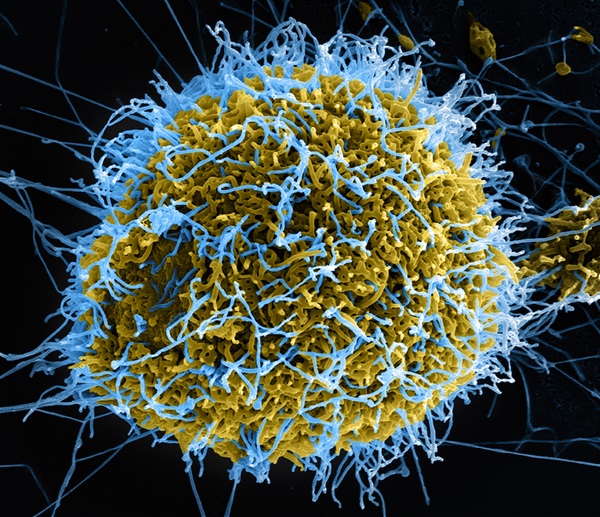
You’ve probably heard of the field of astrobiology, but what about the field of astrovirology?
http://www.astronomy.com/news/2018/01/the-hunt-for-viruses-in-space
----------------------------
Are Viruses the New Frontier for Astrobiology?
2018
https://www.space.com/40714-search-for-alien-life-viruses.html
On Earth, viruses are thought to outnumber cellular life forms by a factor of 10. And our planet is teeming with virions. In fact, a teaspoon of sea water can contain up to 50 million virions.
"It makes sense to be looking for the things that are likely to be the most abundant," said Stedman, who also chairs the Virus Focus Group within NASA’s Astrobiology Institute. "If an alien intelligence came to Earth looking for life, they would probably get a sample of sea water, loaded with virions. The alien life would come to the conclusion that Earth is inhabited by virions."

A three-dimensional representation of a norovirus virion, based on electron microscopic imagery. Should astrobiologists also be considering virions and viruses when looking for life beyond Earth
----------------------------
Trillions upon Trillions of Viruses Fall From the Sky Each Day
https://www.nytimes.com/2018/04/13/science/virosphere-evolution.html
----------------------------
The Reason for the Season: why flu strikes in winter
2014
http://sitn.hms.harvard.edu/flash/2014/the-reason-for-the-season-why-flu-strikes-in-winter/
----------------------------
Feed a Virus, Starve a Bacterium?
2016https://directorsblog.nih.gov/2016/09/13/feed-a-virus-starve-a-bacterium/
Yes, the season of colds and flu is coming. You’ve probably heard the old saying “feed a cold and starve a fever.” But is that sound advice? According to new evidence from mouse studies, there really may be a scientific basis for “feeding” diseases like colds and flu that are caused by viruses, as well as for “starving” certain fever-inducing conditions caused by bacteria.
In the latest work, an NIH-funded research team found that providing nutrition to mice infected with the influenza virus significantly improved their survival. In contrast, the exact opposite proved true in mice infected with Listeria, a fever-inducing bacterium. When researchers forced Listeria-infected mice to consume even a small amount of food, they all died.
Just like humans, when mice and other mammals come down with many infectious illnesses, they often lose their appetites and shun food. In the new study reported in the journal Cell, a team led by Ruslan Medzhitov, a Howard Hughes Medical Institute Investigator at Yale University School of Medicine, New Haven, CT, and former Lurie Prize winner from the Foundation for NIH, set out to explore how the presence or lack of nutrition might influence recovery from infections.
-----------------------------
Most of us have viruses sleeping inside us, and spaceflight wakes them up
2019
https://www.popsci.com/spaceflight-reactivate-dormant-herpes-virus
One more thing to worry about in space.
Human spaceflight seems all the more remarkable when you consider the fact that our bodies didn’t evolve for space. We suffer in major ways as a result of microgravity and living in confined quarters hundreds of miles above the surface of the planet. Even our our immune systems take a hit, leaving us more susceptible to infection and disease as we spend more time in space.
A new study published last month in Frontiers in Microbiology reports that herpes viruses lying dormant inside the body become reactivated in more than half of all astronauts sent into space, potentially exacerbating what is already a high-risk environment. While we’ve yet to run into any kind of worrisome situation resulting from this phenomenon, those concerns loom larger as we set our sights on longer duration missions in orbit and seek to send astronauts back to the moon and on to Mars.
“NASA scientists have been studying the effects of spaceflight on the immune system for over 20 years,” says Satish Mehta, a scientist at the agency’s Johnson Space Center and the senior author of the new study. “It is believed that stressful life situations cause the lowered immunity, which causes viral reactivating.” And there are obviously very few situations that induce more stress on the human body than living and working in space.
Dormant viruses are not new phenomena. Herpes viruses are often never fully eradicated from the body, and are instead subdued by the immune system. The herpes virus that causes chickenpox and shingles (VZV), for example, remains in our spinal cord cells for life. The ones that cause mononucleosis (CMV and EBV) can actually end up renting out space within our own immune cells during childhood.
(Before you freak out that we’re talking about herpes, remember one thing: you probably have herpes of some sort, even if you’re an astronaut. And that’s okay!)
But stress can compromise the immune system. Mehta and his team believe that microgravity, cosmic radiation, and the extreme G forces experienced during launch (among other factors like confined life in a spacecraft and disruptions in circadian rhythms) encourage an increase in hormones like cortisol and adrenaline, which suppress the immune system. The factors that keep dormant viruses at bay are weakened, sometimes for up to 60 days after spaceflight.
We constantly shed viral cells from our body through fluids like urine and saliva, and higher rates showing up in bodily fluids indicate a virus is active. For this particular study, Mehta and his team studied blood, urine, and saliva samples of astronauts who had completed space shuttle missions (between 10 and 16 days in space) and stays on the International Space Station (typically over 180 days in space).
They found that four of the eight herpes viruses known to infect humans reactivated and resurfaced as a result of spaceflight. Specifically, 53 percent of astronauts who underwent space shuttle missions and 61 percent of astronauts who had gone on long stays on the ISS were shedding herpes viruses at much higher rates through their saliva and urine. The four herpes viruses detected include the three aforementioned types, in addition to the HSV type that causes oral and genital herpes.
Viral shedding indicates a reactivation of the virus, but it certainly doesn’t indicate sickness. In fact, the research team found that only six of the 112 astronauts who participated in the study experienced symptoms related to their viruses, and those symptoms were quite minor.
Nevertheless, there’s no question the new findings raise some concerns. Everyone’s body works differently, and minor symptoms in one person could be serious symptoms in another. “During deep space exploration missions, crew members would be constrained to a smaller area for a longer period of time with little or no ability to return to Earth quickly,” says Mehta. “The factors that negatively affect immunity will all be elevated. NASA is focused on understanding how these viruses behave and developing countermeasures to protect astronauts on longer duration missions farther into space.”
That’s much easier said than done, of course. When it comes to these herpes viruses, the ideal countermeasure, says Mehta, is vaccination, but so far this is only available against the VZV variety. Herpes vaccines have never shown much success. Mehta says previous studies have discussed other options like nutritional supplements and better medications, but none of these interventions is beyond the concept phase.
Moreover, the study underscores just how precarious the immune system is in space. Just last fall, it was revealed that superbugs had somehow managed to colonize the ISS. While these pathogens don’t really pose a risk that we don’t already see in hospitals, at least hospitals are equipped to help patients fight off infections. When you’re sick in space, you have to make do with what you already have.
While we’re pouring more resources into studying human immunity in space, we’re limited by the fact that there just aren’t that many astronauts to study. There is, however, one environment on Earth that we could use as a testing ground: Antarctica, where humans who spend the winter for research purposes have experienced depressed immunities, along with herpes virus reactivation. “In Antarctica, crews also experience isolation, stress, an extreme environment, and circadian rhythm misalignment in the form of 24-hour darkness,” says Mehta. These conditions are remarkably similar to spaceflight, and it may be prudent to study people stationed in Antarctica to get a better grasp on viral infections for future astronauts spending months or years in space.
After all, it’s going to be hard to sell tickets for a six-month space ride to Mars if there’s an acute fear of grappling with shingles the entire way over.
----------------------------
Dormant viruses activate during spaceflight
2019
https://phys.org/news/2019-03-dormant-viruses-spaceflight.html
---------------------------
Interstellar Influenza? Space Viruses Could Reveal Alien Life
2018
https://www.livescience.com/61515-astrovirology-viruses-at-large.html
You probably think about viruses only when you're sick, but there's a group of microbiologists who want to change that. In fact, they want you to consider the possibility that viruses may be found in space.
In a recent review, published online Jan. 10 in the journal Astrobiology, a trio of scientists from the U.S. and Japan posited that viruses may be spread across interplanetary space. Those researchers want to convince astrobiologists to devote more time looking for these curious molecular machines.
A virion — the form a virus takes outside of a host — consists of genetic material encapsulated in a protein shell. Some viruses also have an outer lipid layer called an envelope. One way to think of a virion is as a seed or a spore, the authors wrote.
Viruses straddle the definition of life. They lack the machinery to reproduce on their own, so they must infect a host cell and hijack its machinery. This has led to decades of debate over whether viruses should technically be considered living.
But for the review authors, viruses' reproductive methods are enough. Indeed, "when one considers the whole virus replication cycle, it comes close to NASA's working definition of life: 'a self-sustaining chemical system capable of Darwinian evolution,'" the review said.
Semantics aside, if scientists were to identify a virus in space — on a meteor, perhaps — very few people would claim the discovery was not evidence of life in space, the authors wrote.
So why aren't scientists prowling the Martian surface, the lakes of Titan or the geysers of Enceladus for evidence of these tiny "life-forms"?
In part, it's because the technology to do so is still in development, said senior review author Kenneth Stedman, a professor of biology at Portland State University. Currently, scientists are searching for chemical signatures they can use to identify viruses in the fossil record. But if they can't find viruses in really old rocks on Earth, they won't be able to do it in really old rocks on Mars or Titan, he said.
Viruses are not metabolically active on their own, so they produce few by-products. Lipids in the envelopes are the current front-runner for a virus biomarker, since these compounds can survive for hundreds of millions of years, Stedman told Live Science. But scientist have yet to establish that these molecules are unique to viruses, and don’t exist in any cellular organism as well.
Currently, scientists can identify viruses by looking at the structure of their shells using electron microscopes. But it isn't possible to strap these high-powered machines onto a Mars rover, yet. And given the diversity of virus forms on Earth, Stedman said that he doubts scientists would even recognize the shape of an alien virus.
Here on Earth, viruses form a crucial part of life, Stedman said. For one, viruses are everywhere. The oceans alone contain an estimated 10^31 individual virions. That's about 1 million times more than estimates of the number of stars in the observable universe. And viruses are integral in most of the nutrient cycles on our planet.
What's more, viruses and cells have been coevolving basically since life arose on the planet, Stedman said. Cells evolving to resist their viral invaders give rise to new forms and behaviors. And viruses shepherd genes between unrelated cells in what scientists call horizontal gene transfer. While this process has precipitated tremendous diversity of life on Earth, it muddies the water for researchers tracking viral evolution. "If there's any water in the mud, you're in luck," Stedman said.
Scientists do know that viruses use both RNA and DNA, in single- and double-stranded forms, to code their genetic information, Stedman said. All known cellular life uses double-stranded DNA, so some scientists think that viruses may be remnants of ancient life-forms that predate the development of DNA.
This is all to say that "life on Earth would be very different if there were no viruses," Stedman said.
Scientists are currently skilled in identifying only cellular life. In addition to helping scientists learn more about our own origins, devising ways to identify viruses is good practice for recognizing other, novel forms of life we might encounter, according to Stedman. Keeping an open mind when looking for life is crucial, as many environments are quite different than Earth.
"What is life? Are viruses alive? If we find viruses [in space], is it indicative of life? And would this be life as we know it or life as we don't know it?" Stedman asked. "We're hoping to get people thinking about these types of definitions."
---------------------------
The universe is infected with ALIEN DISEASES according to scientists in radical new study
2018
SCIENTISTS believe the key to finding life in the universe could be the discovery of viruses on alien planets according to a radical new study which will begin a “new age” in science and a new discipline, it has been revealed.
https://www.express.co.uk/news/science/908457/Alien-disease-space-NASA-universe-evolution-DNA-viruses-news-latest-microbe-science
------------------------------
Might astronauts bring back a deadly disease from Mars?
https://geneticliteracyproject.org/2015/04/08/might-astronauts-bring-back-a-deadly-disease-from-mars/
----------------------------
NASA Should Start Looking For Viruses On Other Planets, Scientists Suggest
2018
https://www.newsweek.com/nasa-viruses-planets-786150
---------------------------
Our Universe Could Be Littered With Alien Viruses—and We Should Be Looking for Them
2018
https://gizmodo.com/our-universe-could-be-littered-with-alien-viruses-and-w-1822232430
---------------------------
{Many say we should find viruses in space and other planets, this is so that we
could genetically modify these viruses to find a cure for these viruses,
just in case one day some virus could travel to Earth or space colonies.
What would be the risk of taking certain viruses, molds and algae from other planets or space, and having those viruses being released on accident or on purpose. The goal of many viruses is to escape in the open to find a host and to create more viruses. Just look at how many times viruses have escaped through virus protected areas for research}.
-----------------------
NASA’s Been Ordered to Search for Life in Space. They Should Start With Viruses. Really!
2018
The not-quite life forms have a bad rap. But they’re a reliable sign of life, and it would be exciting to find them in space.
https://slate.com/technology/2018/01/nasas-been-ordered-to-search-for-life-in-space-they-should-start-with-viruses.html
-----------------------------
----------------------------
----------------------------
Section 27: Staphylococcus
-----------------------------
-----------------------------
------------------------------
Genetic Pathway in Acquisition and Loss of Vancomycin Resistance in a Methicillin Resistant Staphylococcus aureus (MRSA) Strain of Clonal Type USA300
2012
https://journals.plos.org/plospathogens/article?id=10.1371/journal.ppat.1002505
-------------------------
Genetic manipulation of Staphylococci—breaking through the barrier
2012https://www.ncbi.nlm.nih.gov/pmc/articles/PMC3417578/
RM systems of staphylococci—a historical overview
Electroporation of staphylococci—bypassing the restriction barrier
DC10B—A universal host for constructing plasmid for introduction into staphylococci
E. coli strains that modify plasmid DNA for staphylococci
pMAD/pORI280
pKOR1
pIMAY
Validation of mutations by complementation
Shuttle plasmids
Inducible gene expression
Conclusions and future perspectives
-------------------------
Staphylococcus aureus strains that are hypersusceptible to resistance gene transfer from enterococci.
2007https://www.ncbi.nlm.nih.gov/pubmed/17371826/
Abstract
---------------------------
---------------------------
---------------------------
Section 28: Typhoid
---------------------------
---------------------------
---------------------------
Typhoid outbreak: Genetic cause of extensive drug-resistance found
2018
Results suggest that treatment options are running out for typhoid, and there is an urgent need for preventative strategies including vaccines
https://www.sciencedaily.com/releases/2018/02/180220104117.htm- Summary:
- The genetic cause behind a strain of typhoid's resistance to five classes of antibiotics has been uncovered by scientists at the Wellcome Sanger Institute and their collaborators. There is currently a major outbreak of typhoid fever in Pakistan. This study shows the typhoid strain causing the outbreak acquired an additional piece of DNA to become resistant to multiple antibiotics, including a third-generation antibiotic.
----------------------------
Typhoid outbreak: Genetic cause of extensive drug-resistance found
2018
Results suggest that treatment options are running out for typhoid, and there is an urgent need for preventative strategies including vaccines
https://www.sciencedaily.com/releases/2018/02/180220104117.htm
------------------------
The Role of Typhoid Toxin in Salmonella Typhi Virulence?
2017
https://www.ncbi.nlm.nih.gov/pmc/articles/PMC5482304/
-----------------------------
Typhoid outbreak: Genetic cause of extensive drug-resistance found
2018
https://www.sciencedaily.com/releases/2018/02/180220104117.htm
Results suggest that treatment options are running out for typhoid, and there is an urgent need for preventative strategies including vaccines
Summary:
The genetic cause behind a strain of typhoid's resistance to five classes of antibiotics has been uncovered by scientists at the Wellcome Sanger Institute and their collaborators. There is currently a major outbreak of typhoid fever in Pakistan. This study shows the typhoid strain causing the outbreak acquired an additional piece of DNA to become resistant to multiple antibiotics, including a third-generation antibiotic.
--------------------------------
Evolution and Emergence of Pathogenic Viruses: Past, Present, and Future
2017
https://www.karger.com/Article/FullText/478729
-----------------------------
----------------------------
----------------------------
Section 29: Rabies
----------------------------
----------------------------
-----------------------------
Researchers Use Modified Rabies Viruses To Map The Brain
2016
(The spinal cord of a mouse with the modified rabies virus).
https://www.popsci.com/researchers-have-repurposed-rabies-virus-for-medical-research
---------------------------
Genetically modified tobacco plants produce antibodies to treat rabies
2013https://www.sciencedaily.com/releases/2013/02/130201100244.htm
- Summary:
- Smoking tobacco is bad for your health, but a genetically altered version of the plant might provide an inexpensive cure for the deadly rabies virus. Scientists have produced a monoclonal antibody in transgenic tobacco plants shown to neutralize the rabies virus. This antibody works by preventing the virus from attaching to nerve endings around the bite site and keeping the virus from traveling to the brain.
Identification of Two Classes of Somatosensory Neurons That Display Resistance to Retrograde Infection by Rabies Virus
2017

(Characterization of the prevalence of neuronal subtypes in lumbar DRGs).
http://www.jneurosci.org/content/37/43/10358
---------------------------
"Zombie Virus" Possible via Rabies-Flu Hybrid?
2010
https://news.nationalgeographic.com/news/2010/10/1001027-rabies-influenza-zombie-virus-science/
Highly improbable genetic tweak could create mutant virus.
For instance, rabies—a viral disease that infects the central nervous system—can drive people to be violently mad, according to Samita Andreansky, a virologist at the University of Miami's Miller School of Medicine in Florida who also appears in the documentary.
Combine rabies with the ability of a flu virus to spread quickly through the air, and you might have the makings of a zombie apocalypse.
Unlike movie zombies, which become reanimated almost immediately after infection, the first signs a human has rabies—such as anxiety, confusion, hallucinations, and paralysis—don't typically appear for ten days to a year, as the virus incubates inside the body.
Once rabies sets in, though, it's fatal within a week if left untreated.
If the genetic code of the rabies virus experienced enough changes, or mutations, its incubation time could be reduced dramatically, scientists say.
Many viruses have naturally high mutation rates and constantly change as a means of evading or bypassing the defenses of their hosts.
There are various ways viral mutations can occur, for example through copying mistakes during gene replication or damage from ultraviolet light.
(Related: "New, Fast-Evolving Rabies Virus Found—and Spreading.")
"If a rabies virus can mutate fast enough, it could cause infection within an hour or a few hours. That's entirely plausible," Andreansky said.
Airborne Rabies Would Create "Rage Virus"
But for the rabies virus to trigger a zombie pandemic like in the movies, it would also have to be much more contagious.
Humans typically catch rabies after being bitten by an infected animal, usually a dog—and the infection usually stops there.
Thanks to pet vaccinations, people rarely contract rabies in the United States today, and even fewer people die from the disease. For example, in 2008 only two cases of human rabies infection were reported to the U.S. Centers for Disease Control and Prevention.
(See pictures of infectious animals in National Geographic magazine.)
A faster mode of transmission would be through the air, which is how the influenza virus spreads.
"All rabies has to do is go airborne, and you have the rage virus" like in 28 Days Later, Max Mogk, head of the Zombie Research Society, says in the documentary. The international nonprofit is devoted to "raising the level of zombie scholarship in the Arts and Sciences," according to their website.
To be transmitted by air, rabies would have to "borrow" traits from another virus, such as influenza.
Different forms, or strains, of the same virus can swap pieces of genetic code through processes called reassortment or recombination, said Elankumaran Subbiah, a virologist at Virginia Tech who was not involved in the documentary.
But unrelated viruses simply do not hybridize in nature, Subbiah told National Geographic News.
Likewise, it's scientifically unheard of for two radically different viruses such as rabies and influenza to borrow traits, he said.
"They're too different. They cannot share genetic information. Viruses assemble only parts that belong to them, and they don't mix and match from different families."
---------------------------
A genetically modified rabies vaccine (ERAGS) induces protective immunity in dogs and cattle.
2017https://www.ncbi.nlm.nih.gov/pubmed/28775977
Abstract
PURPOSE:
The current live attenuated rabies vaccine must be replaced with a safer vaccine based on the ERAGS strain to prevent rabies in South Korea. We evaluated the safety and immunogenicity of a new strain in dogs and cattle.MATERIALS AND METHODS:
The ERAGS strain, featuring two mutations altering two amino acids in a glycoprotein of rabies virus, was propagated in NG108-15 cells. We lyophilized the virus in the presence of two different stabilizers to evaluate the utilities of such preparations as novel rabies vaccines for animals. To explore safety and immunogenicity, dogs and cattle were inoculated with the vaccine at various doses via different routes and observed daily for 8 weeks post-inoculation (WPI). Immunogenicity was evaluated using a fluorescent antibody virus neutralization test or enzyme-linked immunosorbent assay.RESULTS:
The two different stabilizers did not differ greatly in terms of maintenance of virus viability in accelerated stability testing. No clinical signs of rabies developed in dogs or cattle inoculated with the vaccines (107.0 FAID50/mL). Dogs and cattle inoculated intramuscularly with 105.0 FAID50/mL exhibited virus neutralization assay titers of 4.6 IU/mL and 1.5 to 0.87 IU/mL at 4 WPI, respectively. All control animals remained rabies virus-seronegative throughout, confirming that no contact transmission occurred between vaccinated and control animals.-------------------------
Baseline mapping of Lassa fever virology, epidemiology and vaccine research and development
https://www.nature.com/articles/s41541-018-0049-5
Abstract
Lassa fever (LF) is a zoonotic disease associated with acute and potentially fatal hemorrhagic illness caused by the Lassa virus (LASV), a member of the family Arenaviridae. It is generally assumed that a single infection with LASV will produce life-long protective immunity. This suggests that protective immunity induced by vaccination is an achievable goal and that cell-mediated immunity may play a more important role in protection, at least following natural infection. Seropositive individuals in endemic regions have been shown to have LASV-specific T cells recognizing epitopes for nucleocapsid protein (NP) and glycoprotein precursor (GPC), suggesting that these will be important vaccine immunogens. The role of neutralizing antibodies in protective immunity is still equivocal as recent studies suggest a role for neutralizing antibodies. There is extensive genetic heterogeneity among LASV strains that is of concern in the development of assays to detect and identify all four LASV lineages. Furthermore, the gene disparity may complicate the synthesis of effective vaccines that will provide protection across multiple lineages. Non-human primate models of LASV infection are considered the gold standard for recapitulation of human LF. The most promising vaccine candidates to date are the ML29 (a live attenuated reassortant of Mopeia and LASV), vesicular stomatitis virus (VSV) and vaccinia-vectored platforms based on their ability to induce protection following single doses, high rates of survival following challenge, and the use of live virus platforms. To date no LASV vaccine candidates have undergone clinical evaluation.
------------------------
Lassa fever vaccine shows promise and reveals new test for immunity
2018
https://www.sciencedaily.com/releases/2018/10/181011090523.htm
A new Lassa fever and rabies vaccine shows lasting immunity and suggests a new way to test for protection
Lassa fever belongs to the same class of hemorrhagic fevers as Ebola. Like Ebola, it has been a major health threat in Western Africa, infecting 100,000-300,000 people and killing 5,000 per year. A new vaccine against both rabies and Lassa has demonstrated effective protection in animal models of disease, according to research publishing in the journal Nature Communications, on October 11th. The research also points to a new way to test for protection against the virus, a finding that could significantly speed vaccine development in humans.-------------------------
A potent Lassa virus antiviral targets an arenavirus virulence determinant
2018https://journals.plos.org/plospathogens/article?id=10.1371/journal.ppat.1007439
-------------------------
Imported Lassa Fever in Germany: Molecular Characterization of a New Lassa Virus Strain
https://wwwnc.cdc.gov/eid/article/6/5/pdfs/00-0504.pdf

We describe the isolation and characterization of a new Lassa virus strain importedinto Germany by a traveler who had visited Ghana, Côte D’Ivoire, and Burkina Faso.This strain, designated “AV,” originated from a region in West Africa where Lassa feverhas not been reported. Viral S RNA isolated from the patient’s serum was amplified and sequenced. A long-range reverse transcription polymerase chain reaction allowed amplification of the full-length (3.4 kb) S RNA. The coding sequences of strain AV differed from those of all known Lassa prototype strains (Josiah, Nigeria, and LP) by approximately 20%, mainly at third codon positions. Phylogenetically, strain AV appearsto be most closely related to strain Josiah from Sierra Leone. Lassa viruses comprise agroup of genetically highly diverse strains, which has implications for vaccinedevelopment. The new method for full-length S RNA amplification may facilitate identification and molecular analysis of new arenaviruses or arenavirus strains.
--------------------------
-------------------------
-------------------------
Section 30: Ebola
---------------------------
---------------------------
----------------------------
Genetically modified rabies virus-vectored Ebola virus disease vaccines are safe and induce efficacious immune responses in mice and dogs.
2017https://www.ncbi.nlm.nih.gov/pubmed/28822816
------------------------
That Ebola Vaccine Is Related To Rabies. Is It Safe?
2015
(Vesicular Stomatitis Virus (VSV) under an electron microscope).
https://www.forbes.com/sites/jvchamary/2015/08/04/ebola-vaccine/#2aefae36672d
------------------------
GMOs lead the fight against Zika, Ebola and the next unknown pandemic
2016https://theconversation.com/gmos-lead-the-fight-against-zika-ebola-and-the-next-unknown-pandemic-60674
------------------------
Scientists unlock mysteries of how Ebola uses people's immune defenses to cause infection
2017https://www.sciencedaily.com/releases/2017/09/170926125128.htm
-------------------------
Experimental Ebola Treatment Produced In Genetically Modified Plants
https://www.iflscience.com/health-and-medicine/experimental-ebola-treatment-produced-genetically-modified-plants/------------------------
GMO tobacco ‘mystery serum’ rescues Ebola virus victims
2014https://geneticliteracyproject.org/2014/08/06/gmo-tobacco-mystery-serum-rescues-ebola-virus-victims/
---------------------
Genetically Modified Cattle With Human DNA Might Hold Ebola Cure
2014https://www.nbcnews.com/storyline/ebola-virus-outbreak/genetically-modified-cattle-human-dna-might-hold-ebola-cure-n287796
---------------------
--------------------
--------------------
Section 31: A Crimean-Congo hemorrhagic fever (CCHF)
----------------------------------------
---------------------
A Crimean-Congo hemorrhagic fever (CCHF) viral vaccine expressing nucleoprotein is immunogenic but fails to confer protection against lethal disease.
2016https://www.ncbi.nlm.nih.gov/pubmed/26309231
-----------------------
---------------------
---------------------
Section 32: Marburg Virus
----------------------
----------------------
------------------------
Ebola, Marburg viruses edit genetic material during infection
2014
https://www.eurekalert.org/pub_releases/2014-11/asfm-emv103114.php
-----------------------
Bacteria, Viruses, Fungi ... All the Words You Need to Know to Understand Microbes
2017

(MRSA bacteria being phagocytized by neutrophil (white blood cell) that is colored blue).

(Embryonic stem cells).
https://invisiverse.wonderhowto.com/news/bacteria-viruses-fungi-all-words-you-need-know-understand-microbes-0175512/
----------------------------
NEXT GENERATION BIOWEAPONS: THE TECHNOLOGY OF GENETIC ENGINEERING APPLIED TO BIOWARFARE AND BIOTERRORISM
https://fas.org/irp/threat/cbw/nextgen.pdf
-----------------------------
---------------------------
---------------------------
Section 33: SARS
---------------------------
---------------------------
-----------------------------
These Are The Zoonotic Diseases The CDC Is Most Concerned About In The US
https://www.iflscience.com/health-and-medicine/these-are-the-zoonotic-diseases-the-cdc-is-most-concerned-about-in-the-us/
Plague
Brucellosis
Zoonotic influenza
West Nile virus
Rabies
Salmonella
Lyme disease
Emerging coronaviruses
Severe Acute Respiratory Syndrome (SARS) and Middle East Respiratory Syndrome (MERS) fall into this category of diseases. SARS is thought to have originally occurred in bats before transferring to other animals, and was first documented in humans in 2002. There were over 8,000 cases worldwide during an epidemic in 2003, notes the World Health Organization.
MERS was first reported in Saudi Arabia in 2012 and spread to several other countries, including the US. It causes severe acute respiratory illness and can be fatal.
----------------------
Genetically modified tomato
https://en.wikipedia.org/wiki/Genetically_modified_food
Vaccines
Tomatoes (along with potatoes, bananas and other plants) are being investigated as vehicles for delivering edible vaccines. Clinical trials have been conducted on mice using tomatoes expressing antibodies or proteins that stimulate antibody production targeted to norovirus, hepatitis B, rabies, HIV, anthrax and respiratory syncytial virus. Korean scientists are looking at using the tomato to express a vaccine against Alzheimer's disease. Hilary Koprowski, who was involved in the development of the polio vaccine, led a group of researchers in developing a tomato expressing a recombinant vaccine to SARS.
---------------------
--------------------
--------------------
Section 34: Coronavirus (COVID-19) & Middle East respiratory syndrome Coronavirus (MERS-CoV)
--------------------
--------------------
---------------------
--------------------------
Coronavirus Investigation News - Race Virus 201 - Pollution Science 101
March 15th, 2022
https://archive.org/details/covid-news_202302
-----
Coronavirus Investigation News – Race Virus 201 – Part 1
March 15th, 2022
https://pollutionscience101.wordpress.com/2023/02/16/coronavirus-investigation-news-race-virus-201/
-----
Coronavirus Investigation News – Race Virus 201 – Part 2
March 15th, 2022
https://pollutionscience101.wordpress.com/2023/02/16/coronavirus-investigation-news-race-virus-201-part-2/
-----
Coronavirus Investigation News – Race Virus 201 – Part 3
March 15th, 2022
https://pollutionscience101.wordpress.com/2023/02/16/coronavirus-investigation-news-race-virus-201-part-3/
-----
Coronavirus Investigation News – Race Virus 201 – Part 4
March 15th, 2022
https://pollutionscience101.wordpress.com/2023/02/16/coronavirus-investigation-news-race-virus-201-part-4/
-----
Coronavirus Investigation News – Race Virus 201 – Part 5
March 15th, 2022
https://pollutionscience101.wordpress.com/2023/02/16/coronavirus-investigation-news-race-virus-201-part-5/
-----
Coronavirus Investigation News – Race Virus 201 – Part 6
March 15th, 2022
https://pollutionscience101.wordpress.com/2023/02/16/coronavirus-investigation-news-race-virus-201-part-6/
-----
Coronavirus Investigation News – Race Virus 201 – Part 7
March 15th, 2022
https://pollutionscience101.wordpress.com/2023/02/16/coronavirus-investigation-news-race-virus-201-part-7/
-----
Coronavirus Investigation News – Race Virus 201 – Part 8
March 15th, 2022
https://pollutionscience101.wordpress.com/2023/02/16/coronavirus-investigation-news-race-virus-201-part-8/
-----
Coronavirus Investigation News – Race Virus 201 – Part 9
March 15th, 2022
https://pollutionscience101.wordpress.com/2023/02/16/coronavirus-investigation-news-race-virus-201-part-9/
-----
Coronavirus Investigation News – Race Virus 201 – Part 10
March 15th, 2022
https://pollutionscience101.wordpress.com/2023/02/16/coronavirus-investigation-news-race-virus-201-part-10/
-----
Coronavirus Investigation News – Race Virus 201 – Part 11
March 15th, 2022
https://pollutionscience101.wordpress.com/2023/02/16/coronavirus-investigation-news-race-virus-201-part-11/
-----
Coronavirus Investigation News – Race Virus 201 – Part 12
March 15th, 2022
https://pollutionscience101.wordpress.com/2023/02/16/coronavirus-investigation-news-race-virus-201-part-12/
------------------------------
---------------------------
MERS coronaviruses from camels in Africa exhibit region-dependent genetic diversity
2018
https://www.ncbi.nlm.nih.gov/pmc/articles/PMC5866576/
----------------------
----------------------
A spike-modified Middle East respiratory syndrome coronavirus (MERS-CoV) infectious clone elicits mild respiratory disease in infected rhesus macaques
2018
https://www.nature.com/articles/s41598-018-28900-1
----------------------
MERS treatment could come from genetically engineered cows
2018
https://geneticliteracyproject.org/2018/01/17/mers-treatment-come-genetically-engineered-cows/
----------------------
Researchers develop new vaccine against deadly Middle East Respiratory Syndrome
2019
Although the virus is initially spread from camel to human, it can then spread from person to person
https://www.eurekalert.org/pub_releases/2019-04/uotm-rdn040919.php
---------------------
A mouse model for MERS coronavirus-induced acute respiratory distress syndrome.
2016
https://www.ncbi.nlm.nih.gov/pubmed/27892925
---------------------
Canine Coronavirus Highly Pathogenic for Dogs
2006 Mar
Canine coronavirus (CCoV) is usually responsible for mild, self-limiting infections restricted to the enteric tract. We report an outbreak of fatal disease in puppies caused by a pathogenic variant of CCoV that was isolated from organs with severe lesions.
https://www.ncbi.nlm.nih.gov/pmc/articles/PMC3291441/
------------------
{Some researchers are now saying that the Coronavirus may have been caused by the Chinese eating dogs. Doctors were experimenting with vaccines for coronavirus in dogs many years before this new Coronavirus outbreak, this goes to show that dogs can have Coronaviruses while these Orientals eat dogs and cats}.
{Would you like some Corona Extra with that Chinese BBQ dog?}
------------------
Chinese officials 'round up and execute villagers' pets to stop the spread of coronavirus' despite WHO experts saying the disease cannot be spread to animals
20 February 2020
https://www.dailymail.co.uk/news/article-8024787/Chinese-officials-continue-beat-dogs-death-fighting-coronavirus.html
------------------
The outbreaks of both the Wuhan coronavirus and SARS started in Chinese wet markets. Photos show what the markets look like.
Jan 23, 2020
A coronavirus that originated in Wuhan, China, has killed 17 people and infected more than 544.
The Huanan Seafood Market in Wuhan is thought to be the starting point for the virus outbreak. It was shuttered on January 1.
At wet markets, meat is sold alongside live animals like dogs, hares, and civets.
On Wednesday, Wuhan authorities banned the trade of live animals at wet markets to lower the risk of a disease outbreak.
https://www.businessinsider.co.za/wuhan-coronavirus-chinese-wet-market-photos-2020-1?r=US&IR=T
------------------
Coronavirus may have originated in lab linked to China's biowarfare program
January 29, 2020
https://www.washingtontimes.com/news/2020/jan/26/coronavirus-link-to-china-biowarfare-program-possi/
------------------
2/25/2020 - Race Virus 101: The Movie - Asians, Race Viruses & CoronaVirus (Eugenics & Dysgenics) - Full Movie (Remastered)
https://archive.org/details/racevirus101Remaster
---------------------
Coronavirus has mutated at least once, second strain detected: study
March 8, 2020
https://www.fox5atlanta.com/news/coronavirus-has-mutated-at-least-once-second-strain-detected-study
---------------------
Mutations can reveal how the coronavirus moves—but they’re easy to overinterpret
Mar. 9, 2020
https://www.sciencemag.org/news/2020/03/mutations-can-reveal-how-coronavirus-moves-they-re-easy-overinterpret
---------------------
Chinese studies link quarantines with coronavirus mutations that may make it more ‘insidious’
March 12, 2020
https://www.scmp.com/news/china/society/article/3074938/chinese-studies-link-quarantines-coronavirus-mutations-may-make
--------------------
Coronavirus in Scotland: New mutations likely in the coming years, warns expert
March 06 2020
https://www.thetimes.co.uk/article/coronavirus-in-scotland-new-mutations-likely-in-the-coming-years-warns-expert-xmwrlzkwt
------------------
Why Germany's Coronavirus Death Rate Is Far Lower Than In Other Countries
March 25, 2020
https://www.npr.org/2020/03/25/820595489/why-germanys-coronavirus-death-rate-is-far-lower-than-in-other-countries?utm_source=pocket-newtab
--------------------
Why Germany's Coronavirus Death Rate Is Far Lower Than In Other Countries
March 25, 2020
https://www.npr.org/2020/03/25/820595489/why-germanys-coronavirus-death-rate-is-far-lower-than-in-other-countries?utm_source=pocket-newtab
--------------------
Germany's devolved logic is helping it win the coronavirus race
April 5, 2020
https://www.theguardian.com/world/2020/apr/05/germanys-devolved-logic-is-helping-it-win-the-coronavirus-race
With 400 public health offices forging ahead with testing, the country is a model for others to emulate
(Germany’s approach to testing has put it way ahead of other countries in terms of numbers).
-------------------------------------------------------------
Coronavirus: Iceland’s mass testing finds half of carriers show no symptoms
https://english.alarabiya.net/en/features/2020/03/25/Coronavirus-Iceland-s-mass-testing-finds-half-of-carriers-show-no-symptoms
Unlike other countries, where people are only tested if they exhibit symptoms of coronavirus or have come into contact with known spreaders, the country is testing thousands of people from the general population who don’t exhibit any symptoms of the virus whatsoever – helping to reveal information about the nature of the pathogen and its symptoms.
40 mutations of the virus
-------------------
Finland's Fascinating Genes
April. 2005
The
people in this land of lakes and forests are so alike that scientists
can filter out the genes that contribute to heart disease, diabetes, and
asthma
http://discovermagazine.com/2005/apr/29-finlands-fascinating-genes
-------------------
Novel coronavirus attacks and destroys T cells, just like HIV
Apr 13 2020
Now,
a team from the United States and China revealed evidence that the
coronavirus disease, caused by the severe acute respiratory syndrome
coronavirus (SARS-CoV-2), attacks the immune system’s T lymphocytes. The
worrying findings highlight the destructive power of the novel
coronavirus, which can destroy the immune system, leaving the patient
unable to fight off the infection.
https://www.news-medical.net/news/20200413/Novel-coronavirus-attacks-and-destroys-T-cells-just-like-HIV.aspx
-----------------
Nano Research for COVID-19
3-31-2020
https://www.ncbi.nlm.nih.gov/pmc/articles/PMC7123821/
In parallel with diagnostic test developments, researchers are examining
different drug formulations to treat patients suffering with COVID-19.
One potential therapy currently undergoing clinical trials is the HIV
drug combination of liponavir–ritonavir. Thus far, the trials have not
shown a significant difference between patients treated with this drug
cocktail and placebo...
-----------------
More about the Viking hypothesis of origin of the delta32 mutation in the CCR5 gene conferring resistance to HIV-1 infection.
2003
https://www.ncbi.nlm.nih.gov/pubmed/14636691
-----------------------------------------------------
Biologists discover why 10% of Europeans are safe from HIV infection
10-Mar-2005
https://www.eurekalert.org/pub_releases/2005-03/uol-bdw031005.php
------------------------------------------------------
What is CCR5 Delta32?
https://www.delta-32.com/ccr5-delta32.html
CCR5-delta32"
is a deletion mutation of a gene which only 1% of the total population
has two copies of this gene and individuals who carry two copies of this
genetic mutation are immune to Smallpox, The Bubonic Plague (Black
Death) and resistant to HIV, the virus that causes AIDS. Up to 20% of
the population carry only one copy of this genetic mutation depending on
your background and although they still run a significant risk of
contracting HIV, the progress of the disease is greatly reduced and can
result in a longer life expectancy.
-------------------------------------
The Geographic Spread of the CCR5 Δ32 HIV-Resistance Allele
2005 Nov
Abstract
https://www.ncbi.nlm.nih.gov/pmc/articles/PMC1255740/
-------------------------------------------------------
COVID-19 takes unequal toll on immigrants in Nordic region
April 24, 2020
https://www.reuters.com/article/us-health-coronavirus-norway-immigrants-idUSKCN2260XW
Across
Europe, little is known about who is affected by the virus because
governments are releasing limited demographic information about the sick
and those who die. But a Reuters examination of government data in
three Nordic countries where more details are available shows that some
immigrant groups are among those affected at higher rates than the
general populace.
“WORRYING” DISPARITY
In
Norway, where 15% of residents were born abroad, 25% who had tested
positive for COVID-19 by April 19 were foreign-born. Somalis, with 425
confirmed cases, are the largest immigrant group testing positive,
accounting for 6% of all confirmed cases — more than 10 times their
share of the population.
Somalis are the most
overrepresented immigrant group among Sweden’s confirmed cases, as well.
Their 283 positive tests account for about 5% of the nearly 6,000 cases
documented between March 13 and April 7. That’s seven times their share
of the population. Iraqis, Syrians and Turks also made up
disproportionately large shares of positive cases.
In
Finland’s capital city of Helsinki, the mayor said it was “worrying”
that almost 200 Somalis had tested positive by mid-April. They accounted
for about 17% of positive cases — 10 times their share of the city’s
population.
More than 100,000 Somalia-born live in the
three countries, mostly in Sweden and Norway, one of the largest Somali
diasporas in the world. Many arrived as refugees of war in the 1990s,
2000s and 2010s. Several factors place them more at risk of getting
sick, public health officials and researchers say.
-----------------------
Coronavirus takes a toll in Sweden's immigrant community
May 9, 2020
https://abcnews.go.com/International/wireStory/coronavirus-takes-toll-swedens-immigrant-community-70593594
The coronavirus has taken a disproportionate toll among Sweden's immigrants
n
Finland, Helsinki authorities warned of a similar over-representation
among Somali immigrants in the capital — some 200 cases, or about 14%,
of all confirmed infections. In Norway, where immigrants make up nearly
15% of the general population, they represent about 25% of confirmed
coronavirus cases.
“I think a pandemic like this one,
or any crisis will hit the most vulnerable people in society the most
wherever in the world, and we see this in many many countries," said
Isabella Lovin, Sweden's deputy prime minister, in an interview with The
Associated Press.
Noting that the virus was spreading
faster in some crowded Stockholm suburbs, Lovin said said the city is
providing short-term accommodation to some people whose relatives are
vulnerable.
Sweden, Norway and Finland recognized early
failings in community outreach in minority languages and are seeking to
fix this. The town of Jarfalla, outside Stockholm, has had high school
students hand out leaflets in Somali, Persian, French and other
languages, urging people to wash their hands and stay home if sick.
With
Sweden's relatively low-key approach to fighting the virus that relies
mainly on voluntary social distancing, there are concerns the message
has not reached everyone in immigrant neighborhoods.
“It’s
important that everyone living here who has a different mother tongue
gets the right information,” said Warda Addallah, a 17-year-old Somali
Swede.
------------------------
Digging into racial disparities in coronavirus cases from Baltimore | ABC News
Apr 12, 2020
https://www.youtube.com/watch?v=1JqD0aU7pPM
------------------------
The coronavirus is infecting and killing black Americans at an alarmingly high rate
April 7, 2020
https://www.washingtonpost.com/nation/2020/04/07/coronavirus-is-infecting-killing-black-americans-an-alarmingly-high-rate-post-analysis-shows/?arc404=true
------------------------
Why black Americans are at higher risk for coronavirus
April 7, 2020
https://www.cnn.com/2020/04/07/us/coronavirus-black-americans-race/index.html
------------------------
‘Those numbers take your breath away’: Covid-19 is hitting Chicago’s black neighborhoods much harder than others, officials say
April 7, 2020
https://www.washingtonpost.com/nation/2020/04/07/chicago-racial-disparity-coronavirus/
------------------------
------------------------
------------------
-----------------------
---------------------
---------------------
Section 35: MRSA
---------------------
---------------------
-----------------------
Distribution of genes encoding resistance to aminoglycoside modifying enzymes in methicillin-resistant Staphylococcus aureus (MRSA) strains.
2017
https://www.ncbi.nlm.nih.gov/pubmed/29132547
Abstract
Today Methicillin-Resistant Staphylococcus aureus (MRSA) have acquired multiple resistance to a wide range of antibiotics including aminoglycosides. So, this study was aimed to investigate the rate of aminoglycoside resistance and the frequency of aminoglycoside resistance mediated genes of aac(Ia)-2, aph(3)-IIIa and ant(4')-Ia among MRSA strains. A total of 467 staphylococci isolates were collected from various clinical samples. S. aureus strains were identified by standard culture and identification criteria and investigating of presence of 16S rRNA and nuc genes. Cefoxitin disk diffusion, and oxacillin-salt agar screening methods were used to detect the MRSA strains with subsequent molecular identification for the presence of mecA gene. Antibiotic susceptibility of MRSA strains against aminoglycoside antibiotics was evaluated by using agar disk diffusion method. Multiplex PCR for the presence of aac(Ia)-2, aph(3)-IIIa and ant(4')-Ia encoding genes for aminoglycosides were performed for MRSA strains. From total staphylococci tested isolates, 262 (56.1%) were identified as S. aureus, of which 161 (61.45%) were detected as MRSA and all comprised mecA gene. The resistance pattern of MRSA strains to aminoglycoside antibiotics were: gentamicin 136 (84.5%); amikacin 125 (77.6%); kanamycin 139 (86.3%); tobramycin 132 (82%); and neomycin 155 (96.3%). The frequency of aac(Ia)-2, aph(3)-IIIa, and ant(4')-Ia genes among MRSA strains, were 64%, 42% and 11.8% respectively. In conclusion, as MRSA strains are of great concern in human infections, the results of present study could provide a useful resource for health sectors for choosing appropriate antibiotics for the effective treatment of infections due to MRSA strains.
----------------------------
High Genetic Similarity of MRSA ST88 Isolated From Pigs and Humans in Kogi State, Nigeria
2018
https://www.frontiersin.org/articles/10.3389/fmicb.2018.03098/full
We determined the prevalence and genetic characteristics of methicillin-resistant Staphylococcus aureus (MRSA) isolated from pigs and humans between September 2013 and February 2015 in Kogi State, a central region in Nigeria. A total of 680 nasal swabs were collected and analyzed from pigs (n = 425) and “pig-contact” humans (n = 55) on 35 farms, and “non-pig-contact” humans (n = 200). MRSA was recovered from 20 (4.7%) pigs on 12 farms and 18 (7.0%) humans. Six (2.4%) of the human isolates were recovered from “pig-contact” humans, of which only three work on farms also harboring MRSA positive pigs. All 38 MRSA were resistant to β-lactams only, belonged to spa type t1603, sequence type (ST) 88, and mecA was associated with a SCCmec IVa element. Four isolates from a pig, a pig-contact human from the same farm, a pig-contact human from a pig farm in a different district, and a non-pig-contact human were subjected to whole genome sequencing (WGS). Core genome SNP analysis revealed high genetic similarity between strains (3–11 SNP differences), despite the temporal (2 year gap) and geographic (165 km) differences between isolates. Furthermore, these Nigerian isolates form a distinct clade when compared to other African MRSA ST88 isolates. All but one porcine strain was positive for scn suggesting a possible human origin and that pigs were either transiently contaminated by humans or result of a very recent human-to-pig transmission event. To our knowledge, this is the first report of genetically confirmed MRSA in pigs in Nigeria, which appear to be a typical CA-MRSA clone present in the human population.
---------------------------
Immunomimetic Designer Cells Protect Mice from MRSA Infection
https://www.cell.com/cell/pdf/S0092-8674(18)30651-2.pdf
----------------------------
---------------------------
---------------------------
Section 36: Genetically Modified Viruses
----------------------------
----------------------------
-----------------------------
Genetically modified viruses: vaccines by design.
2001
https://www.ncbi.nlm.nih.gov/pubmed/11482348
Abstract
Vaccination has been one of the most successful and cost-effective health interventions ever employed. One disease (smallpox) has been eradicated, another (poliomyelitis) should disappear early in the new millennium and a third (measles) should follow shortly after. Conventional vaccines usually depend on one of three development processes, attenuation of virulent organisms (by passage in cell culture and/or experimental animals), killing of virulent organisms (by chemical inactivation) or the purification of immunogenic molecules (either proteins or carbohydrates) from whole organisms. These traditional processes, although serendipitous and poorly understood, have produced effective pharmaceutical products which give excellent protection against diseases such as smallpox, rabies, measles, yellow fever, tetanus and diphtheria. In spite of these successes however, the application of these protocols have failed to produce safe and efficacious vaccines against other infectious diseases which kill or maim tens of millions of people every year. The most important of these are malaria, AIDS, herpes, dengue fever and some forms of viral hepatitis. Consequently, fundamentally new technologies are required to tackle these important infections. One of the most promising has been the development of genetically modified viruses. This process normally involves taking a proven safe and efficacious vaccine virus, such as vaccinia or adenovirus, and modifying its genome to include genes coding for immunogenic proteins from other viruses such as HIV or measles. This review will describe the generation of such novel vaccine vectors and compare their advantages and shortcomings. In addition the literature describing their use as experimental vaccines will also be reviewed.
-------------------------
Genetically modified bacteria
https://en.wikipedia.org/wiki/Genetically_modified_bacteria
Genetically modified bacteria were the first organisms to be modified in the laboratory, due to their simple genetics. These organisms are now used for several purposes, and are particularly important in producing large amounts of pure human proteins for use in medicine.
Other uses
Other uses for genetically modified bacteria include bioremediation, where the bacteria are used to convert pollutants into a less toxic form. Genetic engineering can increase the levels of the enzymes used to degrade a toxin or to make the bacteria more stable under environmental conditions. GM bacteria have also been developed to leach copper from ore, clean up mercury pollution and detect arsenic in drinking water. Bioart has also been created using genetically modified bacteria. In the 1980s artist Jon Davis and geneticist Dana Boyd converted the Germanic symbol for femininity (?) into binary code and then into a DNA sequence, which was then expressed in Escherichia coli. This was taken a step further in 2012, when a whole book was encoded onto DNA. Paintings have also been produced using bacteria transformed with fluorescent proteins.

(This artwork is made with bacteria modified to express 8 different colours of fluorescent proteins).
--------------------------------
Genetically modified virus
https://en.wikipedia.org/wiki/Genetically_modified_virus
Health applications
Gene therapy
Gene therapy uses genetically modified viruses to deliver genes that can cure diseases in human cells.These viruses can deliver DNA or RNA genetic material to the targeted cells. Gene therapy is also used by inactivating mutated genes that are causing the disease using viruses.
Viruses that have been used for gene therapy are, adenovirus, lentivirus, retrovirus and the herpes simplex virus. The most common virus used for gene delivery come from adenoviruses as they can carry up to 7.5 kb of foreign DNA and infect a relatively broad range of host cells, although they have been know to elicit immune responses in the host and only provide short term expression. Other common vectors are adeno-associated viruses, which have lower toxicity and longer term expression, but can only carry about 4kb of DNA. Herpes simplex viruses is a promising vector, have a carrying capacity of over 30kb and provide long term expression, although it is less efficient at gene delivery than other vectors. The best vectors for long term integration of the gene into the host genome are retroviruses, but their propensity for random integration is problematic. Lentiviruses are a part of the same family as retroviruses with the advantage of infecting both dividing and non-dividing cells, whereas retroviruses only target dividing cells. Other viruses that have been used as vectors include alphaviruses, flaviviruses, measles viruses, rhabdoviruses, Newcastle disease virus, poxviruses, and picornaviruses.
Although primarily still at trial stages, it has had some successes. It has been used to treat inherited genetic disorders such as severe combined immunodeficiency rising from adenosine deaminase deficiency (ADA-SCID), although the development of leukemia in some ADA-SCID patients along with the death of Jesse Gelsinger in another trial set back the development of this approach for many years. In 2009 another breakthrough was achieved when an eight year old boy with Leber’s congenital amaurosis regained normal eyesight and in 2016 GlaxoSmithKline gained approval to commercialise a gene therapy treatment for ADA-SCID. As of 2018, there are a substantial number of clinical trials underway, including treatments for hemophilia, glioblastoma, chronic granulomatous disease, cystic fibrosis and various cancers.[8]Although some successes, gene therapy is still considered a risky technique and studies are still undergoing to ensure safety and effectiveness.
Cancer treatment
Another potential use of genetically modified viruses is to alter them so they can directly treat diseases. This can be through expression of protective proteins or by directly targeting infected cells. In 2004, researchers reported that a genetically modified virus that exploits the selfish behaviour of cancer cells might offer an alternative way of killing tumours. Since then, several researchers have developed genetically modified oncolytic viruses that show promise as treatments for various types of cancer.
Vaccines
Most vaccines consist of viruses that have been attenuated, disabled, weakened or killed in some way so that their virulent properties are no longer effective. Genetic engineering could theoretically be used to create viruses with the virulent genes removed. In 2001, it was reported that genetically modified viruses can possibly be used to develop vaccines against diseases such as, AIDS, herpes, dengue fever and viral hepatitis by using a proven safe vaccine virus, such as adenovirus, and modify its genome to have genes that code for immunogenic proteins that can spike the immune systems response to then be able to fight the virus. Genetic engineered viruses should not have reduced infectivity, invoke a natural immune response and there is no chance that they will regain their virulence function, which can occur with some other vaccines. As such they are generally considered safer and more efficient than conventional vaccines, although concerns remain over non-target infection, potential side effects and horizontal gene transfer to other viruses. Another approach is to use vectors to create novel vaccines for diseases that have no vaccines available or the vaccines that are do not work effectively, such as AIDS, malaria, and tuberculosis. Vector-based vaccines have already been approved and many more are being developed.
Heart pacemaker
In 2012, US researchers reported that they injected a genetically modified virus into the heart of pigs. This virus inserted into the heart muscles a gene called Tbx18 which enabled heartbeats. The researchers forecast that one day this technique could be used to restore the heartbeat in humans who would otherwise need electronic pacemakers.
Bio-control
Animals
In Spain and Portugal, by 2005 rabbits had declined by as much as 95% over 50 years due diseases such as myxomatosis, rabbit haemorrhagic disease and other causes. This in turn caused declines in predators like the Iberian lynx, a critically endangered species. In 2000 Spanish researchers investigated a genetically modified virus which might have protected rabbits in the wild against myxomatosis and rabbit haemorrhagic disease. However, there was concern that such a virus might make its way into wild populations in areas such as Australia and create a population boom. Rabbits in Australia are considered to be such a pest that land owners are legally obliged to control them.
Genetically modified viruses that make the target animals infertile through immunocontraception have been created as well as others that target the developmental stage of the animal. There are concerns over virus containment and cross species infection.
Trees
In 2017 researchers genetically modified a virus to express spinach defensin proteins. The virus was injected into orange trees to combat citrus greening disease that had reduced orange production 70% since 2005.
Technological applications
Lithium-ion batteries
In 2009, MIT scientists created a genetically modified virus has been used to construct a more environmentally friendly lithium-ion battery. The battery was constructed by genetically engineering different viruses such as, the E4 bacteriophage and the M13 bacteriophage, to be used as a cathode. This was done by editing the genes of the virus that code for the protein coat. The protein coat is edited to coat itself in iron phosphate to be able to adhere to highly conductive carbon-nanotubes. The viruses that have been modified to have a multifunctional protein coat can be used as a nano-structured cathode with causes ionic interactions with cations. Allowing the virus to be used as a small battery. Angela Blecher, the scientist who led the MIT research team on the project, says that the battery is powerful enough to be used as a rechargeable battery, power hybrid electric cars, and a number of personal electronics. While both the E4 and M13 viruses can infect and replicate within their bacterial host, it unclear if they retain this capacity after being part of a battery.
Safety concerns and regulation
Bio-hazard research limitations
The National Institute of Health declared a research funding moratorium on select Gain-of-Function virus research in January 2015. Questions about a potential escape of a modified virus from a biosafety lab and the utility of dual-use-technology, dual use research of concern (DURC), prompted the NIH funding policy revision.
GMO lentivirus incident
A scientist claims she was infected by a genetically modified virus while working for Pfizer. In her federal lawsuit she says she has been intermittently paralyzed by the Pfizer-designed virus. "McClain, of Deep River, suspects she was inadvertently exposed, through work by a former Pfizer colleague in 2002 or 2003, to an engineered form of the lentivirus, a virus similar to the one that can lead to acquired immune deficiency syndrome, or AIDS." The court found that McClain failed to demonstrate that her illness was caused by exposure to the lentivirus, but also that Pfizer violated whistleblower laws.
--------------------------
Genetically engineered viruses: a medicine of the future
2017
http://sitn.hms.harvard.edu/flash/2017/genetically-engineered-viruses-medicine-future/
One of the top causes for vision loss in people over 60 is Age-related Macular Degeneration (AMD). In more severe cases of wet AMD, a protein called VEGF is over-active. This protein stimulates new blood vessels to grow, leaking blood and fluid into the eye. This fluid build-up damages light-sensing cells, leaving the owner of the eye with progressively larger blind spots.
Current treatments involve injecting a VEGF-binding protein (sFLT01) directly into the eye, thus taking VEGF out of play and preventing the root cause of wet AMD. However, the body does not produce sFLT01 on its own, so treatments must be repeated monthly, leaving the patient open to eye infections and other side effects.
Scientists at Johns Hopkins genetically engineered a common cold virus (AAV2 strain) to deposit a new gene that codes for sFLT01. The virus was chosen because previous studies have shown it was safe to use in the eye. In a small clinical study, four patients showed significant improvement after only one injection and two patients experienced partial improvement. Five patients had AAV2 antibodies present in their system before the study, meaning their immune system was all too happy to destroy the virus before it could be effective. Scientists measured the expression of the gene in patients for 12 months and concluded that higher doses could safely be used to produce more long-lasting results.
While these preliminary results are promising, the scientists admit that a larger and more thorough study is required before proceeding. This study is the first to show that pre-existing antibodies will significantly affect the outcome. Because most people have experienced the common cold, the antibodies for AAV2 and related virus strains are probably widespread. Scientists might have to use less common, and potentially less safe, virus strains to produce an effective treatment for a significant portion of the population.
---------------------------------
Nathan Wolfe: On the Hunt for New Viruses
2014
Virologist Nathan Wolfe on the race to find new diseases and the growing risk of epidemics
https://www.wsj.com/articles/nathan-wolfe-on-the-hunt-for-new-viruses-1418422069
---------------------------
History of genetic engineering
https://en.wikipedia.org/wiki/History_of_genetic_engineering
---------------------------------------------
Genetic engineering
https://en.wikipedia.org/wiki/Genetic_engineering----------------------------
---------------------------
---------------------------
Section 37: Foot and Mouth
---------------------------
----------------------------
-----------------------------
Evaluation of a genetically modified foot-and-mouth disease virus vaccine candidate generated by reverse genetics
15 September 2011
Abstract
Background
Results
The present study describes the generation of a full-length infectious cDNA clone of FMDV vaccine strain and a genetically modified virus with some amino acid substitutions in antigenic sites 1, 3, and 4, based on the established infectious clone. The recombinant viruses had similar growth properties to the wild O/HN/CHA/93 virus. All swine immunized with inactivated vaccine prepared from the O/HN/CHA/93 were fully protected from challenge with the viruses of ME-SA and SEA topotypes and partially protected against challenge with the virus of CHY topotype at 28 days post-immunization. In contrast, the swine inoculated with the genetically modified vaccine were completely protected from the infection of viruses of the three topotypes.
Conclusions
https://bmcvetres.biomedcentral.com/articles/10.1186/1746-6148-8-57
-----------------------
----------------------
----------------------
----------------------
Section 38: Bolivian Hemorrhagic Fever
----------------------
----------------------
----------------------
Bolivian hemorrhagic fever
https://en.wikipedia.org/wiki/Bolivian_hemorrhagic_fever
Weaponization
Bolivian hemorrhagic fever was one of three hemorrhagic fevers and one of more than a dozen agents that the United States researched as potential biological weapons before the nation suspended its biological weapons program. It was also under research by the Soviet Union, under the Biopreparat bureau.
Vaccine Research
Investigational vaccines exist for Argentine hemorrhagic fever and RVF; however, neither is approved by FDA or commonly available in the United States.[citation needed]
The structure of the attachment glycoprotein has been determined by X-ray crystallography and this glycoprotein is likely to be an essential component of any successful vaccine.
------------------
-----------------
-----------------
Section 39: Machupo Virus
----------------------------------
------------------
Rescue of a Recombinant Machupo Virus from Cloned cDNAs and In Vivo Characterization in Interferon (αβ/γ) Receptor Double Knockout Mice
https://jvi.asm.org/content/88/4/1914ABSTRACT
INTRODUCTION
-----------------
Description and characterization of a novel live-attenuated tri-segmented Machupo virus in Guinea pigs
2018
https://www.ncbi.nlm.nih.gov/pmc/articles/PMC5992841/
Abstract
Background
Findings
Conclusions

(Growth properties of the r3MACV).
-------------------------------------
Machupo Virus Glycoprotein Determinants for Human Transferrin Receptor 1 Binding and Cell Entry
2011https://journals.plos.org/plosone/article?id=10.1371/journal.pone.0021398
--------------------------------
------------------------------
------------------------------
Section 40: Valley Fever
------------------------------
------------------------------
---------------------------------
True story: I was hired working for several cannabis clinics in California, my
job was to pick up trucks of dried cannabis trim, inspect the cannabis trim and
drive truckloads of cannabis from Humboldt County, all the way down
to Los Angeles.
There was a black worker that would always breakout in hives and have
shortness of breath after he touched the cannabis trim. For a while, we
thought that it was the pesticides that caused this, but this was not
the case. This worker smoked a lot of cannabis, but when he started to
touch whole trash bags of dried cannabis leaves and trim, or try to just
smell the giant bags of cannabis trim, he would get sick. If too much cannabis
dust hit him, he would have to wipe his body down with alcohol, take a
shower, and put the air conditioner on very cold, even if it was 40 degrees fahrenheit or 50 degrees fahrenheit outside.
We could not figure out what the problem was for years, even doctors could
not figure it out. This guy would also start breaking out if the weather
became too humid, which is why he always liked to run the air conditioner.
We thought maybe after handing years and years of pesticides, my
friends body started to react more.
We thought maybe it was the resin from the plant.
We thought maybe that some people are allergic to plants, such as
different indoor plants people can be allergic to.
One theory I think I finally figured it out, and it has taken
me over 5 years to figure it out. My friends condition is one of
the reasons what fascinated me about this rare medical condition
in this black person.
Keep in mind, if my friend smells just indoor cannabis, it does not
really harm him.
I think I finally figured out what the problem was, and that there is
a mold in the Southwestern United states that has spores and gets
on plants. When my friend opens up a giant trashbag of cannabis, the
mold dust travels in the air and gets on the black person inspecting
the cannabis trim, and the mold them makes him sick. The black worker
has to go take a shower, wash off the mold and lay down for a couple hours
with difficult breathing.
----------------------------
Arthroconidium
https://en.wikipedia.org/wiki/Arthroconidium
Arthroconidia are a type of fungal spore typically produced by segmentation of pre-existing fungal hyphae.

(Arthroconidia of Coccidioides immitis)
These spores are asexual and are generally not as durable and environmentally persistent as, for instance, bacterial endospores or chlamydospores. Some medically significant pathogens, such as Coccidioides immitis, and Coccidioides posadasii, both causative agents of coccidioidomycosis (also known as San Joaquin Valley fever), are transmitted through airborne arthroconidia. The small size of the arthroconidia, 3 to 5 µm, allow them to lodge themselves into the terminal bronchioles of the lung. There, they develop into a thick-walled spherule filled with endospores that cause a pyogenic (pus-causing) inflammation.
------------------------------
Coccidioidomycosis
https://en.wikipedia.org/wiki/Coccidioidomycosis
Coccidioidomycosis, commonly known as cocci, Valley fever, as well as California fever, desert rheumatism, and San Joaquin Valley fever, is a mammalian fungal disease caused by Coccidioides immitis or Coccidioides posadasii. Coccidioidomycosis is endemic in certain parts of Arizona, California, Nevada, New Mexico, Texas, Utah, and northern Mexico.
C. immitis is a dimorphic saprophytic fungus that grows as a mycelium in the soil and produces a spherule form in the host organism. It resides in the soil in certain parts of the southwestern United States, most notably in California and Arizona. It is also commonly found in northern Mexico, and parts of Central and South America. C. immitis is dormant during long dry spells, then develops as a mold with long filaments that break off into airborne spores when it rains. The spores, known as arthroconidia, are swept into the air by disruption of the soil, such as during construction, farming, or an earthquake. Windstorms may also cause epidemics far from endemic areas. In December 1977, a windstorm in an endemic area around Arvin, California led to several hundred cases, including deaths, in non-endemic areas hundreds of miles away.
Coccidioidomycosis
is a common cause of community-acquired pneumonia in the endemic areas of the United States. Infections usually occur due to inhalation of the arthroconidial spores after soil disruption. The disease is not contagious. In some cases the infection may recur or become chronic.

(A skin lesion due to Coccidioides infection).

(Spherule of Coccidioides immitis with endospores).

(Spherule and endospore forms of Coccidioides immitis).

(Mature spherule with endospores of Coccidioides immitis).
------------------------------------------------------
Everything you need to know about valley fever
2017
https://www.medicalnewstoday.com/articles/189430.php
It can lead to fever, chest pain, coughing, and some other symptoms.
Valley fever is caused by the fungus Coccidioides immitis (C. immitis), or by Coccidioides posadasii (C. posadasii). It is also known as coccidioidomycosis, California disease, desert rheumatism, and San Joaquin valley fever.
The fever does not pass between humans. An infected person will not transmit the disease to another person.
In the states where the fungus is active, there are 42.6 cases per 100,000 people every year, on average.
A number of common factors increase the likelihood of contracting valley fever.
Geographical location: the risk is higher in areas where fungus spores are likely, such as Arizona, New Mexico, Texas, Utah, Nevada, and northwestern Mexico. If the person inhales the spores, they can become infected.
In these areas, people whose work involves disturbing soil have a higher risk. Examples are construction, excavation, agricultural work, or archaeological digging.
Ethnicity: Filipinos, Hispanic Amerindians, Native North Americans, and Asians have a higher risk of developing infections with symptoms, compared with Caucasians.
Pregnancy: in areas where valley fever is endemic, there is a higher risk of infection during the third trimester of pregnancy, and immediately after giving birth.
Diabetes: Individuals with diabetes who live in areas where valley fever exists have a higher risk of becoming infected, compared with other people in the same areas.
Weak immune system: People with a weakened immune system have a higher risk of infection and complications. This includes patients with HIV or AIDS, and those receiving steroid medications, chemotherapy, or immunosuppressant drugs. People with cancer may also be more susceptible.
Advanced age: older people are more likely to develop valley fever.
----------------------------
3 Worst states to live in if you have a mold allergy
2018
https://molekule.com/blog/top-10-worst-places-mold-allergy/
According to the data in the Quest Diagnostics study, the top 3 worst states for mold allergies are:
California, with four cities in the top 20 worst cities for allergy sufferers
Ohio, with two cities in the top 20, ranked 8th and 10th
Texas, with two cities in the top 20, ranked 1st and 20th
The southwestern United States was found to be the worst region overall for mold allergy sufferers. It is home to four of the top five worst cities for mold sensitization.
-------------------------------
NSs Protein of Rift Valley Fever Virus Blocks Interferon Production by Inhibiting Host Gene Transcription
2004

Localization and filament formation of wild-type and mutant NSs proteins.
https://jvi.asm.org/content/78/18/9798
------------------------
Rift Valley Fever Virus Strain MP-12 Enters Mammalian Host Cells via Caveola-Mediated Endocytosis
https://jvi.asm.org/content/86/23/12954
------------------------
Efficient Cellular Release of Rift Valley Fever Virus Requires Genomic RNA
2011
https://journals.plos.org/plosone/article?id=10.1371/journal.pone.0018070
Abstract
The Rift Valley fever virus is responsible for periodic, explosive epizootics throughout sub-Saharan Africa. The development of therapeutics targeting this virus is difficult due to a limited understanding of the viral replicative cycle. Utilizing a virus-like particle system, we have established roles for each of the viral structural components in assembly, release, and virus infectivity. The envelope glycoprotein, Gn, was discovered to be necessary and sufficient for packaging of the genome, nucleocapsid protein and the RNA-dependent RNA polymerase into virus particles. Additionally, packaging of the genome was found to be necessary for the efficient release of particles, revealing a novel mechanism for the efficient generation of infectious virus. Our results identify possible conserved targets for development of anti-phlebovirus therapies.
A. BSR-T7/5 cells were transfected with pRdRp or pGn, and the proteins were visualized with anti-RdRp and anti-Gn, respectively (green channel). Cellular resident Golgi apparatus proteins, GS-28 or β-COP were also labeled (red).
------------------------
Rift Valley Fever Virus: A Real Bioterror Threat
https://www.omicsonline.org/rift-valley-fever-virus-a-real-bioterror-threat-2157-2526.1000108.php?aid=523
------------------------
Reverse genetics technology for Rift Valley fever virus: Current and future applications for the development of therapeutics and vaccines
2010
https://www.ncbi.nlm.nih.gov/pmc/articles/PMC2801414/
I. INTRODUCTION
Several bunyaviruses including Rift Valley fever virus (RVFV), Crimean-Congo hemorrhagic fever and Hantaan viruses are responsible for potentially lethal hemorrhagic fevers and therefore are of significant medical and public health importance. Unfortunately, there are no FDA-approved vaccines available for any of these viruses. The real threat posed by RVFV, coupled with the fact that there currently is no effective licensed vaccine for human use, clearly illustrates the need for more RVFV vaccine research and development. A better understanding of bunyavirus-associated pathogenesis remains critical to the development of effective vaccines and antiviral compounds to combat hemorrhagic fevers. Many vaccine candidates and therapeutics developed through traditional methods either fail to provide protection or result in unacceptable adverse effects. This problem clearly requires the application of new technologies, such as reverse genetics systems, for the design and generation of safe and efficacious vaccine candidates and therapeutics based on the use of genetically manipulated viruses.
------------------------
A cluster of cases of severe fever with thrombocytopenia syndrome bunyavirus infection in China, 1996: A retrospective serological study
2018
https://journals.plos.org/plosntds/article?id=10.1371/journal.pntd.0006603
-------------------------
------------------------
------------------------
Section 41: Nipah Virus
------------------------
------------------------
-------------------------
Vaccine Development for Nipah Virus Infection in Pigs
Feb 2019
https://www.frontiersin.org/articles/10.3389/fvets.2019.00016/full
Nipah virus (NiV) causes a severe and often fatal neurological disease in humans. Whilst fruit bats are considered the natural reservoir, NiV also infects pigs and may cause an unapparent or mild disease. Direct pig-to-human transmission was responsible for the first and still most devastating NiV outbreaks in Malaysia and Singapore in 1998–99, with nearly 300 human cases and over 100 fatalities. Pigs can therefore play a key role in the epidemiology of NiV by acting as an “amplifying” host. The outbreak in Singapore ended with the prohibition of pig imports from Malaysia and the Malaysian outbreak was ended by culling 45% of the country's pig population with costs exceeding US$500 million. Despite the importance of NiV as an emerging disease with the potential for pandemic, no vaccines, or therapeutics are currently approved for human or livestock use. In this mini-review, we will discuss current knowledge of NiV infection in pigs; our ongoing work to develop a NiV vaccine for use in pigs; and the pig as a model to support human vaccine development.
Nipah virus is an Emerging Pathogen With the Potential For Pandemic
Nipah virus (NiV) is an enveloped, single stranded, negative sense RNA paramyxovirus, genus Henipavirus. The natural hosts and wildlife reservoirs of NiV are Old World fruit bats of the genus Pteropus. Both Nipah and the related Hendra virus possess a number of features that distinguish them from other paramyxoviruses. Of particular note is their broad host range which is facilitated by the use of the evolutionary conserved ephrin-B2 and –B3 as cellular receptors. The NiV attachment glycoprotein (G) is responsible for binding to ephrin-B2/-B3. Following receptor binding, the G protein dissociates from the fusion (F) protein. Subsequently, the F protein undergoes a series of conformational changes which in turn initiates fusion of the viral and host membrane allowing entry (4). During viral replication, the F protein is synthesized and cleaved into fusion active F1 and F2 subunits. These subunits are subsequently transported back to the cell surface to be incorporated into budding virions, or facilitate fusion between infected and adjacent uninfected cells. This cell-to-cell fusion results in the formation of multinucleated cells called syncytia, and greatly influences the cyopathogenicity of NiV as it allows spread of the virus, even in the absence of viral budding.
NiV infection is currently classed as a stage III zoonotic disease, meaning it can spill over to humans and cause limited outbreaks of person-to-person transmission. NiV outbreaks have been recognized yearly in Bangladesh since 2001 as well as occasional outbreaks in neighboring India (Figure 1). These outbreaks have been characterized by person-to-person transmission and the death of over 70% of infected people. In May 2018, the first ever outbreak in southern India was reported. A total of 19 NiV cases, of which 17 resulted in death, were reported in the state of Kerala. Pteropus giganteus bats from areas around the index case in Kozhikode, Kerala, were tested at the National High Security Animal Diseases Laboratory at Bhopal. Of these, 19% were found to be NiV positive by RT-PCR (12). Characteristics of NiV that increase the risk of it becoming a global pandemic include: humans are already susceptible; many NiV strains are capable of person-to-person transmission; and as an RNA virus, NiV has a high mutation rate. NiV has been found to survive for up to 4 days when subjected to various environmental conditions, including fruit bat urine and mango flesh. Whilst survival time was influenced by fluctuations in both temperature and pH, the ability for NiV to be spread by fomites could play a role in outbreak situations.
-------------------
------------------
------------------
Section 42: Junin Virus
------------------
------------------
-------------------
Researchers have developed successful new treatment against the deadly Junin virus
2016
Summary:
A laboratory-engineered antibody provided complete protection against the deadly Junin virus, an interdisciplinary research team reports. Junin virus, the infectious agent responsible for Argentine hemorrhagic fever, is a virus spread by rodents that has been identified as a high-priority agent by the U.S. Department of Homeland Security.
https://www.sciencedaily.com/releases/2016/04/160404170312.htm
-------------------------------------
Neutralization of Junín virus by single domain antibodies targeted against the nucleoprotein
2018
(VHHs 1.2, 1.69, 2.96 and 2.3 do not colocalize with JUNV N- or GPC-specific Mabs in fixed non-permeabilized Candid#1 infected cells. Vero cells were mock infected or infected with 150 PFU of Candid#1. At 48 h p.i. cells were first incubated with 10 µM of VHHs followed by incubation with a 1:600 dilution of GPC- (a) or N-specific mAbs (b) in a CO2 incubator).
https://www.nature.com/articles/s41598-018-29508-1
-----------------------------------
GM rice delivers antibodies against deadly rotavirus
Researchers have added an antibody to fight rotavirus into the rice genome
A strain of rice genetically engineered to protect against diarrhoeal disease could offer a cost-effective way to protect children in developing countries, according a study published in the Journal of Clinical Investigation yesterday (8 August).
Researchers engineered the rice, called MucoRice-ARP1, by adding an antibody to fight rotavirus originally found in llamas in the rice genome.
Rotavirus is the leading cause of severe diarrhea in young children and infants, killing more than 520,000 people each year, according to the WHO. More than 85 per cent of those deaths occur in impoverished countries in Africa and Asia.
The team fed MucoRice-ARP1 to mice they subsequently infected with rotavirus, and found these mice had significantly less virus than mice fed normal rice.
https://www.nature.com/news/gm-rice-delivers-antibodies-against-deadly-rotavirus-1.13541
------------------------------------
A vaccine candidate for eastern equine encephalitis virus based on IRES-mediated attenuation
2012https://www.ncbi.nlm.nih.gov/pmc/articles/PMC3283035/
-----------------------------
----------------------------
----------------------------
Section 43: Miscellaneous Viruses
----------------------------
----------------------------
-----------------------------
Endocytic Pathways Used by Andes Virus to Enter Primary Human Lung Endothelial Cells
2016https://journals.plos.org/plosone/article?id=10.1371/journal.pone.0164768
Abstract
Andes virus (ANDV) is the major cause of hantavirus pulmonary syndrome (HPS) in South America. Despite a high fatality rate (up to 40%), no vaccines or antiviral therapies are approved to treat ANDV infection. To understand the role of endocytic pathways in ANDV infection, we used 3 complementary approaches to identify cellular factors required for ANDV entry into human lung microvascular endothelial cells. We screened an siRNA library targeting 140 genes involved in membrane trafficking, and identified 55 genes required for ANDV infection. These genes control the major endocytic pathways, endosomal transport, cell signaling, and cytoskeleton rearrangement. We then used infectious ANDV and retroviral pseudovirions to further characterize the possible involvement of 9 of these genes in the early steps of ANDV entry. In addition, we used markers of cellular endocytosis along with chemical inhibitors of known endocytic pathways to show that ANDV uses multiple routes of entry to infect target cells. These entry mechanisms are mainly clathrin-, dynamin-, and cholesterol-dependent, but can also occur via a clathrin-independent manner.Co-localization of ANDV with high molecular weight dextran and transferrin in HMVEC-L
-------------------------
Rescue of Recent Virulent and Avirulent Field Strains of Bluetongue Virus by Reverse Genetics
2012https://journals.plos.org/plosone/article?id=10.1371/journal.pone.0030540
Abstract
Since 1998, Bluetongue virus (BTV)-serotypes 1, 2, 4, 9, and 16 have invaded European countries around the Mediterranean Basin. In 2006, a huge BT-outbreak started after incursion of BTV-serotype 8 (BTV8) in North-Western Europe. More recently, BTV6 and BTV11 were reported in North-Western Europe in 2008. These latter strains are closely related to live-attenuated vaccine, whereas BTV8 is virulent and can induce severe disease in ruminants, including cattle. In addition, Toggenburg orbivirus (TOV) was detected in 2008 in Swiss goats, which was recognized as a new serotype of BTV (BTV25). The (re-)emergency of known and unknown BTV-serotypes needs a rapid response to supply effective vaccines, and research to study this phenomenon. Recently, orbivirus research achieved an important breakthrough by the establishment of reverse genetics for BTV1. Here, reverse genetics for two recent BTV strains representing virulent BTV8 and avirulent BTV6 was developed. For this purpose, extensive sequencing of full-genomes was performed, resulting in the consensus sequences of BTV8/net07 and BTV6/net08. The recovery of ‘synthetic BTV’, respectively rgBTV8 and rgBTV6, completely from T7-derived RNA transcripts was confirmed by silent mutations by which these ‘synthetic BTVs’ could be genetically distinguished from wild type BTV, respectively wtBTV6 and wtBTV8. The in vitro and in vivo properties of rgBTV6 or rgBTV8 were comparable to the properties of their parent strains. The asymptomatic or avirulent properties of rgBTV6 and the virulence of rgBTV8 were confirmed by experimental infection of sheep. Reverse genetics of the vaccine-related BTV6 provides a perfect start to develop new generations of BT-vaccines. Reverse genetics of the virulent BTV8 will accelerate research on the special features of BTV8, like transmission by species of Culicoides in a moderate climate, transplacental transmission, and pathogenesis in cattle.-----------------------
Toward vaccination against the chikungunya virus
2018A live vaccine genetically engineered from a common measles vaccine promises to be effective against the chikungunya virus. Such is the central finding of a recently completed Phase II trial now published in the prestigious journal The Lancet.
https://medicalxpress.com/news/2018-12-vaccination-chikungunya-virus.html
-----------------------
The First Isolation and Whole Genome Sequencing of Murray Valley Encephalitis Virus from Cerebrospinal Fluid of a Patient with Encephalitis
2018https://www.ncbi.nlm.nih.gov/pmc/articles/PMC6024754/
-----------------------
Escherichia virus T4
https://en.wikipedia.org/wiki/Escherichia_virus_T4
Escherichia virus T4 is a species of bacteriophages that infect Escherichia coli bacteria. It is a member of virus subfamily Tevenvirinae (not to be confused with T-even bacteriophages, which is an alternate name of the species) and includes among other strains (or isolates) Enterobacteria phage T2, Enterobacteria phage T4 and Enterobacteria phage T6. T4 is capable of undergoing only a lytic lifecycle and not the lysogenic lifecycle.
----------------------
BIOTECHNOLOGY: GENETICALLY ENGINEERED PATHOGENS
https://apps.dtic.mil/dtic/tr/fulltext/u2/a556597.pdf
-------------------
Biological warfare: Bioterrorism
http://medind.nic.in/iau/t02/i1/iaut02i1p6g.pdf
-------------------
Is All Fair in Biological Warfare? The Controversy over Genetically Engineered Biological Weapons
2009
https://www.jstor.org/stable/27720364?seq=1#page_scan_tab_contents
-------------------
Vaccination Strategies against Highly Pathogenic Arenaviruses: The Next Steps toward Clinical Trials
2013
https://journals.plos.org/plospathogens/article?id=10.1371/journal.ppat.1003212
-------------------
Here's how artificial intelligence could cure disease in the future
https://splinternews.com/heres-how-artificial-intelligence-could-cure-disease-in-1793856745
---------------------
AI accurately predicts effects of genetic mutations in biological dark matter
2018
The new deep learning technique could help researchers identify the genetic origin of diseases and better understand how evolution shaped our genes
https://www.eurekalert.org/pub_releases/2018-07/sf-aap071518.php
---------------------
Scientists Kick Off Synthetic Biology Project to Make Virus-Resistant Super Cells
2018
https://singularityhub.com/2018/05/15/synthetic-biologists-kick-off-new-project-to-program-virus-resistant-super-cells/
---------------------
Artificial intelligence detects the presence of viruses
Jan 2019
https://phys.org/news/2019-01-artificial-intelligence-presence-viruses.html
----------------------
How machine learning could keep dangerous DNA out of terrorists’ hands
Sophisticated algorithms could help DNA-synthesis companies avoid making dangerous organisms on demand.
Jan 2019
https://www.nature.com/articles/d41586-019-00277-9
----------------------
----------------------
LIST OF HUMAN AND ANIMAL PATHOGENS AND TOXINS FOR EXPORT CONTROL DFAT (Department of Foreign Affairs and Trade)
July 2017https://australiagroup.net/en/human_animal_pathogens.html
Viruses
- African horse sickness virus
- African swine fever virus
- Andes virus
- Avian influenza virus[2]
- Bluetongue virus
- Chapare virus
- Chikungunya virus
- Choclo virus
- Classical swine fever virus (Hog cholera virus)
- Crimean-Congo hemorrhagic fever virus
- Dobrava-Belgrade virus
- Eastern equine encephalitis virus
- Ebolavirus: all members of the Ebolavirus genus
- Foot-and-mouth disease virus
- Goatpox virus
- Guanarito virus
- Hantaan virus
- Hendra virus (Equine morbillivirus)
- Japanese encephalitis virus
- Junin virus
- Kyasanur Forest disease virus
- Laguna Negra virus
- Lassa virus
- Louping ill virus
- Lujo virus
- Lumpy skin disease virus
- Lymphocytic choriomeningitis virus
- Machupo virus
- Marburgvirus: all members of the Marburgvirus genus
- Monkeypox virus
- Murray Valley encephalitis virus
- Newcastle disease virus
- Nipah virus
- Omsk hemorrhagic fever virus
- Oropouche virus
- Peste-des-petits-ruminants virus
- Porcine Teschovirus
- Powassan virus
- Rabies virus and other members of the Lyssavirus genus
- Reconstructed 1918 influenza virus
- Rift Valley fever virus
- Rinderpest virus
- Rocio virus
- Sabia virus
- Seoul virus
- Severe acute respiratory syndrome-related coronavirus (SARS-related coronavirus)
- Sheep pox virus
- Sin Nombre virus
- St. Louis encephalitis virus
- Suid herpesvirus 1 (Pseudorabies virus; Aujeszky's disease)
- Swine vesicular disease virus
- Tick-borne encephalitis virus (Far Eastern subtype)
- Variola virus
- Venezuelan equine encephalitis virus
- Vesicular stomatitis virus
- Western equine encephalitis virus
- Yellow fever virus
Bacteria
- Bacillus anthracis
- Brucella abortus
- Brucella melitensis
- Brucella suis
- Burkholderia mallei (Pseudomonas mallei)
- Burkholderia pseudomallei (Pseudomonas pseudomallei)
- Chlamydia psittaci (Chlamydophila psittaci)
- Clostridium argentinense (formerly known as Clostridium botulinum Type G), botulinum neurotoxin producing strains
- Clostridium baratii, botulinum neurotoxin producing strains
- Clostridium botulinum
- Clostridium butyricum, botulinum neurotoxin producing strains
- Clostridium perfringens, epsilon toxin producing types[3]
- Coxiella burnetii
- Francisella tularensis
- Mycoplasma capricolum subspecies capripneumoniae (“strain F38”)
- Mycoplasma mycoides subspecies mycoides SC (small colony)
- Rickettsia prowazekii
- Salmonella enterica subspecies enterica serovar Typhi (Salmonella typhi)
- Shiga toxin producing Escherichia coli (STEC) of serogroups O26, O45, O103, O104, O111, O121, O145, O157, and other shiga toxin producing serogroups[4]
- Shigella dysenteriae
- Vibrio cholerae
- Yersinia pestis
Toxins as follows and subunits thereof:
- Abrin
- Aflatoxins
- Botulinum toxins
- Cholera toxin
- Clostridium perfringens alpha, beta 1, beta 2, epsilon and iota toxins
- Conotoxins
- Diacetoxyscirpenol
- HT-2 toxin
- Microcystins (Cyanoginosins)
- Modeccin
- Ricin
- Saxitoxin
- Shiga toxins (shiga-like toxins, verotoxins, and verocytotoxins)
- Staphylococcus aureus enterotoxins, hemolysin alpha toxin, and toxic shock syndrome toxin (formerly known as Staphylococcus enterotoxin F)
- T-2 toxin
- Tetrodotoxin
- Viscumin (Viscum album lectin 1)
- Volkensin
Fungi
- Coccidioides immitis
- Coccidioides posadasii
Biological agents and pathogens are controlled when they are an isolated live culture of a pathogen agent, or a preparation of a toxin agent which has been isolated or extracted from any source, or material including living material which has been deliberately inoculated or contaminated with the agent. Isolated live cultures of a pathogen agent include live cultures in dormant form or in dried preparations, whether the agent is natural, enhanced or modified.
[2] This includes only those Avian influenza viruses of high pathogenicity as defined by the World Organization for Animal Health (OIE), the European Union (EU), or competent national regulatory bodies.
[3] It is understood that limiting this control to epsilon toxin-producing strains of Clostridium perfringens therefore exempts from control the transfer of other Clostridium perfringens strains to be used as positive control cultures for food testing and quality control.
[4] Shiga toxin producing Escherichia coli (STEC) includes inter alia enterohaemorrhagic E. coli (EHEC), verotoxin producing E. coli (VTEC) or verocytotoxin producing E. coli (VTEC).
[5] Excluding immunotoxins
[6] Excluding botulinum toxins and conotoxins in product form meeting all of the following criteria:
- are pharmaceutical formulations designed for testing and human administration in the treatment of medical conditions;
- are pre-packaged for distribution as clinical or medical products; and
- are authorised by a state authority to be marketed as clinical or medical products .
Warning List
Bacteria
- Clostridium tetani
- Legionella pneumophila
- Yersinia pseudotuberculosis
- Other strains of Clostridium species that produce botulinum neurotoxin
Fungi
- Fusarium langsethiae
- Fusarium sporotrichioides
An agent is covered by this list except when it is in the form of a vaccine. A vaccine is a medicinal product in a pharmaceutical formulation licensed by, or having marketing or clinical trial authorisation from, the regulatory authorities of either the country of manufacture or of use, which is intended to stimulate a protective immunological response in humans or animals in order to prevent disease in those to whom or to which it is administered.
[2] The Australia Group recognizes that this organism is ubiquitous, but, as it has been acquired in the past as part of biological warfare programs, it is worthy of special caution.
[3] It is the intent of Australia Group members to add to the control list strains of species of Clostridium identified as producing botulinum neurotoxin.
Genetic Elements and Genetically-modified Organisms:
Any genetically-modified organism which contains, or genetic element that codes for:- any gene or genes specific to any listed virus; or
- any gene or genes specific to any listed bacterium or fungus, and which
- in itself or through its transcribed or translated products represents a significant hazard to human, animal or plant health, or
- could endow or enhance pathogenicity; or
- any listed toxins or their sub-units.
Technical note:
- Genetically-modified organisms include organisms in which the nucleic acid sequences have been created or altered by deliberate molecular manipulation.
- Genetic elements include, inter alia: chromosomes, genomes, plasmids, transposons, vectors, and inactivated organisms containing recoverable nucleic acid fragments, whether genetically modified or unmodified, or chemically synthesized in whole or in part. For the purposes of the genetic elements control, nucleic acids from an inactivated organism, virus, or sample are considered 'recoverable' if the inactivation and preparation of the material is intended or known to facilitate isolation, purification, amplification, detection, or identification of nucleic acids.
- These controls do not apply to nucleic acid sequences of shiga toxin producing Escherichia coli of serogroups O26, O45, O103, O104, O111, O121, O145, O157, and other shiga toxin producing serogroups, other than those genetic elements coding for shiga toxin, or for its subunits.
- 'Endow or enhance pathogenicity' is defined as when the insertion or integration of the nucleic acid sequence or sequences is/are likely to enable or increase a recipient organism's ability to be used to deliberately cause disease or death. This might include alterations to, inter alia: virulence, transmissibility, stability, route of infection, host range, reproducibility, ability to evade or suppress host immunity, resistance to medical countermeasures, or detectability.
------------------------------
Overview of Potential Agents of Biological Terrorism
https://www.siumed.edu/im/overview-potential-agents-biological-terrorism.html
Infectious Agents as Tools of Mass Casualties
Bioterrorism , National Security and Law
Historical Perspective and Trends Related to Bioterrorism
Chronology of Anti-Bioterrorism (Biosafety).
Potential Biological Weapons Threat Repositories and Sources
The Threat of Biological Weapons
Types of Bioterrorism Attacks
Agents of Bioterrorism Attacks
Category A Agents
Plague
Category B Agents
Q fever
Brucellosis
Glanders and Melioidosis
Category B - Viral Agents of Bioterrorism
Category B - Biological Toxins
Class C Agents for Bioterroism
---------------------
Bioterrorism and the Role of the Clinical Microbiology Laboratory
https://cmr.asm.org/content/29/1/175
---------------------
Vaccine Development for Biothreat Alphaviruses
2010
https://www.omicsonline.org/vaccine-development-for-biothreat-alphaviruses-2157-2526.S1-001.php?aid=2260
Introduction
Venezuelan (VEEV), eastern (EEEV), and western equine encephalitis (WEEV) viruses, members of the genus Alphavirus in the family Togaviridae, are causative agents of debilitative, acute, and sometimes fatal encephalitis in North, Central, and South America. These viruses are maintained in nature in a zoonotic cycle between susceptible nonhuman vertebrate hosts, and hematophagous mosquito vectors. Natural human cases are rare, and occur via the bite of an infected mosquito. Since the discovery of these viruses, several epizootic outbreaks, infecting human and equid livestock populations, have been recognized. Additionally, these viruses pose a threat to public health, and military personnel because of their potential use as bioweapons. This threat is based on virus characteristics favorable to weaponization, and a known history of weaponization. First, these viruses have been proven to be highly infectious by the aerosol route. They are also easy to produce at high titer, have a low infectious dose, and can be lyophilized. VEEV was tested as a biowarfare agent during the U.S. offensive program in the 1950's and 1960's, and may have been weaponized by the former Soviet Union. Because of the potential for weaponization, VEEV, EEEV, and WEEV are classified as category B pathogens by the Centers for Disease Control and Prevention (CDC), and the National Institutes of Health (NIH). Veterinary vaccines utilizing inactivated alphavirus preparations are available and in routine use to control infection in endemic areas. Unlicensed, investigational vaccines for VEEV, EEEV, and WEEV are also in use to protect at-risk laboratory personnel. There are currently no vaccines licensed for general use in the U.S. for prevention or treatment of alphavirus infections.
Alphavirus virions are small, spherical particles ~ 70 nm in diameter. The viral nucleocapsid core is surrounded by a host-derived lipid membrane in which 80 protein spikes composed of trimers of E1/ E2 heterodimers are embedded. The nucleocapsid consists of the capsid protein (C) surrounding the single-strand, positive sense, ~11 kb RNA genome. The genomic RNA is capped, has a polyadenylated tail, and is immediately translated upon entry into the cell cytoplasm. The 5' region of the genome encodes four nonstructural proteins (responsible for viral transcription and replication), while the 3' region codes for five structural proteins (Figure 1). The structural genes are initially expressed as a polyprotein from a 26S subgenomic RNA (Figure 1). Cleavage events (by furin and signalase) produce the mature structural proteins, including C, E1, and E2, as well as E3 and 6K. The E2 glycoprotein is thought to be involved in receptor binding. The E1 glycoprotein has a role in endosomal membrane fusion, and release of the nucleocapsid into the cytoplasm. In response to infection, most neutralizing antibodies are produced targeting the E2 protein. Given that many studies have demonstrated that a neutralizing antibody response correlates with protection against a subcutaneous challenge, E2 is the most common antigen used in vaccine efforts to combat alphavirus infections. Although, neutralizing antibodies against E1 protein are rare, E1 alone has been successfully used as a vaccine antigen capable of protecting against lethal challenge.
--------------------
New Generation of Vaccines Prepared using Genetically Modified Microorganisms
1995
https://www.tandfonline.com/doi/pdf/10.3109/08910609509140159
--------------------
5 Scariest Disease Outbreaks of the Past Century
2013
https://www.livescience.com/41478-scariest-disease-outbreaks.html
----------------------------
Why Killer Viruses Are On The Rise
2017
https://www.npr.org/sections/goatsandsoda/2017/02/14/511227050/why-killer-viruses-are-on-the-rise
---------------------------
Biological warfare
https://en.wikipedia.org/wiki/Biological_warfare
-------------------
Genetic engineering of Francisella tularensis LVS for use as a novel live vaccine platform against Pseudomonas aeruginosa infections
https://www.tandfonline.com/doi/abs/10.1080/21655979.2015.1011033
--------------------
Glycoconjugate vaccine using a genetically modified O antigen induces protective antibodies to Francisella tularensis
https://www.pnas.org/content/116/14/7062
Significance
Despite the role of T cell-mediated events in control of intracellular infections, studies from several laboratories have shown that humoral immunity also plays a critical role. An effective glycoconjugate vaccine to prevent Francisella tularensis infection has many advantages over live-organism vaccines, but protection against pulmonary infection by conjugate vaccines has never been shown. Here, we report a glycoconjugate vaccine that protects against intranasal challenge with F. tularensis. We determined that lipopolysaccharide-specific antibodies induced by a larger sized O antigen exhibit significantly enhanced relative affinity. This observation challenges the paradigm of a direct correlation between the amount of IgG induced by a glycoconjugate and the level of protection conferred, encouraging the development of conjugate vaccines inducing high-affinity antibodies to important pathogens.
Abstract
Francisella tularensis is the causative agent of tularemia, a category A bioterrorism agent. The lipopolysaccharide (LPS) O antigen (OAg) of F. tularensis has been considered for use in a glycoconjugate vaccine, but conjugate vaccines tested so far have failed to confer protection necessary against aerosolized pulmonary bacterial challenge. When F. tularensis OAg was purified under standard conditions, the antigen had a small molecular size [25 kDa, low molecular weight (LMW)]. Using milder extraction conditions, we found the native OAg had a larger molecular size [80 kDa, high molecular weight (HMW)], and in a mouse model of tularemia, a glycoconjugate vaccine made with the HMW polysaccharide coupled to tetanus toxoid (HMW-TT) conferred better protection against intranasal challenge than a conjugate made with the LMW polysaccharide (LMW-TT). To further investigate the role of OAg size in protection, we created an F. tularensis live vaccine strain (LVS) mutant with a significantly increased OAg size [220 kDa, very high molecular weight (VHMW)] by expressing in F. tularensis a heterologous chain-length regulator gene (wzz) from the related species Francisella novicida. Immunization with VHMW-TT provided markedly increased protection over that obtained with TT glycoconjugates made using smaller OAgs. We found that protective antibodies recognize a length-dependent epitope better expressed on HMW and VHMW antigens, which bind with higher affinity to the organism.
--------------------
Genetically engineered mosquitoes resist dengue fever virus
2017
https://www.sciencedaily.com/releases/2017/01/170112141301.htm
---------------------
Genetic modification of Bluetongue virus by uptake of "synthetic" genome segments
2010
https://virologyj.biomedcentral.com/articles/10.1186/1743-422X-7-261
Abstract
Since 1998, several serotypes of Bluetongue virus (BTV) have invaded several southern European countries. In 2006, the unknown BTV serotype 8 (BTV8/net06) unexpectedly invaded North-West Europe and has resulted in the largest BT-outbreak ever recorded. More recently, in 2008 BTV serotype 6 was reported in the Netherlands and Germany. This virus, BTV6/net08, is closely related to modified-live vaccine virus serotype 6, except for genome segment S10. This genome segment is closer related to that of vaccine virus serotype 2, and therefore BTV6/net08 is considered as a result of reassortment. Research on orbiviruses has been hampered by the lack of a genetic modification method. Recently, reverse genetics has been developed for BTV based on ten in vitro synthesized genomic RNAs. Here, we describe a targeted single-gene modification system for BTV based on the uptake of a single in vitro synthesized viral positive-stranded RNA. cDNAs corresponding to BTV8/net06 genome segments S7 and S10 were obtained by gene synthesis and cloned downstream of the T7 RNA-polymerase promoter and upstream of a unique site for a restriction enzyme at the 3'-terminus for run-off transcription. Monolayers of BSR cells were infected by BTV6/net08, and subsequently transfected with purified in vitro synthesized, capped positive-stranded S7 or S10 RNA from BTV8/net06 origin. "Synthetic" reassortants were rescued by endpoint dilutions, and identified by serotype-specific PCR-assays for segment 2, and serogroup-specific PCRs followed by restriction enzyme analysis or sequencing for S7 and S10 segments. The targeted single-gene modification system can also be used to study functions of viral proteins by uptake of mutated genome segments. This method is also useful to generate mutant orbiviruses for other serogroups of the genus Orbivirus for which reverse genetics has not been developed yet.
---------------------
Structural Protein VP2 of African Horse Sickness Virus Is Not Essential for Virus Replication In Vitro
https://jvi.asm.org/content/91/4/e01328-16
-------------------
Enhancing viral vaccine production using engineered knockout vero cell lines – A second look
2018
https://www.ncbi.nlm.nih.gov/pmc/articles/PMC5890396/
-------------------
Genetically-engineered Newcastle Disease Virus for malignant melanoma therapy
2009
https://www.ncbi.nlm.nih.gov/pmc/articles/PMC2882235/
-------------------
Genetic Modification of Oncolytic Newcastle Disease Virus for Cancer Therapy
https://jvi.asm.org/content/90/11/5343
ABSTRACT
Clinical development of a mesogenic strain of Newcastle disease virus (NDV) as an oncolytic agent for cancer therapy has been hampered by its select agent status due to its pathogenicity in avian species. Using reverse genetics, we have generated a lead candidate oncolytic NDV based on the mesogenic NDV-73T strain that is no longer classified as a select agent for clinical development. This recombinant NDV has a modification at the fusion protein (F) cleavage site to reduce the efficiency of F protein cleavage and an insertion of a 198-nucleotide sequence into the HN-L intergenic region, resulting in reduced viral gene expression and replication in avian cells but not in mammalian cells. In mammalian cells, except for viral polymerase (L) gene expression, viral gene expression is not negatively impacted or increased by the HN-L intergenic insertion. Furthermore, the virus can be engineered to express a foreign gene while still retaining the ability to grow to high titers in cell culture. The recombinant NDV selectively replicates in and kills tumor cells and is able to drive potent tumor growth inhibition following intratumoral or intravenous administration in a mouse tumor model. The candidate is well positioned for clinical development as an oncolytic virus.
IMPORTANCE Avian paramyxovirus type 1, NDV, has been an attractive oncolytic agent for cancer virotherapy. However, this virus can cause epidemic disease in poultry, and concerns about the potential environmental and economic impact of an NDV outbreak have precluded its clinical development. Here we describe generation and characterization of a highly potent oncolytic NDV variant that is unlikely to cause Newcastle disease in its avian host, representing an essential step toward moving NDV forward as an oncolytic agent. Several attenuation mechanisms have been genetically engineered into the recombinant NDV that reduce chicken pathogenicity to a level that is acceptable worldwide without impacting viral production in cell culture. The selective tumor replication of this recombinant NDV, both in vitro and in vivo, along with efficient tumor cell killing makes it an attractive oncolytic virus candidate that may provide clinical benefit to patients.
--------------------
Generation of Recombinant Oropouche Viruses Lacking the Nonstructural Protein NSm or NSs
https://jvi.asm.org/content/90/5/2616
Oropouche virus (OROV) is a midge-borne human pathogen with a geographic distribution in South America. OROV was first isolated in 1955, and since then, it has been known to cause recurring outbreaks of a dengue-like illness in the Amazonian regions of Brazil. OROV, however, remains one of the most poorly understood emerging viral zoonoses. Here we describe the successful recovery of infectious OROV entirely from cDNA copies of its genome and generation of OROV mutant viruses lacking either the NSm or the NSs coding region. Characterization of the recombinant viruses carried out in vitro demonstrated that the NSs protein of OROV is an interferon (IFN) antagonist as in other NSs-encoding bunyaviruses. Additionally, we demonstrate the importance of the nine C-terminal amino acids of OROV NSs in IFN antagonistic activity. OROV was also found to be sensitive to IFN-α when cells were pretreated; however, the virus was still capable of replicating at doses as high as 10,000 U/ml of IFN-α, in contrast to the family prototype BUNV. We found that OROV lacking the NSm protein displayed characteristics similar to those of the wild-type virus, suggesting that the NSm protein is dispensable for virus replication in the mammalian and mosquito cell lines that were tested.
--------------------
Establishment of a minigenome system for Oropouche virus reveals the S genome segment to be significantly longer than reported previously
2015
https://www.microbiologyresearch.org/docserver/fulltext/jgv/96/3/513_vir000005.pdf?expires=1557462483&id=id&accname=guest&checksum=07AA5ECCDF35106D08BE3133D7DD0DC5
--------------------
Evolutionary genetics underlying the spread of peste des petits ruminants virus
2014
https://academic.oup.com/af/article/4/1/14/4638679
Peste des petits ruminants (PPR) constitutes one of the major hurdles to the improvement of small-ruminant production in countries where it is endemic, directly affecting the poor, the main keepers of those species. Despite the existence of highly effective vaccines for more than 25 years, this disease remains a worrying and emerging cause of morbidity and mortality in endemic and high-risk regions of Africa, the Middle East, and Asia
Biology and Epidemiology of PPR
Peste des petits ruminants (PPR) is one of the most widespread and devastating infectious disease of domestic and wild small ruminants. Since its first description in Côte d'Ivoire in 1942, the disease has steadily progressed over time across Africa, the Middle East, and Asia. When the infection results in overt and acute disease, the most common outcome is death with case fatality rates that may exceed 90% in naïve populations. As well as imposing a major disease burden, the economic impact of PPR is considerable, with effects on food security and animal products. It is in the list of animal diseases, which on identification, has to be notified to the World Organization for Animal Health (OIE).
The causative agent of this disease, peste des petits ruminants virus (PPRV), is an enveloped, single-stranded negative-sense RNA virus belonging to the genus Morbillivirus within the family Paramyxoviridae where it groups together with measles (MV), canine distemper (CDV), and rinderpest (RPV) viruses.
--------------------
6 Genetically Engineered Viruses You Never Knew About
2014
https://topyaps.com/6-genetically-engineered-viruses-you-never-knew-about/
6. Enhancing lithium-air battery performance:
Genetically engineered viruses have been able to greatly enhance the performance of lithium-air battery by creating nano-wires. The nano-wires, which approximately match the size of RBCs, can function as electrodes with much larger surface areas than that of normally used electrodes. The virus built nano-wires have a rough and spiky surface that dramatically increases their surface area thus improving the rate of charging and discharging. In addition they are cross linked, providing greater stability to the electrode.
5. AIDS Virus to the rescue:
You can marvel at the way scientists have maneuvered the AIDS virus which has been responsible for more than 30 million deaths, to cure metachromatic leukodystrophy and Wiskott-Aldrich syndrome in children. After 16 years of research, scientists genetically modified the HIV virus to be a carrier for a therapeutic gene which allows the cells to restore the missing protein, the deficiency of which causes these genetic disorders. The combination of modified virus and stem cells are directly inserted into the patient’s bone marrow, which facilitates further production.
4. Gene VI:
The viral Gene VI was found in most of the genetically modified food crops. People who are unaware of this lurking danger in the attractive, modified food products will be more susceptible to viral infections, including colds, hepatitis, cancer and even AIDS after consumption. Though the biotech industry has ignored the warnings, saying that their genetically modified products are harmless, you might notice that Gene VI has shown dangerous effects in recently conducted animal studies. The Gene VI has shown effects of organ damage, sterility, infant mortality, tumors and accelerated aging in rats.
3. Pexa-Vec or JX-594:
Scientists have partially disabled cowpox virus to create a genetically engineered virus known as Pexa-vec or JX-594 to target and destroy cancer cells. Further, the virus is known to initiate and anti-tumor immune response in the body. Pexa-Vec is an oncolytic and immunotherapeutic vaccnia virus that helps in control of hepatocellular carcinoma (HCC) in addition to activating a patient’s own immune defense against the cancer.
2. Source of energy:
It could be quite shocking (pun intended) to know that researchers have come up with a device that can generate energy by harnessing the electro-mechanical properties of genetically engineered viruses. The sheets of viruses generate enough electricity to light up a small LCD when pressure is applied to squish them. At present the current produced is small because only a thin layer of the virus deforms is used; yet they can be useful for small scale applications like pacemakers.
1. Pain killers:
A genetically modified virus is used to carry the prepro-beta-endorphin gene into the nerve cells to tackle chronic pain which can debilitate people causing a lot of trauma. The Endorphin gene activates the opiate receptors in the never cells, imitating painkillers like morphine, minus their side-effects. The virus is injected directly into the spinal fluid for maximum effects. In the tests conducted on rats suffering from neuropathic pain, the therapy worked for 3 months keeping them pain free. This could be a blessing to those people who are suffering chronic pain throughout their lives.
----------------------------
Genetically Engineered Pig Models for Human Diseases
2015
https://www.ncbi.nlm.nih.gov/pmc/articles/PMC4460601/
-------------------
Production of transgenic pig as an Alzheimer’s disease model using a multi-cistronic vector system
2017
https://journals.plos.org/plosone/article?id=10.1371/journal.pone.0177933
----------------------------
The Superpowers of Genetically Modified Pigs
2018
https://www.the-scientist.com/notebook/the-superpowers-of-genetically-modified-pigs-64513
Scientists have engineered swine that pollute less, fend off disease, and produce more meat, but you won’t find them outside experimental farms . . . yet.
Several years ago, at a lab near a massive experimental farm in Guangdong Province in China, scientists prepped a whopping 4,008 genetically modified pig embryos for implantation into just 16 sows. It’s a numbers game these livestock engineers are familiar with; knowing that many embryos will not survive the procedure—and those that do may not make it through gestation or life beyond the womb—the researchers overproduce embryos and hope for the best.
Normally, a sow will have a litter of 10 piglets from 15–20 fertilized eggs, says Jinzeng Yang of the University of Hawai‘i at Manoa. “When we do transgenic nuclear transfer, you need to do 200 eggs. You don’t know how many are alive, how many are dead. If you are lucky you get two or three [piglets]. If not, you get nothing.”
As a member of the team developing these pigs, Yang kept his fingers crossed. About seven years ago, he had traveled to the lab in China to help set up the micromanipulator the researchers used for somatic cell nuclear transfer—the insertion of a genetically manipulated nucleus into an enucleated egg. Producing so many transgenic embryos is a labor-intensive process, but China has made considerable investments in livestock technology, and now the country has some of the largest and most advanced facilities in the world, says Yang. Whereas a transgenic livestock facility in the US might have around 20–50 sows, the experimental farm in China has hundreds.
And for good reason. For this project, Yang’s colleagues were trying something scientifically ambitious: introducing genes for three microbial enzymes into the pigs’ genomes that were intended to help the animals metabolize their feed more efficiently, while producing less nitrogen and phosphorus waste. Those enzymes, ß-glucanase, xylanase, and phytase, break down matter that pigs don’t otherwise digest; the researchers engineered them to be produced in the modified pigs’ salivary glands. See “Designer Livestock”
The endeavor was a success. Months after the embryos’ implantations, 33 live piglets were born, eight of which survived to sexual maturity. In a paper published in eLife a few months ago, the scientists report that the transgenic pigs indeed produced less nitrogen and phosphorus in their feces, had a faster growth rate, and boosted their feed conversion—the proportion of food that turns into meat.
Most impressive among the genetically engineered improvements, according to Christine Tait-Burkard of the University of Edinburgh, was the reduction in pollutants in the pigs’ poop. “Waste is starting to pose a real problem,” she says. “If we can tackle things like that through breeding or editing, it would have very good environmental benefits.”
But perhaps an even more pressing problem in the swine industry is one that Tait-Burkard is working on: porcine reproductive and respiratory syndrome virus (PRRSV). It’s a nasty infection that causes huge economic losses across Europe, Asia, and North America. “It can kill basically all the suckling pigs on a farm in a week,” she says. Her group and others have homed in on a particular receptor, CD163, on macrophages in the pigs’ immune system that the virus grabs hold of and uses to infect the cells. One team from Kansas State University reported this year that disabling the CD163 gene via CRISPR-Cas9 in sows could protect developing fetuses, which have a functioning CD163, from a viral infection in utero.
-------------------
2A self-cleaving peptide-based multi-gene expression system in the silkworm Bombyx mori
2015
https://www.nature.com/articles/srep16273
-------------------
An mRNA Vaccine Protects Mice against Multiple Tick-Transmitted Flavivirus Infections
2018
https://www.cell.com/cell-reports/pdfExtended/S2211-1247(18)31873-4
-------------------
Genetic Detection and Characterization of Lujo Virus, a New Hemorrhagic Fever–Associated Arenavirus from Southern Africa
2009
https://journals.plos.org/plospathogens/article?id=10.1371/journal.ppat.1000455
Abstract
Lujo virus (LUJV), a new member of the family Arenaviridae and the first hemorrhagic fever–associated arenavirus from the Old World discovered in three decades, was isolated in South Africa during an outbreak of human disease characterized by nosocomial transmission and an unprecedented high case fatality rate of 80% (4/5 cases). Unbiased pyrosequencing of RNA extracts from serum and tissues of outbreak victims enabled identification and detailed phylogenetic characterization within 72 hours of sample receipt. Full genome analyses of LUJV showed it to be unique and branching off the ancestral node of the Old World arenaviruses. The virus G1 glycoprotein sequence was highly diverse and almost equidistant from that of other Old World and New World arenaviruses, consistent with a potential distinctive receptor tropism. LUJV is a novel, genetically distinct, highly pathogenic arenavirus.
---------------------
Reverse Genetics Recovery of Lujo Virus and Role of Virus RNA Secondary Structures in Efficient Virus Growth
https://jvi.asm.org/content/86/19/10759
---------------------
The Genomes of Sheeppox and Goatpox Viruses
2002
https://www.ncbi.nlm.nih.gov/pmc/articles/PMC136203/
Sheeppox and goatpox are endemic throughout southwest and central Asia, the Indian subcontinent, and northern and central Africa. In contrast, LSDV occurs largely in central and southern Africa and is absent in Asia. Sheeppox and goatpox exhibit similar clinical signs that are typical of generalized poxviral diseases, including pyrexia, cutaneous lesions, and notably the development of lung lesions. Transmission of sheeppox and goatpox is efficient and suspected to occur via aerosol and insect vector.
------------------
West Nile and St. Louis encephalitis viral genetic determinants of avian host competence
2018
https://www.ncbi.nlm.nih.gov/pmc/articles/PMC5831645/
------------------
The Specter of New and Deadlier Smallpox
https://www.nytimes.com/2002/10/14/opinion/the-specter-of-a-new-and-deadlier-smallpox.html?mtrref=undefined&gwh=D823878F5B7DEADC969255350DCD320B&gwt=pay
------------------
A paper showing how to make a smallpox cousin just got published
2018
https://www.sciencemag.org/news/2018/01/paper-showing-how-make-smallpox-cousin-just-got-published-critics-wonder-why
-----------------
Smallpox: a disease and a weapon
2004
Introduction
Smallpox as a Bioweapon
History of smallpox as a weapon
History of production of smallpox biological weapons
Testing the smallpox biological weapon and its potential effectiveness
Potential methods of release and consequences of smallpox attack
Creation of genetically engineered orthopoxviruses may result in new smallpox weapons
Conclusions and recommendations
References
https://www.ijidonline.com/article/S1201-9712(04)00130-4/fulltext
------------------
{It is debated if soldiers put smallpox on the blankets for Indians.
We can see that just a handshake would be enough to spread
many diseases. We see how disease also killed many South Americans}.
--------------------
How Europeans brought sickness to the New World
2015
https://www.sciencemag.org/news/2015/06/how-europeans-brought-sickness-new-world
-------------------------------
European Slaughter of Indigenous Americans May Have Cooled the Planet
Feb 2019
https://www.livescience.com/64723-great-dying-little-ice-age.html
The Europeans killed so many indigenous Americans during the 16th century — through warfare and by causing disease and famine — that it actually cooled the planet during the Little Ice Age, a new study suggests.
Essentially, once these tens of millions of people died in North, Central and South America, they could no longer farm. The forest then crept in, taking over farmland and doing what plants and trees do best: breathe in carbon dioxide (CO2). This process decreased the amount of CO2 in the atmosphere, leading to widespread cooling, the researchers said.
--------------------------------
The Impact of European Diseases on Native Americans
2001
https://www.encyclopedia.com/science/encyclopedias-almanacs-transcripts-and-maps/impact-european-diseases-native-americans
------------------------------
Megadeath in Mexico
Epidemics followed the Spanish arrival in the New World, but the worst killer may have been a shadowy native—a killer that could still be out there.
http://discovermagazine.com/2006/feb/megadeath-in-mexico
Acuña-Soto sent the text of the original Latin manuscript to a friend, a physician working with the Centers for Disease Control in Washington, D.C., who was also a Greek and Latin scholar. The new translation he got back described cocolitzli in terms that did not match any Old World disease:
The fevers were contagious, burning, and continuous, all of them pestilential, in most part lethal. The tongue was dry and black. Enormous thirst. Urine of the colors of sea-green, vegetal green, and black, sometimes passing from the greenish color to the pale. Pulse was frequent, fast, small, and weak—sometimes even null. The eyes and the whole body were yellow. This stage was followed by delirium and seizures. Then, hard and painful nodules appeared behind one or both ears along with heartache, chest pain, abdominal pain, tremor, great anxiety, and dysentery. The blood that flowed when cutting a vein had a green color or was very pale, dry, and without serosity. . . . Blood flowed from the ears and in many cases blood truly gushed from the nose. . . . This epidemic attacked mainly young people and seldom the elder ones.
"This was certainly not smallpox," Acuña-Soto says. "If they described something real, then it appeared to be a hemorrhagic fever."
Hemorrhagic fevers are viral diseases with names that evoke justifiable dread—Ebola, Marburg, Lassa. They strike with sudden intensity, rarely respond to treatment, kill at high rates, then vanish as mysteriously as they came. They are called hemorrhagic because victims bleed, hemorrhaging in their capillaries, beneath the skin, often from the mouth, nose, and ears. The bleeding doesn't kill, but the breakdown of the nervous system does. At first there is fever, fatigue, and dizziness, but within a few days the person falls into delirium and finally a coma.
All types of hemorrhagic viruses share traits. They are extremely simple, composed only of RNA enveloped in a fatty membrane, and they all must develop first in an animal host—often rodents or bats—and are spread by insects such as ticks or mosquitoes. A bite, direct exposure to rodent feces or urine, or indirect exposure through windblown particles can pass the virus to humans.
If cocolitzli had been caused by a hemorrhagic virus, Acuña-Soto realized, the Spanish could not have brought it with them. Such diseases do not readily pass from one person to another, so the virus must have been native.
This raised two questions. First, were people prepared to absolve the Spanish of responsibility for one of the great evils of the colonial era? The destruction of ancient Mexico's culture by the Spanish invaders is an integral part of every Mexican's understanding of the country's history. The miseries of the plague years are taken as object lessons in the evils of colonialism. "My grandmother wrote histories, and the terrible things that the Spanish did were always a part of them," says Acuña-Soto. The second question was rooted in science: If the Spanish didn't bring about the cocolitzli, what did?
The only way to find out was to look at each wave of the epidemic and compare it with the others, looking at what happened before, during, and after. As Acuña-Soto painstakingly plotted out these details, he found that, as they say on the TV show CSI: Crime Scene Investigation, the killer had a pattern. The common factor was rain.
For the Aztecs, as for any agricultural society, rainfall was so important that it was well recorded in the codices. The Valley of Mexico in which the Aztecs lived was not easy land to farm. At 7,000 feet, and surrounded by volcanic peaks more than twice that high, the land undulates with often steep hills. The rains, only 30 to 40 inches a year, come between May and October. There are frequent late and early frosts that can kill maize crops. Roughly half a million people lived in the densely populated basin at the heart of the Aztec kingdom, and they depended on the harvest. It is no surprise that the codices all bear witness through evocative pictographs of heavy rains, frosts, or—more telling—catastrophic droughts.
Acuña-Soto saw that each of the cocolitzli epidemics appeared to be preceded by several years of drought. He also found that the epidemics didn't happen during the drought. They appeared only in the wet periods that followed. That was the crucial clue he had missed: It was raining when people got sick.
--------------------
Native American disease and epidemics
https://en.wikipedia.org/wiki/Native_American_disease_and_epidemics
-----------------------
Vaccines for Venezuelan equine encephalitis
2010
https://www.ncbi.nlm.nih.gov/pmc/articles/PMC2764542/
Disease in humans
VEEV infection has an incubation period of 2–10 days, which results typically in non-specific flu-like symptoms. Severe encephalitis is a less common outcome of VEEV infection in comparison to EEEV and WEEV infection, although VEEV-associated encephalitis is a more common outcome in children. Neurological disease, including disorientation, ataxia, mental depression, and convulsions can be detected in up to 14% of infected individuals, especially children, although the human case-fatality rate is low (<1%).
-------------------
Cracking the Code of Deadly Venezuelan Equine Encephalitis
2016
https://globalbiodefense.com/2016/11/29/cracking-code-deadly-venezuelan-equine-encephalitis/
--------------
Identification of Genetically Modified Maraba Virus as an Oncolytic Rhabdovirus
2010
https://www.cell.com/molecular-therapy-family/molecular-therapy/fulltext/S1525-0016(16)31093-0
Introduction
Replicating viruses are being evaluated in the lab and the clinic as therapies against cancer. Our interest was to identify new oncolytic agents that could be used to complement our existing arsenal of viruses, and to use as a tool to better understand the oncolytic effect with relation to immunology, host and viral genetics.
There are >250 known rhabdoviruses currently divided into six genera;
all classically identified by their bullet shaped virion. The archetypal rhabdoviruses are rabies and vesicular stomatitis virus (VSV), the most studied of the virus family. Although these viruses share similar morphologies, they are very different in their life cycle, host range, and pathology. In fact members of the rhabdovirus family have been shown to infect all organisms except bacteria (i.e., mammals, reptiles, fish, insects, fungi, and plants).
However, with the exception of lyssaviruses such as rabies, rhabdoviruses are rarely associated with disease in humans. These relatively simple viruses have a number of properties that make them attractive as potential oncolytic agents. For example, antibodies to these rhabdoviruses will be rare in most populations of the world. Humoral immunity is known to be a limiting factor in administering viral therapeutics.
Thus a lack of pre-existing antibody may improve the efficacy of the initial dosing, as well as allow for serial dosing with antigenically distinct strains. Many rhabdoviruses replicate very rapidly and to high titer in mammalian cells. Their small virion size is amenable to filtration through 0.2-µm pore. These two properties together significantly ease production and purification challenges commonly faced with virus-based therapeutics. Rhabdoviruses are amenable to genetic manipulation allowing for transgene insertion, structure/function guided mutagenesis to engineer improvements, or introduction of reporter genes to help track virus in vivo. Finally, the rhabdovirus life cycle occurs entirely within the cytoplasm of infected cells and never proceeds via a DNA intermediate so there is no opportunity for genotoxicity. Based on these properties, we propose that the Rhabdoviridae are a compelling oncolytic virus platform.
------------
Genetically engineered vesicular stomatitis virus in gene therapy: application for treatment of malignant disease.
2002

Histopathological analysis of tumors. Tumors from C57BL/6 and BALB/c mice were removed 7 days after receiving intratumoral injections of heat-inactivated VSV (A), VSV-IL-4 (B), or VSV-TK (C). The left panels indicate large areas of cell death in B16(F10) tumors treated with VSV-TK and VSV-IL-4. The right panels emphasize increased infiltration of eosinophils in D1-DMBA3 tumors injected with VSV-IL-4.
https://www.ncbi.nlm.nih.gov/pubmed/11752178
-----------
-----------
------------------
Section 44: Anthrax
------------------
-----------
-----------
Genetically modified anthrax lethal toxin safely delivers whole HIV protein antigens into the cytosol to induce T cell immunity
2000
https://www.ncbi.nlm.nih.gov/pmc/articles/PMC16664/
----------
How To Weaponize Anthrax?
2004

https://www.journalagent.com/ejm/pdfs/EJM_9_1_13_16.pdf
----------
A silent bomb: The risk of anthrax as a weapon of mass destruction
2003
https://www.pnas.org/content/100/8/4355
-----------
Botulinum toxin: Bioweapon & magic drug
2010
https://www.ncbi.nlm.nih.gov/pmc/articles/PMC3028942/
----------------
Capillary morphogenesis gene 2 inhibits growth of breast cancer cells and is inversely correlated with the disease progression and prognosis.
2014
https://www.ncbi.nlm.nih.gov/pubmed/24667935
BACKGROUND:
Capillary morphogenesis gene 2 (CMG2) also known as anthrax toxin receptor 2 was identified as a gene being up-regulated in capillary morphogenesis. It has been shown to be involved in cell adhesion and motility which are critical functions for cancerous cells to disseminate. The present study aimed to examine the expression of CMG2 in breast cancer and its implication in the disease progression.
-----------------------------
Anthrax toxin receptor 2 is expressed in murine and tumor vasculature and functions in endothelial proliferation and morphogenesis.
2010
https://www.ncbi.nlm.nih.gov/pubmed/19901963
---------------------------
The dark sides of capillary morphogenesis gene 2.
2012
https://www.ncbi.nlm.nih.gov/pubmed/22215446
Abstract
Capillary morphogenesis gene 2 (CMG2) is a type I membrane protein involved in the homeostasis of the extracellular matrix. While it shares interesting similarities with integrins, its exact molecular role is unknown. The interest and knowledge about CMG2 largely stems from the fact that it is involved in two diseases, one infectious and one genetic. CMG2 is the main receptor of the anthrax toxin, and knocking out this gene in mice renders them insensitive to infection with Bacillus anthracis spores. On the other hand, mutations in CMG2 lead to a rare but severe autosomal recessive disorder in humans called Hyaline Fibromatosis Syndrome (HFS). We will here review what is known about the structure of CMG2 and its ability to mediate anthrax toxin entry into cell. We will then describe the limited knowledge available concerning the physiological role of CMG2. Finally, we will describe HFS and the consequences of HFS-associated mutations in CMG2 at the molecular and cellular level.
----------------------------
Capillary morphogenesis gene 2 regulates adhesion and invasiveness of prostate cancer cells
2014
https://www.ncbi.nlm.nih.gov/pmc/articles/PMC4049711/
--------------------------
---------------
-----------
-----------
Section 45: Clostridium
-----------
-----------
----------------
----------------------------
Engineering Clostridium Strain to Accept Unmethylated DNA
2010https://journals.plos.org/plosone/article?id=10.1371/journal.pone.0009038
-----------
A genetically engineered vaccine against the alpha-toxin of Clostridium perfringens protects mice against experimental gas gangrene
1993https://www.sciencedirect.com/science/article/pii/0264410X9390051X
-------------
A novel approach to generate a recombinant toxoid vaccine against Clostridium difficile
2013
https://www.microbiologyresearch.org/docserver/fulltext/micro/159/7/1254_mic066712.pdf?expires=1557466816&id=id&accname=guest&checksum=B83DA3556CD972BB4C8D75EAD5DB89C4
-------------
New class of precision antimicrobials redefines role of Clostridium difficile S-layer in virulence and viability
2017https://stm.sciencemag.org/content/9/406/eaah6813
-------------
ClosTron-mediated engineering of Clostridium.
Abstract
https://www.ncbi.nlm.nih.gov/pubmed/21815105
--------
Genetic engineering of Clostridium thermocellum DSM1313 for enhanced ethanol production
2016
https://www.ncbi.nlm.nih.gov/pmc/articles/PMC4896269/
----------
Botulinum toxin
https://en.wikipedia.org/wiki/Botulinum_toxin#Side_effects
Botulinum toxin (BTX) is a neurotoxic protein produced by the bacterium Clostridium botulinum and related species. It prevents the release of the neurotransmitter acetylcholine from axon endings at the neuromuscular junction and thus causes flaccid paralysis. Infection with the bacterium causes the disease botulism. The toxin is also used commercially in medicine, cosmetics and research.
------------
-----------
-----------
Section 46: Rickettsiae
-----------
-----------
------------
Pathogenic Rickettsiae as Bioterrorism Agents
2007
https://academic.oup.com/cid/article/45/Supplement_1/S52/357709
Abstract
Because of their unique biological characteristics, such as environmental stability, small size, aerosol transmission, persistence in infected hosts, low infectious dose, and high associated morbidity and mortality, Rickettsia prowazekii and Coxiella burnetii have been weaponized. These biological attributes would make the pathogenic rickettsiae desirable bioterrorism agents. However, production of highly purified, virulent, weapon-quality rickettsiae is a daunting task that requires expertise and elaborate, state-of-the art laboratory procedures to retain rickettsial survival and virulence. Another drawback to developing rickettsial pathogens as biological weapons is their lack of direct transmission from host to host and the availability of very effective therapeutic countermeasures against these obligate intracellular bacteria.
The Challenge Of Rickettsial Pathogens
Rickettsioses are a good example of diseases whose importance is not adequately appreciated, except by patients. Even today, many cases of rickettsial diseases will never be diagnosed. Rickettsial diseases are widely distributed throughout the world as zoonotic cycles in foci of endemicity, with sporadic and often seasonal outbreaks developing. However, from time to time, these infections have reemerged in epidemic form in human populations (e.g., infection with R. prowazekii was responsible for >30 million cases of typhus during and immediately after World War I, causing ∼3 million deaths). The recent outbreak of louseborne typhus in refugee camps in Burundi, which involved thousands of cases in humans (with the associated mortality rate exceeding 10%), is a reminder that rickettsial diseases can reemerge in epidemic forms as a result of the catastrophic breakdown of social conditions. In contrast to the explosive nature of outbreaks of louseborne typhus, sporadic but limited outbreaks of louseborne typhus and other rickettsial diseases have been reported throughout the world. In the United States, drastic increases in the number of cases of murine typhus in the 1940s, Rocky Mountain spotted fever (RMSF) in the late 1970s, and human ehrlichioses in the 1990s attest to the potential emergence of these infections in at-risk populations.
--------------
Directed Mutagenesis of the Rickettsia prowazekii pld Gene Encoding Phospholipase D
https://iai.asm.org/content/77/8/3244
------------
Identification and Characterization of Novel Small RNAs in Rickettsia prowazekii
2016https://www.frontiersin.org/articles/10.3389/fmicb.2016.00859/full
-------------
-------------
-------------
Section 47: Tularemia
-------------
-------------
-------------
Francisella tularensis: Taxonomy, Genetics, and Immunopathogenesis of a Potential Agent of Biowarfare
2007

A whole mount electron micrograph of F. tularensis subsp. holarctica strain 1547 fixed in glutaraldehyde with ruthenium red and stained with monoclonal antibody XE8 and secondary antibodies conjugated to colloidal gold to show the putative surface capsule. Magnification: ×15,000.
https://www.ncbi.nlm.nih.gov/pmc/articles/PMC1945232/
----------------------
Genetic Manipulation of Francisella Tularensis
2010
https://www.ncbi.nlm.nih.gov/pmc/articles/PMC3095392/

Abstract
Francisella tularensis is a facultative intracellular pathogen that causes the disease tularemia. F. tularensis subsp. tularensis causes the most severe disease in humans and has been classified as a Category A select agent and potential bioweapon. There is currently no vaccine approved for human use, making genetic manipulation of this organism critical to unraveling the genetic basis of pathogenesis and developing countermeasures against tularemia. The development of genetic techniques applicable to F. tularensis have lagged behind those routinely used for other bacteria, primarily due to lack of research and the restricted nature of the biocontainment required for studying this pathogen. However, in recent years, genetic techniques, such as transposon mutagenesis and targeted gene disruption, have been developed, that have had a dramatic impact on our understanding of the genetic basis of F. tularensis virulence. In this review, we describe some of the methods developed for genetic manipulation of F. tularensis.
---------------------
Dog Gives Woman Fatal Tularemia, A Potential Bioterrorism Agent
2017
https://www.acsh.org/news/2017/08/24/dog-gives-woman-fatal-tularemia-potential-bioterrorism-agent-11741
In its latest weekly report, the CDC details the case of a woman from Arizona who died from tularemia, a rare disease that she acquired from her dog.
Tularemia is caused by the bacterium Francisella tularensis. Because it naturally infects rabbits and rodents, there isn't much we can do to eradicate it. Humans can become sick (with symptoms that can be flu-like) when they come into contact with an infected animal or are bitten by a tick or insect that is acting as a vector. Only about 125 Americans are diagnosed with tularemia annually.
Tularemia: An Agent of Bioterrorism
Though it is a naturally occurring disease, bioterrorism analysts have long feared the deployment of tularemia as a biological weapon. Ken Alibek, a Soviet defector, claimed that the Soviets infected Nazi soldiers with tularemia in 1942, helping them win the Battle of Stalingrad.
That account, however, is hotly disputed by genetics professor Erhard Geissler. He argues, rather convincingly, that the evidence suggests that tularemia played only a very minor role in Nazi war casualties. He believes that any cases of tularemia were the result of natural infection, not biological warfare.
Still, the threat that a microbe could be weaponized to terrify soldiers and citizens is very realistic. Even if such a weapon would have a limited impact in terms of death and injury, the psychological scar upon a nation could be substantial. For whatever reason, humans find invisible microbes to be scarier than bullets and bombs. All one needs to consider is how the U.S. responded to the anthrax attacks of 2001 and the Ebola outbreak of 2014. (One research article estimated the cost of decontamination alone following the anthrax attack at $320 million.)
If a terrorist wished to weaponize tularemia, how would he go about it? There are two possibilities.
First, a terrorist could aerosolize the bacteria. A small nebulizer (a medical device that creates aerosols) could be placed in highly trafficked areas, such as a transportation hub or mall. According to one journal article, as few as 10 bacteria are sufficient to cause infection if inhaled. Left untreated, this form of tularemia would kill 30 to 60% of those it infected. Thus, aerosolization could be an efficient method of dissemination.
Second, a terrorist could infect rodents with tularemia and then release them into a city. Rodents could then spread them to other rodents, creating a large reservoir from which humans could become infected.
How Realistic Is Bioterrorism?
Realistic enough. Given that it's already happened, we must anticipate that a terrorist will try it again, and governments should prepare for that eventuality. Intelligence analysts often classify such acts as "high impact, low probability" -- which means that a bioterrorist attack is not likely to occur, but if it does, it will be very disruptive. (Once again, the disruption is more likely to be psychological and economic rather than in terms of body count.)
As a result, public officials must always keep in the back of their minds that rare disease outbreaks may not be accidental.
---------------------
Tularemia, Biological Warfare, and the Battle for Stalingrad (1942-1943)
https://watermark.silverchair.com/milmed-166-10-837.pdf?token=AQECAHi208BE49Ooan9kkhW_Ercy7Dm3ZL_9Cf3qfKAc485ysgAAAlMwggJPBgkqhkiG9w0BBwagggJAMIICPAIBADCCAjUGCSqGSIb3DQEHATAeBglghkgBZQMEAS4wEQQMyExFEmOTY-wpUzJ9AgEQgIICBqTzompv-z7mZIsLRhtLVOoWMsjade9r92GFljSwq41C0ML0TwRU9T-0k2E16VgjOVKCWPdjb51-E5fsnpVqH6vWvLtDBENkznV5n3ePU_rIaxE6vZdje3o0ffLS-utVtBMmbcNoc0yswyn-9JkcM82yJc3gfglew1S5lQe6jRLmZYQs0FlzgpXhf-7nZkrfYpodBDYArw0Pr8LPYmhbTUeAsfNW-gUsZsgyQ8OFO10Y3Gt8wtNNhBRkYiA5TkT5B9a1e8Cb4-oYyefRq7yoOcoEjaPIUdCqvAaru91yyBf6a3SEQKGZaRqmSF1W58qoC3Py0OPXm6nBNmNpbUNAK_p5dLQ7KN2_YyAb54k12DegOs3j8D_ic9fvv5ohnIb4qV_SMFiOeEtn-crmWlRUtblQvOzbttbgo1eYpeL2g5qAI_x3bg1OXwca4ooFxOUVLnlzwMZpm38lp7BmYshy5q5XpvsZETM2Ru4G0ZfeaPh3uF_tNny0S2zbcZgFYllA91KS_OhxTgM1uzjKm3RxvoeJZIaO3mbBv92ItByIINTtO_jthFrwwVX26MzvUdula-D6_ZSir5ma6IdQWrrT7WUp7KquOZbCOXDnAUkycG--pTHi2EEwb7Ymt_KZZjbIBndsIvSYV7XrMj_kZFEj0sL0HftfdlGJL9sTWPwlCgB4DcaZURs8
--------------------
Tularemia

Another culture of Francisella tularensis
https://en.wikipedia.org/wiki/Tularemia
----------
DNA Markers Distinguish Between Harmless and Deadly Bacteria
2016
https://www.infectioncontroltoday.com/bioterrorism/dna-markers-distinguish-between-harmless-and-deadly-bacteria

Scanning electron micrograph of a murine macrophage infected with Francisella tularensis strain LVS. Macrophages were dry-fractured by touching the cell surface with cellophane tape after critical point drying to reveal intracellular bacteria. Bacteria (colorized in blue) are located either in the cytosol or within a membrane-bound vacuole. Courtesy of NIAID
----------------------
------------------------
----------------------
Section 48: Haplogroup
---------------------
-----------------------
---------------------
Human Y-chromosome DNA haplogroup
https://en.wikipedia.org/wiki/Human_Y-chromosome_DNA_haplogroup
In human genetics, a human Y-chromosome DNA haplogroup is a haplogroup defined by mutations in the non-recombining portions of DNA from the Y chromosome (called Y-DNA). Mutations that are shared by many people are called single-nucleotide polymorphisms (SNPs).
The human Y-chromosome accumulates roughly two mutations per generation. Y-DNA haplogroups represent major branches of the Y-chromosome phylogenetic tree that share hundreds or even thousands of mutations unique to each haplogroup.
Contents
1 Naming convention
2 Phylogenetic structure
3 Major Y-DNA haplogroups
3.1 Haplogroups A & B
3.2 Haplogroup CT (P143)
3.3 Haplogroup C (M130)
3.4 Haplogroup D (M174)
3.5 Haplogroup E (M96)
3.6 Haplogroup F (M89)
3.7 Haplogroup G (M201)
3.8 Haplogroup H (M69)
3.9 Haplogroup I (M170)
3.10 Haplogroup J (M304)
3.11 Haplogroup K (M9)
3.12 Haplogroups L & T (K1)
3.13 Haplogroup K2 (K-M526)
3.14 Haplogroups K2a, K2a1, NO & NO1
3.15 Haplogroup N
3.16 Haplogroup O
3.17 Haplogroups K2b1, M & S
3.18 Haplogroup P (K2b2)
3.19 Haplogroup Q M242
3.20 Haplogroup R (M207)
4 Chronological development of haplogroups
--------------------
Haplotype
https://en.wikipedia.org/wiki/Haplotype
A haplotype (haploid genotype) is a group of alleles in an organism that are inherited together from a single parent. However, there are other uses of this term. First, it is used to mean a collection of specific alleles (that is, specific DNA sequences) in a cluster of tightly linked genes on a chromosome that are likely to be inherited together—that is, they are likely to be conserved as a sequence that survives the descent of many generations of reproduction. A second use is to mean a set of linked single-nucleotide polymorphism (SNP) alleles that tend to always occur together (i.e., that are associated statistically). It is thought that identifying these statistical associations and few alleles of a specific haplotype sequence can facilitate identifying all other such polymorphic sites that are nearby on the chromosome. Such information is critical for investigating the genetics of common diseases; which in fact have been investigated in humans by the International HapMap Project. Thirdly, many human genetic testing companies use the term in a third way: to refer to an individual collection of specific mutations within a given genetic segment; (see short tandem repeat mutation).
The term 'haplogroup' refers to the SNP/unique-event polymorphism (UEP) mutations that represent the clade to which a collection of particular human haplotypes belong. (Clade here refers to a set of haplotypes sharing a common ancestor.) A haplogroup is a group of similar haplotypes that share a common ancestor with a single-nucleotide polymorphism mutation. Mitochondrial DNA passes along a maternal lineage that can date back thousands of years.
------------------
Haplogroup
https://en.wikipedia.org/wiki/Haplogroup
A haplotype is a group of alleles in an organism that are inherited together from a single parent, and a haplogroup (haploid from the Greek: ἁπλούς, haploûs, "onefold, simple" and English: group) is a group of similar haplotypes that share a common ancestor with a single-nucleotide polymorphism mutation. More specifically, a haplogroup is a combination of alleles at different chromosomes regions that are closely linked and that tend to be inherited together. As a haplogroup consists of similar haplotypes, it is usually possible to predict a haplogroup from haplotypes. Haplogroups pertain to a single line of descent. As such, membership of a haplogroup, by any individual, relies on a relatively small proportion of the genetic material possessed by that individual.
-----------------
Population genetics
https://en.wikipedia.org/wiki/Population_genetics
Population genetics is a subfield of genetics that deals with genetic differences within and between populations, and is a part of evolutionary biology. Studies in this branch of biology examine such phenomena as adaptation, speciation, and population structure.
-----------------
Haplogroup DE
https://en.wikipedia.org/wiki/Haplogroup_DE
Haplogroup DE is a human Y-chromosome DNA haplogroup. It is defined by the single nucleotide polymorphism (SNP) mutations, or UEPs, M1(YAP), M145(P205), M203, P144, P153, P165, P167, P183.
DE is unique because it is distributed in several geographically distinct clusters. An immediate subclade, haplogroup D, is normally found only in eastern Asia, and the other immediate subclade, haplogroup E, is common in Africa, Europe and the Middle East.
-----------------
Correlations between mitochondrial DNA haplogroup D5 and chronic hepatitis B virus infection in Yunnan, China
2018
https://www.nature.com/articles/s41598-018-19184-6
Abstract
Mitochondrial abnormality is frequently reported in individuals with hepatitis B virus (HBV) infection, but the associated hosts’ mitochondrial genetic factors remain obscure. We hypothesized that mitochondria may affect host susceptibility to HBV infection. In this study, we aimed to detect the association between chronic HBV infection and mitochondrial DNA in Chinese from Yunnan, Southwest China. A total of 272 individuals with chronic HBV infection (CHB), 310 who had never been infected by HBV (healthy controls, HC) and 278 with a trace of HBV infection (spontaneously recovered, SR) were analysed for mtDNA sequence variations and classified into respective haplogroups. Haplogroup frequencies were compared between HBV infected patients, HCs and SRs. Haplogroup D5 presented a higher frequency in CHBs than in HCs (P = 0.017, OR = 2.87, 95% confidence interval [CI] = (1.21–6.81)) and SRs (P = 0.049, OR = 2.90, 95% CI = 1.01–8.35). The network of haplogroup D5 revealed a distinct distribution pattern between CHBs and non-CHBs. A trend of higher viral load among CHBs with haplogroup D5 was observed. Our results indicate the risk potential of mtDNA haplogroup D5 in chronic HBV infection in Yunnan, China.
---------------
Mitochondrial DNA Haplogroup Confers Genetic Susceptibility to Nasopharyngeal Carcinoma in Chaoshanese from Guangdong, China
2014
https://journals.plos.org/plosone/article?id=10.1371/journal.pone.0087795
Abstract
Recent studies have shown association of mtDNA background with cancer development. We analyzed mitochondrial DNA (mtDNA) control region variation of 201 patients with nasopharyngeal carcinoma (NPC) and of 201 normal controls from Chaoshan Han Chinese to discern mtDNA haplogroup effect on the disease onset. Binary logistic regression analysis with adjustment for gender and age revealed that the haplogroup R9 (P = 0.011, OR = 1.91, 95% CI = 1.16–3.16), particularly its sub-haplogroup F1 (P = 0.015, OR = 2.43, 95% CI = 1.18–5.00), were associated significantly with increased NPC risk. These haplogroups were further confirmed to confer high NPC risk in males and/or individuals ≥40 years of age, but not in females or in subjects <40 years old. Our results indicated that mtDNA background confers genetic susceptibility to NPC in Chaoshan Han Chinese, and R9, particularly its sub-haplogroup F1, is a risk factor for NPC.
---------------
Please ignore mtDNA and Y chromosomal haplogroups
2013
http://blogs.discovermagazine.com/gnxp/2013/05/please-ignore-mtdna-and-y-chromosomal-haplogroups/#.XNZjoKR7ldg
---------------
Mitochondrial DNA ancestry, HPV infection and the risk of cervical cancer in a multiethnic population of northeastern Argentina
2018
https://www.ncbi.nlm.nih.gov/pmc/articles/PMC5766133/
Abstract
Background
Misiones Province in northeastern Argentina is considered to be a region with a high prevalence of HPV infection and a high mortality rate due to cervical cancer. The reasons for this epidemiological trend are not completely understood. To gain insight into this problem, we explored the relationship between mitochondrial DNA (mtDNA) ancestry, HPV infection, and development of cervical lesions/cancer in women from the city of Posadas in Misiones Province.
---------------
European mitochondrial DNA haplogroups and liver fibrosis in HIV and hepatitis C virus coinfected patients.
2011
https://www.ncbi.nlm.nih.gov/pubmed/21673559
CONCLUSION:
The mtDNA haplogroups HV and H were associated with slower fibrosis progression, and the haplogroup U was associated with faster fibrosis progression in HIV/HCV coinfected patients. These data suggest that mtDNA haplogroup may play a significant role in liver fibrogenesis during HCV infection.
---------------
Relationship between European Mitochondrial Haplogroups and Chronic Renal Allograft Rejection in Patients with Kidney Transplant
2014
http://www.medsci.org/v11p1129.htm
---------------
Mitochondrial haplogroup H is related to CD4+ T cell recovery in HIV infected patients starting combination antiretroviral therapy
2018
https://link.springer.com/article/10.1186/s12967-018-1717-y
---------------
Haplotypes in CCR5-CCR2 , CCL3 and CCL5 are associated with natural resistance to HIV-1 infection in a Colombian cohort
2017
http://www.scielo.org.co/scielo.php?script=sci_arttext&pid=S0120-41572017000200267
---------------
Correlation of mutations of the SH2D1A gene and Epstein-Barr virus infectionwith clinical phenotype and outcome in X-linked lymphoproliferative disease
http://www.bloodjournal.org/content/bloodjournal/96/9/3118.full.pdf?sso-checked=true
------------
Production of genetically modified Epstein-Barr virus-specific cytotoxic T cells for adoptive transfer to patients at high risk of EBV-associated lymphoproliferative disease.
1995
https://www.ncbi.nlm.nih.gov/pubmed/7633844
Abstract
EBV-induced lymphoproliferative disease (EBV-LPD) is a disorder most commonly associated with the immunocompromise that follows allogeneic organ transplantation. In patients receiving T cell-depleted bone marrow from HLA-mismatched or HLA-matched unrelated donors, the incidence of EBV-LPD is particularly high, ranging from 5 to 30%. Administration of EBV-specific cytotoxic T lymphocytes may be one means of preventing and treating this disease. We now describe a method that allows the routine and timely preparation of large numbers of such cells to allow their safe administration to bone marrow transplant recipients. We also describe how these cells may be genetically marked before infusion, to determine their fate and disposition in vivo.
-----------
Host Genetic Factors in Susceptibility to Herpes Simplex Type 1 Virus Infection: Contribution of Polymorphic Genes at the Interface of Innate and Adaptive Immunity
2012
http://www.jimmunol.org/content/188/9/4412
-------------
Mitochondrial DNA haplogroups are associated with severe sepsis and mortality in patients who underwent major surgery
2014
https://www.journalofinfection.com/article/S0163-4453(14)00201-1/pdf
Objective
To analyse whether mitochondrial DNA (mtDNA) haplogroups are associated with severe sepsis and mortality after major surgery.
Methods
We performed a case-control study on 240 cardiac or abdominal surgery patients developing severe sepsis (Case-group) and 267 cardiac or abdominal surgery patients without severe sepsis and with systemic inflammatory response syndrome (SIRS, Control-group). Furthermore, a longitudinal substudy was performed for analysing the survival in septic patients. Only European white patients within the N macro-cluster were included.
Conclusions
European mitochondrial haplogroups might be related to the onset of severe sepsis in patients who underwent major cardiac surgery, but not in patients underwent major abdominal surgery. Besides, mtDNA haplogroups could have influence on mortality in septic patients.
-------------
Mitochondrial DNA haplogroups and risk of transient ischaemic attack and ischaemic stroke: a genetic association study
2010
https://www.thelancet.com/journals/laneur/article/PIIS1474-4422%2810%2970083-1/fulltext
Background
Genetic factors have a role in the pathogenesis of ischaemic stroke, but the main genes involved have yet to be defined. Mitochondrial mechanisms have been implicated in the pathophysiology of acute stroke, but the role of mitochondrial DNA (mtDNA) has not been comprehensively studied. We investigated whether there is an association between mtDNA haplotypes and incidence of stroke.
Methods
The major European mtDNA haplogroups were identified in two independent subpopulations (n=950) from a study of occurrence of transient ischaemic attack (TIA) and ischaemic stroke and were compared with those of patients with acute coronary syndromes from the same populations (n=340) and with those of independent population controls (n=2939).
Findings
The presence of mtDNA sub-haplogroup K was significantly less frequent in patients with TIA or stroke than in controls in both subpopulations separately and in a pooled analysis (odds ratio 0·54, 95% CI 0·39–0·75, p<0·00001). This association remained highly significant after adjustment for multiple haplogroup comparisons. The association was significant for patients with TIA and stroke separately and was independent of known risk factors, but was not found for patients with acute coronary events. The mtDNA sub-haplogroup K was present in 8·7% of the total UK population controls and therefore confers a 4·0% (95% CI 2·2–5·7) reduction in population attributable risk of TIA and stroke.
-------------
Live birth derived from oocyte spindle transfer toprevent mitochondrial disease
2017
https://www.rbmojournal.com/article/S1472-6483(17)30041-X/pdf
Introduction
Mitochondria provide energy for most eukaryotic cells and are as-sembled with proteins encoded by both nuclear and mitochondrialDNA (mtDNA). At least 1 in 5000 people in the general population hasone mutation in mtDNA, which can cause mitochondrial dysfunction and maternally inherited diseases. When bothwild type (normal) and mutant mitochondrial genomes co-exist, a con-dition called heteroplasmy, the severity of the symptoms is associatedwith the level of mtDNA mutation load. Leigh syndrome is a devastating childhood disease caused by mitochondrialdeficiency. About 20–25% of Leigh syndrome cases are caused bymtDNA mutations. The mtDNA 8993T>G mu-tation, one of the most common of such mutations, impairs the functionof the F0 portion of ATPase causing ATP-synthetic defects. In cells harbouring 8993T>G mutation, mitochondrial ATP synthesis is reducedby 50–70%, thereby causing failure of the mitochondrial respiratory chain. Patients with Leigh syndrome oftendevelop regression of both mental and motor skills leading to dis-ability and rapid progression to death, often owing to seizures andrespiratory failure. WhenmtDNA 8993T>G mutation load is less than 30%, the carrier is expected to be asymptomatic. A large cohort study showed that theprobability of having severe symptoms, i.e., pathological phenotype, is low until the mutant load (heteroplasmy level) reaches 60–70% forthe 8993T>G mutation, indicating a high toler-ance threshold for mutation load.
----------------------
Archaeogenetics of the Near East
https://en.wikipedia.org/wiki/Archaeogenetics_of_the_Near_East
The archaeogenetics of the Near East is the study of the genetics of past human populations (archaeogenetics) in the Ancient Near East using DNA from ancient remains. Researchers use Y-DNA, mtDNA and other autosomal DNAs to identify haplogroups and haplotypes in ancient populations of Egypt, Persia, Mesopotamia, Anatolia, Arabia, the Levant and other areas.
----------------
Haplogroup B-M60
Haplogroup B (B-M60) is a human Y-chromosome DNA haplogroup common to paternal lineages in Africa. It is a primary branch of the haplogroup BT.
https://en.wikipedia.org/wiki/Haplogroup_B-M60
-------------
Y-DNA haplogroups by ethnic group
https://en.wikipedia.org/wiki/Y-DNA_haplogroups_by_ethnic_group
------------
Y-DNA haplogroups in populations of South Asia
https://en.wikipedia.org/wiki/Y-DNA_haplogroups_in_populations_of_South_Asia
------------
Genetics and archaeogenetics of South Asia
https://en.wikipedia.org/wiki/Genetics_and_archaeogenetics_of_South_Asia
-------------
Genetic variation in South Asia: assessing the influences of geography, language and ethnicity for understanding history and disease risk
2009
https://academic.oup.com/bfg/article/8/5/395/180098
Disease Genetics
Families from South Asia have been invaluable in understanding genetic basis of several Mendelian disorders. The high rate of consanguineous marriages, large family sizes and contemporary inbreeding among tribes, clans and ethnicities make them suitable for linkage analyses. The genetic basis of several single gene disorders leading to syndromic and non-syndromic blindness, deafness, thalassemias, skeletal, hair, skin and nail disorders has been unraveled in families and populations from this region. Several of these mutations are family- or population-specific. The immediate benefits of these analyses include genetic testing to exclude such disease in the unborn child. However, the real challenge is to translate these findings into public health education programs that will benefit these families and communities.
Determination of HLA frequencies at higher allelic resolution than achieved by serological methods through DNA based genotyping in ethnic groups from this region and their association with diseases like malaria, tuberculosis, leprosy and rheumatic heart fever have identified several high risk alleles. Genetic association studies using single, or a few genetic variants, have also been carried out but their associations have not been replicated across populations possibly due to ascertainment bias, choice of markers, insufficient statistical power, population stratification, or differences in linkage-disequilibrium patterns in patients and controls.
-------------
Molecular phylogenetics
https://en.wikipedia.org/wiki/Molecular_phylogenetics
Molecular phylogenetics is the branch of phylogeny that analyzes genetic, hereditary molecular differences, predominately in DNA sequences, to gain information on an organism's evolutionary relationships. From these analyses, it is possible to determine the processes by which diversity among species has been achieved. The result of a molecular phylogenetic analysis is expressed in a phylogenetic tree. Molecular phylogenetics is one aspect of molecular systematics, a broader term that also includes the use of molecular data in taxonomy and biogeography.
------------
List of Y-chromosome haplogroups in populations of the world
https://en.wikipedia.org/wiki/List_of_Y-chromosome_haplogroups_in_populations_of_the_world
The following articles are lists of human Y-chromosome DNA haplogroups found in populations around the world.
Y-DNA haplogroups by ethnic group
Y-DNA haplogroups in populations of Europe
Y-DNA haplogroups in populations of the Near East
Y-DNA haplogroups in populations of North Africa
Y-DNA haplogroups in populations of Sub-Saharan Africa
Y-DNA haplogroups in populations of the Caucasus
Y-DNA haplogroups in populations of South Asia
Y-DNA haplogroups in populations of East and Southeast Asia
Y-DNA haplogroups in populations of Central and North Asia
Y-DNA haplogroups in populations of Oceania
Y-DNA haplogroups in indigenous peoples of the Americas
List of haplogroups of historic people
-------------
Inheritance of coronary artery disease in men: an analysis of the role of the Y chromosome
2012
https://www.thelancet.com/journals/lancet/article/PIIS0140-6736(11)61453-0/fulltext
------------
Association of Y chromosome haplogroup I with HIV progression, and HAART outcome
2009
https://www.ncbi.nlm.nih.gov/pmc/articles/PMC2885350/
------------
Conversion table for Y chromosome haplogroups
https://en.wikipedia.org/wiki/Conversion_table_for_Y_chromosome_haplogroups
-------------
Genetic history of North Africa
https://en.wikipedia.org/wiki/Genetic_history_of_North_Africa
--------------
Genetic history of Europe
https://en.wikipedia.org/wiki/Genetic_history_of_Europe
--------------
Archaeogenetics of the Near East
https://en.wikipedia.org/wiki/Archaeogenetics_of_the_Near_East
------------
Genetic studies on Jews
https://en.wikipedia.org/wiki/Genetic_studies_on_Jews
Contents
1 Recent studies
2 Maternal lineages
3 Paternal lineage, Y chromosome
3.1 Y-DNA of Ashkenazi Jews
3.2 Y-DNA of Sephardi Jews
3.2.1 Y-DNA of Jews from North Africa
3.2.2 Y-DNA of Portuguese Jews
3.2.3 Y-DNA of Oriental Jews
3.2.4 Y-DNA of Roman Jews
3.3 Y-DNA of Kurdish Jews
3.4 Y-DNA of the Jews of Yemen
3.5 Y-DNA of Mountain Jews
3.6 Y-DNA of Jews from Ethiopia
3.7 Y-DNA of Indian Jews
3.8 Priestly Families
3.8.1 Cohanim
3.8.2 Levites
4 Maternal line: Mitochondrial DNA
4.1 Mt-DNA of Ashkenazi Jews
4.2 Mt-DNA of Jews from North Africa
4.3 Mt-DNA of Jews from the Iberian Peninsula
4.4 Mt-DNA of Jews from Iraq
4.5 Mt-DNA of Jews from Libya
4.6 Mt-DNA of Jews from Tunisia
4.7 Mt-DNA of Jews from Ethiopia
4.8 Mt-DNA of the Jews of Turkey
4.9 Mt-DNA of the Jews of Georgia
4.10 Mt-DNA of the Mountain Jews
4.11 Mt-DNA of Jews from Yemen
4.12 Mt-DNA of Bukharan and Persian Jews
4.13 Mt-DNA of Moroccan Jews
4.14 Mt-DNA of Indian Jews
5 Autosomal DNA
6 Comparison with the genetic inheritance of non-Jewish populations
6.1 Levantines
6.2 Samaritans
6.3 Lembas
6.4 Inhabitants of Spain, Portugal, and Ibero-America
-------------
Y-chromosomal Aaron
https://ipfs.io/ipfs/QmXoypizjW3WknFiJnKLwHCnL72vedxjQkDDP1mXWo6uco/wiki/Y-chromosomal_Aaron.html
Y-chromosomal Aaron is the name given to the hypothesized most recent common ancestor of many of the patrilineal Jewish priestly caste known as Kohanim (singular "Kohen", "Cohen", or Kohane). In the Torah (known as the Old Testament of the Christian Bible), this ancestor is identified as Aaron, the brother of Moses.
The original scientific research was based on the hypothesis that a majority of present-day Jewish Kohanim share a pattern of values for 6 Y-STR markers, which researchers named the Cohen Modal Haplotype (CMH).
Additional early research using 12 Y-STR markers indicated that about half of contemporary Jewish Kohanim shared Y-chromosomal J1 M267, specifically haplogroup J-P58 (also called J1c3). Other Kohanim groups share a different ancestry, including haplogroup J2a (J-M410).
Molecular phylogenetic research published in 2013 and 2016 for haplogroup J-M267 places the Y-chromosomal Aaron within subhaplogroup Z18271, age estimate 2638-3280 years Before Present (yBP).
Responses
The finding led to excitement in religious circles, with some seeing it as providing some proof of the historical veracity of part of the Bible or other religious convictions.
Following the discovery of the very high prevalence of 6/6 CMH matches amongst Cohens, other researchers and analysts were quick to look for it. Some groups have taken the presence of this haplotype as indicating possible Jewish ancestry, although the chromosome is not exclusive to Jews. It is widely found among other Semitic peoples of the Mideast.
Some persons said that the 6/6 matches found among male Lemba of Southern Africa confirmed their oral history of descent from Jews and connection to Jewish culture.(Thomas MG et al. 2000);
Some researchers suggested that 4/4 matches found in non-Jewish Italians might be a genetic inheritance from Jewish slaves. They were deported from ? by Emperor Titus in large numbers after the fall of the Temple in AD 70. Some men were put to work building the Colosseum in Rome. They were the start of a substantial Jewish community in Rome that developed in the early centuries of the Common Era.
Critics such as Avshalom Zoosmann-Diskin suggested that the paper's evidence was being overstated in terms of showing Jewish descent among these distant populations.
------------
Prehistoric Caucasus
https://en.wikipedia.org/wiki/Prehistoric_Caucasus#Genetic_history
-------------
Genetic genealogy
https://en.wikipedia.org/wiki/Genetic_genealogy
-------------------
How Science and Genetics are Reshaping the Race Debate of the 21st Century
http://sitn.hms.harvard.edu/flash/2017/science-genetics-reshaping-race-debate-21st-century/
-------------------
Fury at DNA pioneer's theory: Africans are less intelligent than Westerners
Celebrated
scientist attacked for race comments: "All our social policies are
based on the fact that their intelligence is the same as ours - whereas
all the testing says not really"
https://www.independent.co.uk/news/science/fury-at-dna-pioneers-theory-africans-are-less-intelligent-than-westerners-394898.html
-------------------
What DNA Says About Human Ancestry—and Bigotry
http://web.mit.edu/racescience/in_media/what_dna_says_about_human/
-------------------
DNA Discoverer: Blacks Less Intelligent Than Whites
One of the world's most eminent scientists has created a racial firestorm in Britain.
James
D. Watson, 79, co-discoverer of the DNA helix and winner of the 1962
Nobel Prize in medicine, told the Sunday Times of London that he was
"inherently gloomy about the prospect of Africa" because "all our social
policies are based on the fact that their intelligence is the same as
ours — whereas all the testing says not really."
He
recognized that the prevailing belief was that all human groups are
equal, but that "people who have to deal with black employees find this
not true."
http://www.foxnews.com/story/2007/10/18/dna-discoverer-blacks-less-intelligent-than-whites.html
---------------------
Mysterious ‘ghost’ populations had multiple trysts with human ancestors
Feb. 20, 2020
https://www.sciencemag.org/news/2020/02/mysterious-ghost-populations-had-multiple-trysts-human-ancestors
----------------------
Who were the ghost people of Africa? DNA reveals ancient Africans bred with new unknown race of humans just 50,000 years ago
13 February 2020
The researchers studied the genetic material of 405 people from West Africa
They discovered mystery genetic material, which they have termed 'ghost DNA'
It suggests that humans mixed with an unknown group about 50,000 years ago
https://www.dailymail.co.uk/sciencetech/article-7997861/New-study-shows-ghost-DNA-modern-day-population-west-Africa.html
--------------------------------
The 'Ghosts' of 2 Unknown Extinct Human Species Have Been Found in Modern DNA
17 JULY 2019
https://www.sciencealert.com/two-unknown-species-of-ancient-extinct-hominids-have-been-identified-in-modern-dna
--------------------------------
Aboriginal Australians, Pacific Islanders carry DNA of unknown human species, research analysis suggests
---------------------
Close inbreeding and low genetic diversity in Inner Asian human populations despite geographical exogamy
2018
https://www.nature.com/articles/s41598-018-27047-3
-----------------------
“Chinese Are a Subspecies”
All Look Same?
http://alllooksame.com/chinese-are-a-subspecies/
---------------------
IQ by Country (Map)
https://commons.wikimedia.org/wiki/File:IQ_by_Country.png
-------------------------
{To continue this article view our following publications}.
--------
3/5/2019 - Race Dysgenics: Evolution, Dysgenic De-evolution, Eugenics & Genetic Modification - The History of the Lineage of Man - https://racedysgenics.blogspot.com
4/4/2019 - The Rockefeller Dynasty Investigation 2020 - The Eugenics Investigation - https://rockefellerdynastyinvestigation.blogspot.com/
04/19/2018 The Dysgenics Investigation - Race, Science & the Human Genome Project - The Eugenics Investigation (Akoniti) - DysgenicsInvestigation.blogspot.com
8/15/2017 - Genetically Modified Vaccines Investigated - The Eugenics Investigation (MonsantoInvestigation.com) - GMOvaccinesinvestigated.blogspot.com
July 7th, 2017 - Genetically Modified Humans & Viruses - The Eugenics Investigation - GMOhumansandviruses.blogspot.com
Feb/18/2014 - The DuPont investigation - http://dupontinvestigation.blogspot.com
-------------------------------------------------------------------------------------------------------------
TheInvestigations@email.com